View Article

  • Formulation and Evaluation of Herbal Hydrogel Patch Containing Aloe vera and Curcuma longa for Wound Healing

  • Department of pharmacy, Shree Dev Bhoomi Institute of Education Science and Technology, Pondha, Dehradun, Uttarakhand-248007

Abstract

The present study focuses on the formulation and evaluation of herbal hydrogel patches containing Aloe vera and Curcuma longa for wound healing applications. Herbal hydrogel patches were prepared using chitosan as the polymeric matrix along with Carbopol 934 through a polymerization technique. Three different formulations (F1, F2, and F3) were developed by varying the concentrations of polymer and herbal extracts. The prepared hydrogel patches were evaluated for various physicochemical parameters including physical appearance, thickness, weight uniformity, folding endurance, surface pH, moisture content, moisture uptake, drug content uniformity, skin irritation, and in-vitro drug permeation studies. All formulations exhibited satisfactory appearance, flexibility, and skin compatibility. Among the developed formulations, F2 demonstrated balanced physicochemical characteristics, satisfactory mechanical strength, appropriate moisture retention, and sustained drug release behavior. The incorporation of Aloe vera and Curcuma longa provided beneficial wound healing properties such as anti-inflammatory, antioxidant, antimicrobial, and tissue-regenerating effects. The study concluded that the developed herbal hydrogel patch could serve as a promising and patient-friendly wound dressing system for effective wound management and sustained topical delivery of herbal constituents.

Keywords

Herbal Hydrogel Patch, Wound Healing, Transdermal, Chitosan Hydrogel, Controlled Drug Release, Herbal Formulation, Biomedical Hydrogel, Topical Drug Delivery, Skin Regeneration, Moist Wound Healing

Introduction

× Popup Image

Transdermal drug delivery system

Transdermal drug delivery system plays a crucial role in innovative drug delivery systems. Transdermal patches (also known as skin patches) utilize a specialized membrane to regulate the rate at which the liquid medication stored in the patch's reservoir can permeate the skin and enter the bloodstream [1]. Certain medications need to be mixed with agents, such as alcohol, that enhance their capacity to penetrate the skin for effective use in a skin patch. Transdermal drug delivery is a non-invasive technique for administering medications systemically by applying a drug formulation onto intact skin. The drug first penetrates the stratum corneum and subsequently moves through the deeper layers of the epidermis and dermis, without accumulating in the dermal layer [2]. Transdermal delivery not only facilitates a controlled and consistent administration of medication, but it also enables the continuous introduction of drugs with brief biological half-lives, thereby preventing the pulsed entry into systemic circulation that frequently leads to unwanted side effects [3]. Consequently, various types of novel drug delivery systems, including transdermal drug delivery systems, controlled release systems, and transmucosal delivery systems, have been developed. The transdermal drug delivery method offers several significant benefits, such as the reduction of hepatic first-pass metabolism, an increase in therapeutic efficacy, and the maintenance of stable plasma drug levels.[4]

The typical components utilized in the formulation of Transdermal Drug Delivery Systems (TDDS) are outlined as follows.

  1. Drug: The drug is in direct contact with the release liner. For example: Nicotine, Methotrexate, and Estrogen.
  2. Liners: These protect the patch during storage. For example: polyester film.
  3. Adhesive: This component ensures the patch adheres to the skin for the systemic delivery of the drug. For example: Acrylates, Polyisobutylene, Silicones.
  4. Permeation enhancers: These regulate the release of the drug. For example: Terpenes, Terpenoids, Pyrrolidones, and solvents such as alcohol, Ethanol, and Methanol. Surfactants include Sodium Lauryl sulfate, Pluronic F127, and Pluronic F68.
  5. Backing layer: This protects the patch from external environmental factors. For example: Cellulose derivatives, polyvinyl alcohol, Polypropylene, and Silicon rubber.[5]

HYDROGELS

Hydrogels are three-dimensional hydrophilic polymer networks capable of absorbing and retaining substantial amounts of water or biological fluids. Their structural resemblance to natural living tissue makes them highly biocompatible and suitable for biomedical applications. Hydrogels maintain a moist wound environment, which is critical for optimal healing as it enhances epithelial cell migration, reduces dehydration of tissue, and minimizes the formation of scabs [6]. Due to their soft, elastic nature and high permeability to oxygen, nutrients, and bioactive compounds, hydrogels serve as ideal carriers for therapeutic agents such as herbal extracts, antibiotics, and growth factors. Their ability to provide controlled and sustained drug release at the wound site further enhances their role In advanced wound care management [7].

Advantages of Hydrogels [08,09]

  1. Excellent moisture retention: Maintains a moist environment essential for wound healing.
  2. High biocompatibility: Non-toxic and gentle on skin tissues.
  3. Cooling and soothing effect: Provides comfort and reduces inflammation.
  4. Permeability to oxygen: Enhances cell growth and tissue regeneration.
  5. Controlled drug release: Allows sustained and targeted delivery of herbal or pharmaceutical agents.
  6. Flexibility and elasticity: Easily conforms to wound shape and body movement.
  7. Transparency: Allows monitoring of the wound without removing the dressing.

Disadvantages of Hydrogels [10]

  1. Low mechanical strength: Can tear easily without polymer reinforcement.
  2. Limited absorption capacity: Not suitable for heavily exuding wounds.
  3. Risk of dehydration: Can dry out if not properly stored or sealed.
  4. Higher production cost: More expensive than traditional dressings like gauze.
  5. Potential microbial contamination: High water content may support microbial growth if not sterilized properly.

WOUND HEALING

Wound healing is a complex physiological process involving a sequence of overlapping phases: haemostasis, inflammation, proliferation, and remodelling. Effective wound care aims to restore the structural and functional integrity of the skin. Topical and transdermal systems play a crucial role in wound management by providing a moist environment, preventing microbial infiltration, accelerating tissue regeneration, and enabling controlled delivery of therapeutic agents [11].

Natural bioactive compounds such as Aloe vera, curcuma longa and other herbal extracts are widely incorporated into hydrogel patches due to their soothing, anti-inflammatory, antioxidant, and collagen-stimulating properties. Hydrogels, because of their high-water content, softness, biocompatibility, and oxygen permeability, are particularly suitable for wound healing applications. They maintain a moist wound environment, absorb exudates, and allow sustained drug release, thereby promoting faster epithelialization and improved healing outcomes [12].

METHODOLOGY

Preparation of Herbal Hydrogel Patch

  • Required quantity of chitosan was weighed accurately.
  • Chitosan was dispersed in 1% acetic acid solution with continuous stirring until a homogeneous solution was formed [13].
  • Nitrogen gas was purged through the solution to remove dissolved oxygen.
  • In another beaker, AMPS was dissolved in distilled water at room temperature.
  • APS and SHS initiators were added into the AMPS solution with continuous stirring [14].
  • Aloe vera extract and Curcuma longa extract were added into the polymeric solution and mixed uniformly.
  • The monomer-initiator solution was added dropwise into the chitosan solution with continuous stirring [15].
  • Cross-linking agent was added slowly into the mixture while stirring continuously.
  • The prepared solution was poured into labelled petri dishes [16].
  • Petri dishes were placed in a water bath sequentially at [17]:
    1. 40°C for 2 hours
    2. 50°C for 2 hours
    3. 60°C for 20 hours
  • After polymerization, prepared hydrogel patches were removed from petri dishes [18].
  • Patches were washed with water: ethanol solution (70:30) to remove unreacted materials.
  • Washed patches were dried in a vacuum oven at 40°C for 48 hours.
  • Dried hydrogel patches were stored in airtight containers for further evaluation [19].

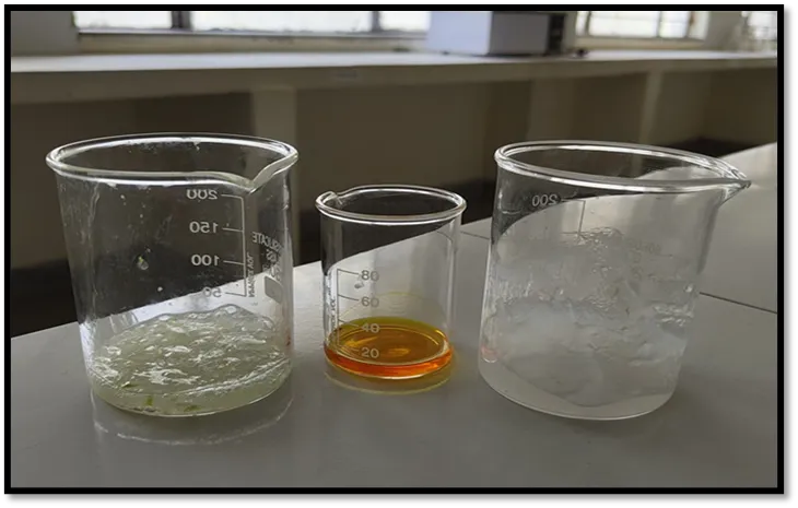
Fig1 : Preparation of components &polymer base for hydrogel patch

TABLE NO.1 - FORMULATION TABLE [20,21,22,23,24]


S.No


Ingredients


F1


F2


F3


Role


1


Chitosan


1.5g


2g


2.5g


Pollymmeric Matrix Forming Agent


2


Aloe vera extract


0.5g


1g


1.5g


Wound Healing, Moisturizing Agent


3


Curcuma longa

extract


0.25g


0.5g


0.75g


Anti-inflammatory


4


Acetic acid


40ml


50ml


60ml


Solvent for chitosan


5


Carbopol 934


0.1g


0.2g


0.3g


Gelling agent


6


Ammonium persulfate


0.1g


0.2g


0.3g


Initiator


7


Sodium hydrogen sulfite


0.1g


0.2g


0.3g


Co-initiator


8


Cross-linking agent


0.05g


0.1g


0.15g


Cross-linking agent


9


Distilled water


40ml


35ml


40ml


Vehicle


10


Water: Ethanol

(70:30)


q.s


q.s


q.s


Washing solution

Evaluation of Hydrogel Patches: -

  1. Physical Appearance: All the transdermal patches were visually inspected for colour, clarity, flexibility and smoothness [25].
  2. Thickness: The thickness of the film was measured at three different points using digital thickness gauge and the average thickness was calculated. The experiment was performed in triplicate (n=3) [26].
  3. Weight uniformity: in hydrogel patches refers to the consistency in mass across multiple patc hes obtained from the same batch. It is a critical quality parameter in transdermal and topical dru g delivery systems because it reflects the uniform distribution of the polymer matrix and any acti ve pharmaceutical ingredients (APIs) throughout the patch. Patches with poor weight uniformity may lead to inconsistent drug release, dosing errors, and variable therapeutic effects [27].
  4. Folding Endurance: For each formulation, three randomly selected patches were used. For weight variation test, 3 films from each batch were weighed individually and the average weight was calculated. Folding endurance of the film was determined by repeatedly folding a small strip of film (2cm x 2cm) at the same place till it broke. The number of times, the film could be folded at the same place without breaking, gave the value of folding endurance [28].
  5. Percentage Moisture Content: The prepared films were weighed individually and kept in a desiccator containing fused calcium chloride at room temperature for 24 hrs. After 24 hrs the films were reweighed and the percentage moisture content was determined by using the given formula. Percentage moisture content = initial weight − final weight final weight [29].
  6. Percentage Moisture: Uptake ×100 The weighed films were kept in a desiccator at room temperature for 24 hrs containing saturated solution of potassium chloride in order to maintain 84% RH. After 24 hrs the films were reweighed and the percentage moisture uptake was determined by using the given formula. Percentage Moisture Uptake = final weight − initial weight initial weight [30].
  7. Drug Content Uniformity: The uniformity of drug content of the transdermal film was determined, based on dry weight of drug and polymer used, by means of a UV/VIS spectrophotometer method. A specified area (2cm²) of patch was cut and dissolved in 10 ml of phosphate buffer pH 7.4. Then the solution was transferred in a volumetric flask and the volume made up to 10 ml. Appropriate dilutions were made using phosphate buffer pH 7.4, filtered and analysed for drug content at 290 nm by using UV spectrophotometer and determine the % drug content. Drug Content Uniformity = test absorbance standard absorbance [31].
  1. In-vitro Drug Permeation Study: In-vitro permeation studies were performed by using a modified Franz diffusion cell with a receptor compartment capacity of 25ml. The treated synthetic cellophane membrane was mounted between the donor and receptor compartment of the diffusion cell. The formulated patches were cut into the size of 4cm² and placed over the cellophane membrane and the receptor compartment of the diffusion cell was filled with pH 7.4 phosphate buffer.[32]

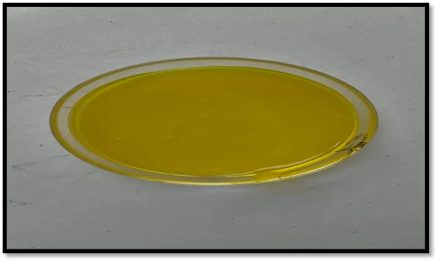
Fig 2 : Prepared herbal hydrogel patch containing aloe vera and curcuma longa.

TABLE NO.2 - RESULT DISCUSSION TABLE

EVALUATION PARAMETR


F1


F2


F3


Physical Appearance


Smooth &flexible


Smooth &flexible


Smooth, flexible&

transparent

Thickness

0.48mm

0.42mm

0.51mm

Weight uniformity

220mg

223mg

248mg

Folding endurance

240

258

285

Surface pH

6.5

6.5

6.8

Bio adhesive property

Good adhesion on skin surface

Good adhesion on skin surface

Good adhesion on skin surface

Skin irritation test

No irritation

No irritation

No irritation

RESULT

Three different formulations of herbal hydrogel patches containing Aloe vera and Curcuma longa were prepared and evaluated for their physicochemical properties and wound healing suitability. All formulations produced uniform hydrogel films with acceptable appearance and consistency. The prepared patches were smooth in texture, flexible in nature, and free from visible surface imperfections. Among the three formulations, F1 showed comparatively lower thickness and moderate flexibility, whereas F3 produced thicker patches with comparatively higher moisture absorption capacity. Formulation F2 exhibited balanced physical characteristics with satisfactory flexibility and uniform texture. The thickness values of the formulations ranged between 0.36 ± 0.02 mm and 0.51 ± 0.04 mm, while weight variation studies confirmed acceptable uniformity among the prepared patches. Folding endurance analysis demonstrated that all formulations possessed sufficient mechanical strength for handling and topical application. Moisture content and moisture uptake studies indicated that the hydrogel patches were capable of maintaining hydration, which is considered beneficial for wound healing applications. Surface pH values remained within the acceptable skin pH range, suggesting good dermatological compatibility. Drug content estimation confirmed uniform incorporation of the herbal constituents within the polymeric matrix. Swelling behavior was observed to increase gradually with higher polymer concentration, particularly in formulation F3. In-vitro permeation studies revealed prolonged release of active components from all formulations, with F2 showing a more controlled and sustained release profile compared to F1 and F3. No visible signs of irritation or discomfort were observed during skin compatibility testing. Based on the obtained evaluation results, formulation F2 was considered the most suitable formulation due to its balanced physicochemical and release characteristics.

DISCUSSION

The present investigation focused on the formulation and evaluation of three different batches of herbal hydrogel patches containing Aloe vera and Curcuma longa for wound healing applications. The prepared formulations, namely F1, F2, and F3, were successfully developed using the hydrogel polymerization method and evaluated for various physicochemical parameters. All prepared formulations exhibited satisfactory appearance, flexibility, and uniformity without visible cracks or air entrapment. Comparative evaluation revealed noticeable variations among the three batches due to differences in polymer and herbal extract concentrations. Formulation F1 showed comparatively lower thickness and swelling behavior, which may be attributed to the lower concentration of polymeric components. In contrast, formulation F3 demonstrated higher swelling capacity and moisture uptake due to increased polymer concentration, although the patch appeared comparatively thicker.  Among all formulations, F2 exhibited balanced physicochemical properties with appropriate thickness, satisfactory flexibility, acceptable moisture retention, and good mechanical strength. Folding endurance results confirmed that the prepared patches possessed adequate elasticity for topical application and handling. Drug content analysis indicated uniform distribution of herbal constituents throughout the hydrogel matrix in all formulations. The surface pH of each formulation remained close to normal skin pH, suggesting compatibility with topical administration and minimal risk of irritation. In-vitro permeation studies demonstrated sustained release of active constituents from the hydrogel system. Formulation F2 showed a more controlled and prolonged drug release profile compared to F1 and F3, which may improve therapeutic effectiveness and reduce the frequency of application. Overall, the findings suggest that formulation F2 possessed the most suitable characteristics for wound healing applications.

CONCLUSION

The present study successfully developed and evaluated herbal hydrogel patches containing Aloe vera and Curcuma longa for wound healing purposes. Three different formulations, F1, F2, and F3, were prepared and assessed for their physicochemical characteristics, mechanical properties, moisture behavior, drug content uniformity, and in-vitro drug release profile. All formulations demonstrated acceptable physical appearance, flexibility, and compatibility for topical application. Comparative evaluation indicated that variation in polymer and herbal extract concentration influenced the overall properties of the hydrogel patches. Formulation F1 exhibited lower swelling and drug release behavior, whereas formulation F3 showed comparatively higher swelling capacity and moisture absorption due to increased polymer concentration. Among all prepared formulations, F2 was found to be the optimized formulation because it exhibited balanced thickness, satisfactory folding endurance, appropriate moisture retention, uniform drug distribution, and sustained drug release characteristics. The formulation also maintained a skin-compatible pH and showed no visible signs of irritation, indicating good dermatological safety. The combined incorporation of Aloe vera and Curcuma longa contributed beneficial wound healing properties such as anti-inflammatory, antimicrobial, antioxidant, and tissue-regenerating activities. The hydrogel system further supported prolonged retention of moisture and controlled release of active constituents at the wound site.

Overall, the developed herbal hydrogel patch may serve as a promising and patient-friendly wound dressing system for effective wound management. Further pharmacological and clinical investigations may be performed in the future to establish its therapeutic potential on a larger scale.

REFERENCES

  1. Nejatzadeh-Barandozi, F., Naghavi, M. R., Hassani, M. E., Mostofi, Y., Mousavi, R. S., & Tahmasebi Enferadi, S. (2012). Diversity of Iranian Aloe vera genotypes based on aloenin contents and some morphological traits. The Journal of Horticultural Science and Biotechnology, 87(6), 673-677.
  2. Surjushe, A., Vasani, R., & Saple, D. G. (2008). Aloe vera: A short review. Indian Journal of Dermatology, 53(4), 163-166.
  3. Hamman, J. H. (2008). Composition and applications of Aloe vera leaf gel. Molecules, 13(8), 1599-1616.
  4. Eshun, K., & He, Q. (2004). Aloe vera: A valuable ingredient for the food, pharmaceutical and cosmetic industries. Critical Reviews in Food Science and Nutrition, 44(2), 91-96.
  5. Choi, S., & Chung, M. H. (2003). A review on the relationship between Aloe vera components and their biologic effects. Seminars in Integrative Medicine, 1(1), 53-62.
  6. Gupta, S. C., Patchva, S., & Aggarwal, B. B. (2013). Therapeutic roles of curcumin: Lessons learned from clinical trials. The AAPS Journal, 15(1), 195-218.
  7. Hewlings, S. J., & Kalman, D. S. (2017). Curcumin: A review of its effects on human health. Foods, 6(10), 92.
  8. Amalraj, A., Pius, A., Gopi, S., & Gopi, S. (2017). Biological activities of curcuminoids and related mechanisms of action: A review. Journal of Traditional and Complementary Medicine, 7(2), 205-233.
  9. Menon, V. P., & Sudheer, A. R. (2007). Antioxidant and anti-inflammatory properties of curcumin. Advances in Experimental Medicine and Biology, 595, 105-125.
  10.  Peppas, N. A., Bures, P., Leobandung, W., & Ichikawa, H. (2000). Hydrogels in pharmaceutical formulations. European Journal of Pharmaceutics and Biopharmaceutics, 50(1), 27-46.
  11. Mogosanu, G. D., & Grumezescu, A. M. (2014). Natural and synthetic polymers for wounds and burns dressing. International Journal of Pharmaceutics, 463(2), 127-136.
  12. Boateng, J. S., Matthews, K. H., Stevens, H. N., & Eccleston, G. M. (2008). Wound healing dressings and drug delivery systems: A review. Journal of Pharmaceutical Sciences, 97(8), 2892-2923.
  13. Peppas, N. A., Bures, P., Leobandung, W., & Ichikawa, H. (2000). Hydrogels in pharmaceutical formulations. European Journal of Pharmaceutics and Biopharmaceutics, 50(1), 27-46. https://doi.org/10.1016/S0939-6411(00)00090-4
  14. Dash, M., Chiellini, F., Ottenbrite, R. M., & Chiellini, E. (2011). Chitosan—A versatile semi-synthetic polymer in biomedical applications. Progress in Polymer Science, 36(8), 981-1014. https://doi.org/10.1016/j.progpolymsci.2011.02.001
  15. Hamman, J. H. (2010). Chitosan-based polyelectrolyte complexes as potential carrier materials in drug delivery systems. Marine Drugs, 8(4), 1305-1322. https://doi.org/10.3390/md8041305
  16. George, M., & Abraham, T. E. (2006). Polyionic hydrocolloids for the intestinal delivery of protein drugs: Alginate and chitosan—A review. Journal of Controlled Release, 114(1), 1-14. https://doi.org/10.1016/j.jconrel.2006.04.017
  17. Kokabi, M., Sirousazar, M., & Hassan, Z. M. (2007). PVA-clay nanocomposite hydrogels for wound dressing. European Polymer Journal, 43(3), 773-781. https://doi.org/10.1016/j.eurpolymj.2006.11.030
  18. Boateng, J. S., Matthews, K. H., Stevens, H. N., & Eccleston, G. M. (2008). Wound healing dressings and drug delivery systems: A review. Journal of Pharmaceutical Sciences, 97(8), 2892-2923. https://doi.org/10.1002/jps.21210
  19. Mogosanu, G. D., & Grumezescu, A. M. (2014). Natural and synthetic polymers for wounds and burns dressing. International Journal of Pharmaceutics, 463(2), 127-136.
  20. Bala, R., & Sharma, S. (2011). Formulation optimization and evaluation of transdermal patches: A review. International Journal of Pharmaceutical Sciences and Research, 2(12), 3056-3068.
  21. Saroha, K., Yadav, B., & Sharma, B. (2011). Transdermal patches: A successful tool in transdermal drug delivery system. Der Pharmacia Sinica, 2(5), 17-29.
  22. Franz, T. J. (1975). Percutaneous absorption: On the relevance of in vitro data. Journal of Investigative Dermatology, 64(3), 190-195. https://doi.org/10.1111/1523-1747.ep12533356
  23. Prausnitz, M. R., & Langer, R. (2008). Transdermal drug delivery. Nature Biotechnology, 26(11), 1261-1268. https://doi.org/10.1038/nbt.1504
  24. Barry, B. W. (2001). Novel mechanisms and devices to enable successful transdermal drug delivery. European Journal of Pharmaceutical Sciences, 14(2), 101-114. https://doi.org/10.1016/S0928-0987(01)00167-1
  25. Patel, D., Chaudhary, S. A., Parmar, B., & Bhura, N. (2012). Transdermal drug delivery system: A review. The Pharma Innovation Journal, 1(4), 66-75.
  26. Mishra, R. K., Ramasamy, K., & Ahmad, A. (2011). Herbal drug delivery system: An updated review. International Journal of Pharmaceutical Sciences and Research, 2(3), 363-371.
  27. Park, K. (2014). Controlled drug delivery systems: Past forward and future back. Journal of Controlled Release, 190, 3-8. https://doi.org/10.1016/j.jconrel.2014.03.054
  28. Patel, D., Chaudhary, S. A., Parmar, B., & Bhura, N. (2012). Transdermal drug delivery system: A review. The Pharma Innovation Journal, 1(4), 66-75.
  29. Bala, R., Pawar, P., Khanna, S., & Arora, S. (2013). Orally dissolving strips: A new approach to oral drug delivery system. International Journal of Pharmaceutical Investigation, 3(2), 67-76. https://doi.org/10.4103/2230-973X.114897
  30. Mishra, R., Amin, A., & Mishra, B. (2009). Development and characterization of matrix type transdermal patches of metoprolol tartrate. Acta Pharmaceutica, 59(4), 413-423. https://doi.org/10.2478/v10007-009-0033-8
  31. Arora, P., Mukherjee, B., & Singhal, P. (2002). Design, development, physicochemical and in vitro and in vivo evaluation of transdermal patches containing diclofenac dimethylammonium salt. Journal of Pharmaceutical Sciences, 91(9), 2076-2089. https://doi.org/10.1002/jps.10184
  32. Gupta, R., Mukherjee, B., & Tiwari, A. (2011). Development and evaluation of matrix-type transdermal patches of diclofenac sodium. International Journal of Pharmaceutical Sciences and Drug Research, 3(4), 300-305

Reference

  1. Nejatzadeh-Barandozi, F., Naghavi, M. R., Hassani, M. E., Mostofi, Y., Mousavi, R. S., & Tahmasebi Enferadi, S. (2012). Diversity of Iranian Aloe vera genotypes based on aloenin contents and some morphological traits. The Journal of Horticultural Science and Biotechnology, 87(6), 673-677.
  2. Surjushe, A., Vasani, R., & Saple, D. G. (2008). Aloe vera: A short review. Indian Journal of Dermatology, 53(4), 163-166.
  3. Hamman, J. H. (2008). Composition and applications of Aloe vera leaf gel. Molecules, 13(8), 1599-1616.
  4. Eshun, K., & He, Q. (2004). Aloe vera: A valuable ingredient for the food, pharmaceutical and cosmetic industries. Critical Reviews in Food Science and Nutrition, 44(2), 91-96.
  5. Choi, S., & Chung, M. H. (2003). A review on the relationship between Aloe vera components and their biologic effects. Seminars in Integrative Medicine, 1(1), 53-62.
  6. Gupta, S. C., Patchva, S., & Aggarwal, B. B. (2013). Therapeutic roles of curcumin: Lessons learned from clinical trials. The AAPS Journal, 15(1), 195-218.
  7. Hewlings, S. J., & Kalman, D. S. (2017). Curcumin: A review of its effects on human health. Foods, 6(10), 92.
  8. Amalraj, A., Pius, A., Gopi, S., & Gopi, S. (2017). Biological activities of curcuminoids and related mechanisms of action: A review. Journal of Traditional and Complementary Medicine, 7(2), 205-233.
  9. Menon, V. P., & Sudheer, A. R. (2007). Antioxidant and anti-inflammatory properties of curcumin. Advances in Experimental Medicine and Biology, 595, 105-125.
  10.  Peppas, N. A., Bures, P., Leobandung, W., & Ichikawa, H. (2000). Hydrogels in pharmaceutical formulations. European Journal of Pharmaceutics and Biopharmaceutics, 50(1), 27-46.
  11. Mogosanu, G. D., & Grumezescu, A. M. (2014). Natural and synthetic polymers for wounds and burns dressing. International Journal of Pharmaceutics, 463(2), 127-136.
  12. Boateng, J. S., Matthews, K. H., Stevens, H. N., & Eccleston, G. M. (2008). Wound healing dressings and drug delivery systems: A review. Journal of Pharmaceutical Sciences, 97(8), 2892-2923.
  13. Peppas, N. A., Bures, P., Leobandung, W., & Ichikawa, H. (2000). Hydrogels in pharmaceutical formulations. European Journal of Pharmaceutics and Biopharmaceutics, 50(1), 27-46. https://doi.org/10.1016/S0939-6411(00)00090-4
  14. Dash, M., Chiellini, F., Ottenbrite, R. M., & Chiellini, E. (2011). Chitosan—A versatile semi-synthetic polymer in biomedical applications. Progress in Polymer Science, 36(8), 981-1014. https://doi.org/10.1016/j.progpolymsci.2011.02.001
  15. Hamman, J. H. (2010). Chitosan-based polyelectrolyte complexes as potential carrier materials in drug delivery systems. Marine Drugs, 8(4), 1305-1322. https://doi.org/10.3390/md8041305
  16. George, M., & Abraham, T. E. (2006). Polyionic hydrocolloids for the intestinal delivery of protein drugs: Alginate and chitosan—A review. Journal of Controlled Release, 114(1), 1-14. https://doi.org/10.1016/j.jconrel.2006.04.017
  17. Kokabi, M., Sirousazar, M., & Hassan, Z. M. (2007). PVA-clay nanocomposite hydrogels for wound dressing. European Polymer Journal, 43(3), 773-781. https://doi.org/10.1016/j.eurpolymj.2006.11.030
  18. Boateng, J. S., Matthews, K. H., Stevens, H. N., & Eccleston, G. M. (2008). Wound healing dressings and drug delivery systems: A review. Journal of Pharmaceutical Sciences, 97(8), 2892-2923. https://doi.org/10.1002/jps.21210
  19. Mogosanu, G. D., & Grumezescu, A. M. (2014). Natural and synthetic polymers for wounds and burns dressing. International Journal of Pharmaceutics, 463(2), 127-136.
  20. Bala, R., & Sharma, S. (2011). Formulation optimization and evaluation of transdermal patches: A review. International Journal of Pharmaceutical Sciences and Research, 2(12), 3056-3068.
  21. Saroha, K., Yadav, B., & Sharma, B. (2011). Transdermal patches: A successful tool in transdermal drug delivery system. Der Pharmacia Sinica, 2(5), 17-29.
  22. Franz, T. J. (1975). Percutaneous absorption: On the relevance of in vitro data. Journal of Investigative Dermatology, 64(3), 190-195. https://doi.org/10.1111/1523-1747.ep12533356
  23. Prausnitz, M. R., & Langer, R. (2008). Transdermal drug delivery. Nature Biotechnology, 26(11), 1261-1268. https://doi.org/10.1038/nbt.1504
  24. Barry, B. W. (2001). Novel mechanisms and devices to enable successful transdermal drug delivery. European Journal of Pharmaceutical Sciences, 14(2), 101-114. https://doi.org/10.1016/S0928-0987(01)00167-1
  25. Patel, D., Chaudhary, S. A., Parmar, B., & Bhura, N. (2012). Transdermal drug delivery system: A review. The Pharma Innovation Journal, 1(4), 66-75.
  26. Mishra, R. K., Ramasamy, K., & Ahmad, A. (2011). Herbal drug delivery system: An updated review. International Journal of Pharmaceutical Sciences and Research, 2(3), 363-371.
  27. Park, K. (2014). Controlled drug delivery systems: Past forward and future back. Journal of Controlled Release, 190, 3-8. https://doi.org/10.1016/j.jconrel.2014.03.054
  28. Patel, D., Chaudhary, S. A., Parmar, B., & Bhura, N. (2012). Transdermal drug delivery system: A review. The Pharma Innovation Journal, 1(4), 66-75.
  29. Bala, R., Pawar, P., Khanna, S., & Arora, S. (2013). Orally dissolving strips: A new approach to oral drug delivery system. International Journal of Pharmaceutical Investigation, 3(2), 67-76. https://doi.org/10.4103/2230-973X.114897
  30. Mishra, R., Amin, A., & Mishra, B. (2009). Development and characterization of matrix type transdermal patches of metoprolol tartrate. Acta Pharmaceutica, 59(4), 413-423. https://doi.org/10.2478/v10007-009-0033-8
  31. Arora, P., Mukherjee, B., & Singhal, P. (2002). Design, development, physicochemical and in vitro and in vivo evaluation of transdermal patches containing diclofenac dimethylammonium salt. Journal of Pharmaceutical Sciences, 91(9), 2076-2089. https://doi.org/10.1002/jps.10184
  32. Gupta, R., Mukherjee, B., & Tiwari, A. (2011). Development and evaluation of matrix-type transdermal patches of diclofenac sodium. International Journal of Pharmaceutical Sciences and Drug Research, 3(4), 300-305

Photo
Neha Sodiyal
Corresponding author

Shree Dev Bhoomi Institute of Education Science and Technology, Pondha, Dehradun, Uttarakhand-248007

Photo
Prithvi raj
Co-author

Shree Dev Bhoomi Institute of Education Science and Technology, Pondha, Dehradun, Uttarakhand-248007

Photo
Shivanand Patil
Co-author

Shree Dev Bhoomi Institute of Education Science and Technology, Pondha, Dehradun, Uttarakhand-248007

Prithvi Raj, Neha Sodiyal*, Shivanand Patil, Formulation and Evaluation of Herbal Hydrogel Patch Containing Aloe vera and Curcuma longa for Wound Healing, Int. J. of Pharm. Sci., 2026, Vol 4, Issue 6, 4416-4425. https://doi.org/ 10.5281/zenodo.20736577

More related articles
A Review of Medicinal Plants with Antiulcer Activi...
Swapnali Darandale, Dr. R. B Pandhare, Priyanka Darandale...
Formulation and Evaluation of Herbal Tooth Powder...
Dr. Vijaykumar Kale, Vaibhav Narwade, Dr. Mahesh Thakare, Gauri B...
Formulation and Evaluation of Herbal Cough Syrup C...
Abdul Ansari, Abrar Khan, Ratan Pitale, Vishal Bhondave, Vaibhav...
Title of the Manuscript: Formulation and Evaluation of Herbal Mouthwash...
Pathan Kamran Ayubkhan, Dr. Ganesh Tolsadwad, Sunil Dongre, Sardar Parvin Mahamad, Mulla Samir Ismai...
Targeting Oxidative Stress and Inflammation in Nephrotoxicity: Evidence from Eup...
K. Saimahesh , Preethi S.M , likitha V , Niharika C.P , Keshava murthy S.G ...
Related Articles
Synthesis, Characterization and Biological Activity Prediction of Chalcone Deriv...
Padmavati Biradar, P.N.Sable, Rutuja Kulkarni , Tanushree Kadam , Senerita Kamble ...
Formulation and Assessment of Herbal Liquid Sindoor with Bixa Orellana...
Rajat Das, Jyochhana Priya Mohanty, Anushree Paul, Ashik Subba, Anwesh Pandey, Anushka Pradhan...
Formulation & Evaluation of Polyherbal Papaya Churna for Supportive Management o...
Varsharani Patil, Kedar Kale, Sanket Kamble, Mrunali Patil, Sakshi Jagtap, Kirti Andewad...
Comparative Study of Dossier Compilation & Submission Process of Drug Product in...
Sakshi Swami, Pratima Shinde, Anjali Dudhate, Dr. Kranti Satpute, Nirbhay Chalmale...
A Review of Medicinal Plants with Antiulcer Activity: Emphasis on Cordia obliqua...
Swapnali Darandale, Dr. R. B Pandhare, Priyanka Darandale...
More related articles
A Review of Medicinal Plants with Antiulcer Activity: Emphasis on Cordia obliqua...
Swapnali Darandale, Dr. R. B Pandhare, Priyanka Darandale...
Formulation and Evaluation of Herbal Tooth Powder...
Dr. Vijaykumar Kale, Vaibhav Narwade, Dr. Mahesh Thakare, Gauri Bhange, Pradip Raut...
Formulation and Evaluation of Herbal Cough Syrup Containing Natural Antitussive ...
Abdul Ansari, Abrar Khan, Ratan Pitale, Vishal Bhondave, Vaibhav Shinde, R. G Pimple, Dr. Ramesh In...