

We use cookies to ensure our website works properly and to personalise your experience. Cookies policy
Department of Pharmaceutics, Bharath institute of pharmacy college, Mangalpally, Hyderabad, Ranga reddy, Telangana - 501510, India.
The objective of the present study was to formulate and evaluate Mango leaf extract loaded phytosomes for improving the bioavailability and therapeutic potential of its bioactive constituents. Mango leaf extract, rich in mangiferin, flavonoids, and phenolic compounds, possesses significant antioxidant and antidiabetic properties; however, its clinical application is limited by poor absorption and low bioavailability. To address these challenges, phytosomes were developed using phosphatidylcholine through the solvent evaporation technique.The prepared formulations were characterized for physicochemical properties including particle size, entrapment efficiency, Fourier Transform Infrared Spectroscopy (FTIR), Scanning Electron Microscopy (SEM), in-vitro drug release, and stability studies. FTIR analysis confirmed the compatibility of the extract with formulation components and the successful formation of the phytosomal complex. SEM studies revealed the formation of discrete spherical vesicles with smooth surface morphology. The formulated phytosomes exhibited particle sizes in the nanometric range, with the optimized formulation (F4) showing a particle size of 244 nm and an entrapment efficiency of 85.26%.In-vitro release studies demonstrated a sustained drug release pattern, with the optimized formulation releasing 98.88% of the entrapped extract over a period of 8 hours. Stability studies performed under accelerated and room-temperature storage conditions indicated that the optimized formulation maintained its physicochemical characteristics and drug release behavior throughout the study period. The enhanced entrapment efficiency, nanoscale particle size, and controlled release profile suggest improved delivery of the phytoconstituents present in Mango leaf extract.
PHYTOSOMES
Phytoconstituents derived from medicinal plants have been used for centuries in the prevention and treatment of various diseases. Herbal medicines contain biologically active constituents such as flavonoids, polyphenols, alkaloids, terpenoids, tannins, glycosides and other phenolic compounds that contribute to their therapeutic effects. Despite their significant pharmacological potential, many plant-derived compounds exhibit limitations such as poor aqueous solubility, low lipid solubility, instability, poor absorption and limited bioavailability. These drawbacks often restrict their therapeutic effectiveness and necessitate the development of suitable drug delivery approaches to improve the performance of herbal bioactive compounds.Phospholipids play a major role in the formation of phytosomes because they possess both hydrophilic and lipophilic portions. The polar groups of the phospholipid interact with the polar functional groups of the phytoconstituent through hydrogen bonding and other intermolecular interactions, while the lipid portion provides improved compatibility with biological membranes. As a result, the phytoconstituent becomes more efficiently incorporated into the lipid environment, producing a complex with enhanced membrane permeability and stability.
The major advantage of phytosomal technology is its ability to improve the bioavailability of poorly absorbed herbal constituents. Many flavonoids and polyphenolic compounds show considerable pharmacological activity but are poorly absorbed following oral administration because of their inadequate lipid solubility and rapid metabolism. Conversion of these compounds into phytosomal complexes can improve their physicochemical properties, intestinal absorption and overall systemic availability. Therefore, phytosomes are considered a promising approach for improving the therapeutic efficacy of herbal drugs. Phytosomes may also provide improved protection of sensitive phytoconstituents against environmental and physiological degradation. The association of the active constituent with phospholipids can enhance its stability and facilitate better transport across biological membranes. In addition, phytosomal formulations may exhibit improved distribution of the active constituent into tissues, thereby increasing the possibility of achieving the desired therapeutic response at lower or more effective doses.
The preparation of phytosomes can be carried out using different techniques depending on the nature of the extract, phospholipid and selected solvent system. Commonly employed methods include solvent evaporation, anti-solvent precipitation, thin-film hydration and mechanical dispersion techniques. Among these, solvent evaporation is frequently used because it is relatively simple and suitable for preparing phospholipid–phytoconstituent complexes. The prepared phytosomes are subsequently characterized using various physicochemical and pharmaceutical evaluation techniques.
MATERIALS
Mango Leaf Extract was procured from Synpharma Research Labs, Hyd, Soya lecithin, Dichloromethane, Cholesterol and Ethanol were obtained from Synpharma Research Labs, Hyderabad. Other chemicals and the reagents used were of analytical grade.
METHODOLOGY
FTIR Studies
Drug polymer interactions were studied by FT-IR spectroscopy. One to 2mg of extract, polymer and physical mixtures of samples were weighed and mixed properly to a uniform mixture. A small quantity of the powder was compressed into a thin semi-transparent pellet by applying pressure. The IR spectrum of the pellet from 400-4000cm1 was recorded taking air as the reference and compared to study any interference.
Preparation of Mango Leaf Extract Leaf for Extraction
The fresh leaves were cleaned out first with tape water to remove dusts and dirt then washed with distilled water twice. The cleaned leaves were dried in oven at 45 C for 3 to 5 days to get a constant weight dry mass. The dry leaves then powdered using grinding mill to a particle size of about 0.5 mm. The powdered dried leaves then packed in polyethylene bag and stored in sealed container at dark cool room until use.
Extraction of Dried Powdered Mango Leaf Extract Leaf:
The dried powdered leaves were subjected to extraction using ethanol as solvent by Soxhlet extraction method. About 50 g of powdered material was packed in a Soxhlet apparatus and extracted with 70% ethanol for 6–8 hours until complete extraction was achieved. The obtained extract was filtered and concentrated using a rotary evaporator or water bath at controlled temperature below 50°C to remove the solvent. The concentrated extract was then dried to obtain a semisolid or dry mass and stored in an airtight container under refrigerated conditions until further use for Phytosomes preparation.
FORMULATION DEVELOPMENT
Table 1: Formulation Development of Phytosomes
| Ingredients | F1 | F2 | F3 | F4 |
|---|---|---|---|---|
| Extract (mg) | 100 | 100 | 100 | 100 |
| Soya lecithin(mg) | 100 | 200 | 300 | 400 |
| Cholesterol (mg) | 50 | 50 | 50 | 50 |
| Dichloromethane(ml) | 10 | 10 | 10 | 10 |
| Ethanol(ml) | 5 | 5 | 5 | 5 |
Preparation of Phytosomes - Dissolve the weighed amount of phosphatidylcholine in Dichloromethane in a round-bottom flask. Heat gently (30–40 °C) if needed to solubilize. Dissolve the calculated amount of mango leaf extract in ethanol and add to the PC solution. For improved complexation, add the extract slowly while stirring. Optionally add a small amount of cholesterol to improve membrane rigidity. Attach the flask to a rotary evaporator and evaporate the solvent under reduced pressure at 40–45 °C to form a thin lipid film on the flask wall. Keep the flask under vacuum for 30–60 min to remove residual solvent. Hydrate the thin film with phosphate buffer pH 7.4 at room temperature with gentle rotation to yield a suspension. Sonicate the suspension (bath sonicator) in an ice bath for 5–10 min (pulse mode) to reduce particle size and obtain uniform vesicles. Store the phytosomal suspension at 4 °C for further characterization.
CHARACTERIZATION
Particle Size
The particle size of the Phytosomes was determined using Particle Size Analyzer (PSA) with the dynamic light scattering (DLS) method. The measurements were performed using Horiba Scientific SZ-100, with the sample diluted 10 times in aqueous medium at room temperature.
Zeta-Potential:
The sample was diluted with distilled water (1:100 (V/V)) and zeta potential was determined using Malvern zetasizer (Nano ZS, Malvern Instruments, United Kingdom). Measurement was based on the electrophoretic mobility of the particles, which was converted to the zeta potential by inbuilt software based on the Helmholtz-Smoluchowski equation.
SEM Analysis
The shape, surface characteristics, and size of the phytosomes were observed by scanning electron microscopy. Once again, 0.2 g of the phytosomes in a glass tube was diluted with 10 ml of pH 7.4 phosphate buffer. The phytosomes were mounted on an aluminium stub using double-sided adhesive carbon tape. Then the vesicles were sputter-coated with gold palladium (Au/Pd) using a vacuum evaporator (Edwards) and examined using a scanning electron microscope (Hitachi 3700N, Germany) equipped with a digital camera, at 10 kV accelerating voltage.
Yield of Phytosomes
The practical yield of phytosomes refers to the percentage of product actually obtained after preparation compared to the total theoretical amount of drug and phospholipid used in the formulation process.
It is calculated using the following formula:
Practical Yield (%)=Weight of dried phytosomes obtained/Total weight of drug + phospholipid taken×100
In Vitro Diffusion Profile
In vitro release study of the formulated phytosomes was carried out by using diffusion cell through membrane as a dialysis membrane. Diffusion cell with inner diameter 24mm was used for the study. 1 mL formulation was placed in donor compartment and freshly prepared pH 7.4 phosphate buffer was placed in receptor compartment. Dialysis membrane was mounted in between donor and receptor compartment. The position of the donor compartment was adjusted so that the membrane just touches the diffusion medium. The whole assembly was placed on the thermostatically controlled magnetic stirrer. The temperature of the medium was maintained at 37°C ± 0.5°C. 1mL of sample was withdrawn from receiver compartment after 12 hrs and same volume of fresh medium was replaced. The withdrawn samples were diluted to 10mL in a volumetric flask with phosphate buffer and analysed by UV spectrophotometer.
Stability Studies
The main objective of the stability testing is to provide evidence on how the quality of the drug product varies with time under the influence of temperature and humidity. The stability study for the phytosomes formulation was done as per ICH guidelines in a stability chamber for a period of 3 months.
RESULTS & DISCUSSION
FT-IR Spectrum of Mango leaf extract
FT-IR Spectra of Mango leaf extract and polymers were recorded. All these peaks have appeared in formulation and physical mixture, indicating no chemical interaction between Mango leaf extract and polymers. It also confirmed that the stability of drug during process.
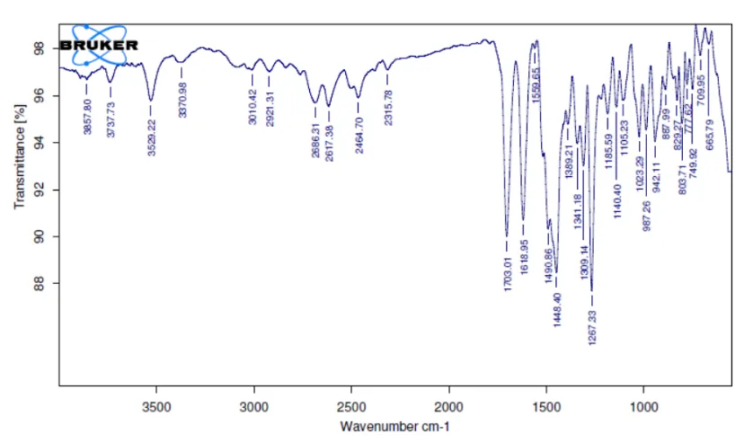
Fig-: 1 FT-IR Sample for Mango Leaf Extract
Table-: 2 Characteristic Peaks and frequency of Mango Leaf Extract
| S. No. | Characteristic Peaks | Frequency range (cm-1) |
Frequency (cm-1) |
|---|---|---|---|
| 1 | OH stretching | 3500-3200 | 3410 |
| 2 | OH Bending | 1440-1260 | 1384 |
| 3 | C-H stretching | 3000-2850 | 2924 |
| 4 | C-N stretching | 1360-1250 | 1280ss |
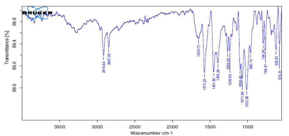
Fig-:2 FT-IR Sample for Optimized Formulation
Table-:3 Characteristic Peaks and Frequency of Optimized Formulation
| S.No. | Characteristic Peaks | Frequency range (cm-1) |
Frequency (cm-1) |
|---|---|---|---|
| 1 | OH stretching | 2916-2847 | 3000 |
| 2 | OH Bending | 1440-1260 | 1380 |
| 3 | C-N stretching | 1360-1250 | 1278 |
Evaluation Parameters:
Determination of Vesicle Morphology and Size
The morphological characteristics of formulated phytosomes were carried by using Scanning electron microscopy (SEM). A small drop of Phytosomal suspension was placed between two rivets fixed on a gold plated copper sample holder. The whole system was slushed under vacuum in liquid nitrogen. The sample was heated to -850C for 30 min to sublime the surface moisture. Finally the sample was coated with gold and allowed the SEM to capture the images at a temperature of - 1200c and voltage of 5kV.
Particle Size Analysis
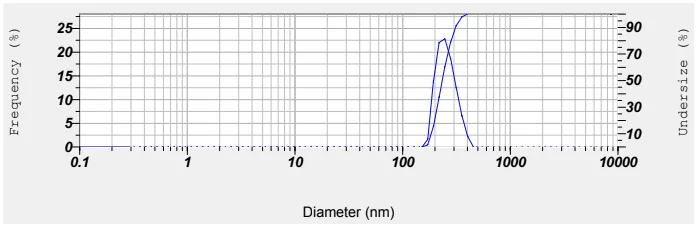
Figure 3: Particle Size Analysis of Phytosomes
DISCUSSION:
SEM analysis demonstrated spherical vesicles with smooth morphology. The particle size analysis revealed vesicles in the range of 244–295 nm, which is optimal for dermal delivery and ensures uniform drug distribution. Among the formulations, F4 showed the smallest particle size (244 nm), which is expected to favor better permeation and stability.
SEM Analysis
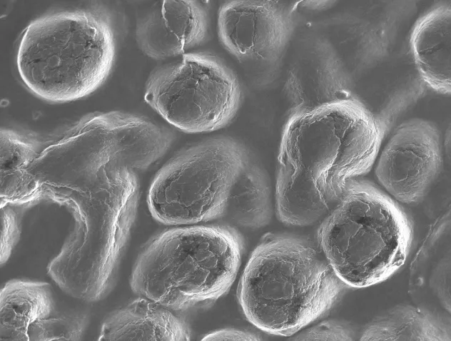
Figure 4: SEM Analysis of Phytosomes
Zeta Potential
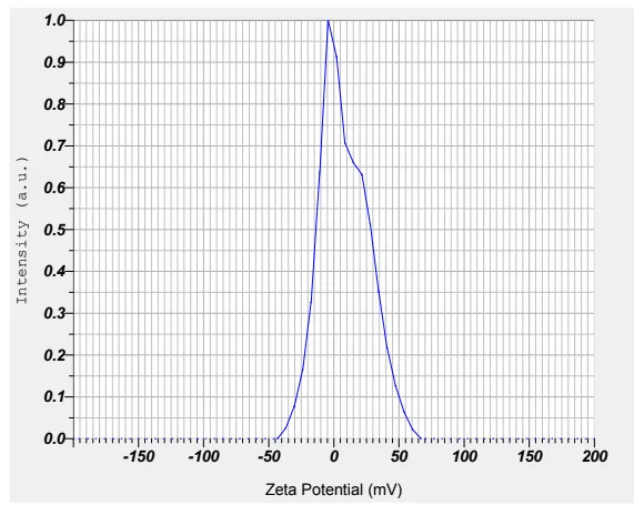
Figure 5: Zeta Potential of Phytosomes
DISCUSSION:
The zeta potential values ranged between –20 to –29 mV, confirming moderate stability of phytosomal dispersions due to sufficient electrostatic repulsion. F3 (–29 mV) and F4 (–25 mV) showed the most stable vesicles, reducing the chances of aggregation during storage.
Table 4: Evaluation Studies of Particle Size and Zeta Potential Phytosomes
| F. No | Particle size (nm) | Zeta potential(mV) |
|---|---|---|
| F1 | 258 | -20 |
| F2 | 261 | -23 |
| F3 | 278 | -29 |
| F4 | 244 | -25 |
Entrapment Efficiency:
Table 5: Drug Entrapment Efficiency
| F. code | Drug entrapment efficiency |
|---|---|
| F1 | 77.96 |
| F2 | 75.50 |
| F3 | 81.21 |
| F4 | 85.26 |
[Figure - see the formatted PDF]
Figure 6: Drug Entrapment Efficiency
Drug entrapment efficiency varied between 77.50% to 85.26%, with F4 showing the highest efficiency. Higher entrapment efficiency ensures maximum therapeutic benefit with controlled release.
In Vitro Release Study:
Phosphate buffer pH 7.4 was used as medium for the release studies and good linearity was observed in the plotted standard graph with a correlation coefficient of 0.998. The drug release profiles of Phytosomes containing different ratios of synthetic polymer. It was cleared from the release profiles of formulations, that the drug release was governed by polymer nature and content.
Table 6: In Vitro Drug Release Profiles of Phytosomal Gel (F1-F4)
| Time (hr) | F1 | F2 | F3 | F4 |
|---|---|---|---|---|
| 0 | 0 | 0 | 0 | 0 |
| 1 | 27.82 | 28.10 | 24.97 | 28.15 |
| 2 | 37.89 | 38.96 | 35.16 | 37.48 |
| 3 | 46.98 | 48.82 | 49.87 | 53.69 |
| 4 | 76.28 | 77.81 | 75.98 | 79.52 |
| 6 | 82.30 | 86.39 | 82.10 | 89.35 |
| 8 | 92.35 | 93.26 | 96.37 | 98.88 |
[Figure - see the formatted PDF]
Figure 7: In Vitro Drug Release Studies of F1-F4 Formulations
Drug release profiles showed sustained release over 8 hours, with maximum release of 98.88% observed for F4. The release was dependent on the polymer composition, with optimized ratios providing controlled and prolonged drug delivery.
Stability Studies :
Table 7: Results of Stability Studies of Optimized Formulation F-4
| Parameters | Initial | 1st Month | 2nd Month | 3rd Month | Limits as per Specifications | |
|---|---|---|---|---|---|---|
| F-4 | 250C/60%RH % Release |
98.88 | 97.85 | 95.58 | 94.37 | Not less than 85 % |
| F-4 | 300C/75% RH % Release |
98.88 | 97.52 | 95.26 | 94.22 | Not less than 85 % |
| F-4 | 400C/75% RH % Release |
98.88 | 97.43 | 94.13 | 94.32 | Not less than 85 % |
SUMMARY AND CONCLUSION
The present study successfully formulated and evaluated a phytosomes containing mango leaf extract with the aim of enhancing its bioavailability therapeutic efficacy. The phytosomes were prepared using phosphatidylcholine, demonstrating good entrapment efficiency, optimal particle size, and stable zeta potential, indicating a well-established complex between the Mango leaf extract and phospholipids. The present study focuses on the formulation and evaluation of Mango leaf extract loaded phytosomes to enhance its therapeutic efficacy and bioavailability.
Characterization through FTIR confirmed successful phytosomes formation. Phytosomes of Mango leaf extract were prepared using the Solvent evaporation method with phosphatidylcholine as a carrier, forming stable Phyto complexes. SEM analysis demonstrated spherical vesicles with smooth morphology. Among the formulations, F4 showed the smallest particle size (244 nm), which is expected to favor better permeation and stability. In vitro drug release studies revealed a sustained release profile.
Drug entrapment efficiency varied between 75.50% to 85.26%, with F4 showing the highest efficiency. Higher entrapment efficiency ensures maximum therapeutic benefit with controlled release. Drug release profiles showed sustained release over 8 hours, with maximum release of 98.88% observed for F4. The release was dependent on the polymer composition, with optimized ratios providing controlled and prolonged drug delivery. Stability testing at different storage conditions (25 °C/60% RH, 30 °C/75% RH, and 40 °C/75% RH) over 3 months demonstrated no significant reduction in drug release (remaining above 94%). This indicates that the optimized phytosomes is physically and chemically stable under accelerated storage conditions. Overall, the Mango leaf extract phytosomes offers a promising natural formulation for topical application, potentially providing enhanced bioavailability therapeutic efficacy and neuroprotective activity.
REFERENCES

P. V. Pavan Kumar, B. Saikiran, A. Pavan Kumar, B. Harika, Formulation and Evaluation of Mango Leaf Extract Loaded Phytosomes for Enhanced Antioxidant and Antidiabetic Activity, Int. J. of Pharm. Sci., 2026, Vol 4, Issue 11, 1357-1366. https://doi.org/10.5281/zenodo.24003481
 10.5281/zenodo.24003481