

We use cookies to ensure our website works properly and to personalise your experience. Cookies policy
1B. Pharmacy, School of pharmaceutical Science, Vels institute of science technology and advanced studies, Chennai, Tamil Nadu, India.
*2Department of Pharmacology, School of pharmaceutical Science, Vels institute of science technology and advanced studies, Chennai, Tamil Nadu India.
Vitiligo is the name given to an auto immune skin disease marked by presence of white colored spots on the skin as a result of loss of melanocytes. Prevalence of vitiligo among the world population is about 1-2%. Different kinds of therapeutic interventions for the disease include topical corticosteroids, phototherapy, laser and graft surgeries. However, each of the aforementioned methods of treatment has certain side effects and does not always produce positive results. Hence, it is vital to develop an alternative therapy based on plant material. The aim of the current research is to develop quercetin and curcumin loaded soap formulations for stimulation of melanogenesis as a therapy for vitiligo. Quercetin and curcumin known for their antioxidant, anti-inflammatory and immunomodulatory properties were formulated in glycerine soap formulation using optimization technique by trial and error. Optimized formulation (F3) was evaluated for cytotoxicity and mitochondria membrane potential in 3t3-l1 fibroblast cell line using MTT and Rhodamine-123 assays. Results show that the soap formulation does not possess cytotoxicity at 100 µg/ml concentrations and there were changes in mitochondria membrane potential. In other words, the changes in cellular activity in terms of melanogenesis mechanisms were identified. The results prove that the developed quercetin and curcumin loaded soap formulation is safe for topical application and could serve as an alternative therapy for vitiligo. Further in vivo experiments are required.
The skin disease known as vitiligo is idiopathic by nature. The condition is characterized by white appearance of skin due to macules which do not contain any melanocytes. Information regarding the existence of vitiligo can be found in Vedic religious literature which predates Hinduism as well as in the Egyptian documents dating back more than 3000 years ago. Phototherapy in order to treat vitiligo was practiced in ancient Egypt and ancient India [1]. Descriptions of disease which resembled vitiligo can be traced to 1.500 BC in the Hindu religious literature [2]. Vitiligo was mentioned by Aulus Cornelius Celsus in his medical writings under the name De Medicina. According to experts, the name vitiligo is originated from the word "Vitium" which means defect or blemish [3]. In Ebers's papyrus written in 1500 BC two skin pigmentation diseases were described, one of which is similar to leprosy and other to vitiligo [4]. In India's Athar Veda the disease was known as "Kilas" meaning white disease [5].
Vitiligo: Automatic vitiligo refers to the chronic immune-related condition resulting in creation of depigmented skin areas. The attack of the immune system on skin cells called melanocytes results in their destruction causing whitening of skin [6]. The disease progresses with time and eventually pigmented cells become disappeared from progressively larger areas of skin. The skin lesions from vitiligo might remain unchanged or might heal in some cases [8]. The first symptom of vitiligo is the occurrence of small pale skin spots which accompany further development of the disease. New skin spots from vitiligo appear close to the old ones or far away from them on the skin [9][10].
Symptoms: The skin disorder begins with improper pigmentation of the skin on areas of hands, exposed face and genitalia. Early graying of hair occurs not only in the scalp but also in the eyebrows, eyelashes and all facial hair [7]. Moreover, the face, wrists, elbows, knees, hands, ankles, etc.… are the vulnerable areas for vitiligo. The skin affected by vitiligo becomes very sensitive to sunburns. The hair follicles affected by vitiligo become white due to disappearance of melanocytes from this area [11][12].
Causes: One out of ten people having vitiligo has the family history of this skin disease. Occasionally, vitiligo becomes worse due to skin injury or conditions which are related to emotional or stressful factors [13]. The condition of vitiligo results in development of various autoimmune diseases such as Addison disease, thyroid disease, pernicious anemia and type 1 diabetes [14].
Epidemiology: Vitiligo affects both male and female population equally although women more often have this skin condition than men and over 60 percent of vitiligo patients detect their symptoms before the age of 30 [15]. Prevalence of vitiligo among population is equal to 0.5 percent to 1 percent although expert research demonstrates that this disease may affect 1.5 percent of people [16]. Worldwide prevalence of vitiligo incidence is 1-2%. Thirty percent of patients having vitiligo have their condition together with their family members who have the same skin problem [17]. Various studies confirm that the vitiligo affects people similarly regardless of their age [18].
Types: There are two types of vitiligo like generalized vitiligo and segmental vitiligo [19]. The disease of vitiligo can occur all over the body. Universal vitiligo indicates involvement of the disease all over the body [20].
Non-segmental vitiligo can also be called as bilateral or generalized vitiligo because it causes formation of white spots in symmetric patches all over the body. Segmental vitiligo is also known as unilateral or localized vitiligo as it causes white spots in only one part of your body. Segmental vitiligo is less frequent than non-segmental vitiligo but affects children more frequently [20].
Therapy: There are various treatments including medicines and therapies like phototherapy, laser therapy and surgery. The most common therapeutic treatments include topical corticosteroids, topical calcineurin inhibitors and phototherapy [17].
Topical Medications: The use of corticosteroid creams which are anti-inflammatory drugs has the properties to prevent vitiligo from progression and activating melanocytes. The patients may suffer some side effects of skin thinning, formation of stretch marks and acne formation [21].
Phototherapy: Phototherapy works effectively when combined with other treatment for the therapy of vitiligo. The process of phototherapy involves exposure of skin to UV A or B rays from specific lamps. Psoralen medicine has to be taken before undergoing phototherapy. PUVA is one of the techniques of phototherapy treatment. Phototherapy treatment increases the risk of skin cancer [22].
Laser Therapy: It is the very efficient method of treatment which ensures the rapid and quick procedure in safe ways. It is very effective in the treatment of the cases when there is a positive reaction to Koebner in the patient with vitiligo [23].
Surgical Therapy: Skin grafting surgery is the procedure in which the healthy skin is taken from another area of the body as a cover for damaged and lost tissues. It is used for treating the vitiligo with the help of covering up the white spots on the skin [22], [23].
Drug profile
Quercetin
Quercetin is found in various fruits, vegetables, seeds, and leaves; hence, capers, red onions, and kale have a high quantity of quercetin in them. It has a bitter taste and functions as a food additive [24], [25]. It is an antioxidant substance [26]. Its application in children is not fully justified for prescription as the remedy [28].
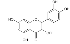
Figure 1: Structure of Quercetin
Properties
Table 1: Quercetin Profile [26], [27]
| Property name | Property value |
|---|---|
| Molecular formula | C15h10o7 |
| Color | Bright yellow yellow color |
| Taste | Bitter teste |
| Odour | Characteristic rubbery odour |
| Appearance | Powdered needle yellow needle |
| Molecular weight | 302.23g/mol |
| Boiling point | Not determined |
| Melting point | 601 – 603of |
| Iupac name | 2-(3,4-dihydroxyphenyl)-3,5,7- trihydroxychromen-4-one |
Curcumin
Polyphenolic groups present in curcumin make it a diarylheptanoid of the curcuminoid pigment class [29][30]. Diferuloylmethane is the chemical structure of curcumin which is an active compound of the yellow spice turmeric. It was aimed to analyze the use of curcumin as a medicinal drug that can be used for the treatment of different disorders like mucositis, rectal cancer, prostate cancer, chronic schizophrenia, and mild cognitive impairment [32].
Figure 2: Chemical Structure of Curcumin-Keto Group Figure 3: Chemical Structure of Curcumin-Enol Group
Properties
Table 2: Profile of Curcumin [31]
| Property name | Property value |
|---|---|
| Molecular formula | C12h20o6 |
| Color | Orange yellow natural yellow |
| Taste | Somewhat bitter spicy earthy flavor |
| Odour | Pungent aroma distinct earthy |
| Appearance | Bright yellow to yellow orange crystalline powder |
| Molecular weight | 368.4 g/mol |
| Boiling point | 591.40c |
| Melting point | 1830c |
| Iupac name | (1e,6e)-1,7-bis(4-hydroxy-3-methoxyphenyl)hepta-1,6-diene-3,5-dione |
MATERIALS AND METHODS
Chemicals: Quercetin, curcumin, ethanol, soap base, bees wax, coconut oil, lavender oil, 3T3-l1, Dulbecco's modified eagle medium, 10% (v/v) heat inactivated fetal bovine serum, 100 u/ml penicillin, 100 μg/ml streptomycin, rhodamine 123 (10 μg/ml), cationic fluorescent dye, HCL, NaCl (5%), methylene blue reagent, chloroform, ether.
Formulation procedure: The experiment involved formulation of an optimal soap by using trial-and-error technique by using quercetin as the active component. Quercetin and curcumin were dissolved in the desired quantity of ethanol. The mixture was stirred thoroughly till it dissolves. Glycerin soap base was cut into pieces. Double boiling technique was employed to melt the soap base. Temperature of the soap base should be maintained in between 60-70 degree centigrade. Melted soap base was mixed with the quercetin and curcumin solution. Coconut oil, lavender oil and bees wax (previously melted) were added to the soap base mixture. The mixture was stirred and poured into the soap molds. Air bubbles were eliminated from the soap molds by tapping the mold gently.
Formula composition table (Table 3)
| Ingredients | F1 | F2 | F3 | F4 |
|---|---|---|---|---|
| Quercetin | 0.5g | 0.5g | 1g | 1g |
| Curcumin | 0.25g | 0.25g | 0.5g | 0.5g |
| Ethanol | 2.5ml | 5ml | 2.5ml | 5ml |
| Coconut oil | 7.5ml | 75ml | 7.5ml | 7.5ml |
| Lavender oil | 2.5ml | 2.5ml | 2.5ml | 2.5ml |
| Bees wax | 5g | 5g | 5g | 5g |
| Soap base | 31.75g | 29.25g | 31g | 28.5g |
In vitro cell line studies
Cell culture: 3T3-l1 fibroblast cell lines were supplied by National Center for Cell Science, Pune, India. Cells were cultured in DMEM media containing 10% FBS and 1% antibiotic solution. Culturing was done in a humidified incubator maintained at 37°c with 5% CO2 and 95% air. Cultures media were replenished every other day.
Determination of cell viability: The effect of the toxicity of the formulation was determined by its effect on the fibroblast cell lines. 1 x 10⁵ cells per well fibroblasts were seeded in 96-well microplates and cultured until confluency of 70-80% at 37°c in 5% CO2 atmosphere. Media was changed and cells were exposed to different concentrations (25, 50, 75 and 100 μg/ml) of quercetin soap formulation for 24 hours. Morphological changes in untreated and treated cells were observed using inverted microscope. Cells were washed with PBS and 20 μl of MTT (5 mg/ml in PBS) was added per well. Incubation was done at 37°c for 3 hours. Dissolution of formazan crystals in 100 μl of DMSO and absorbance was measured at 570 nm. The formazan formation in MTT assay is directly proportional to the mitochondrial activity and therefore, cell viability.
Cell viability (%) = (Absorbance of sample/Absorbance of control) x 100 [33]
Analysis of mitochondrial membrane potential: Changes in levels of MMP (Δψm) can be used as the indicator of apoptosis, mitochondrial dysfunction and health of the cells. Rhodamine 123, a cationic fluorochrome dye, can be used to determine the level of Δψm. Cells were exposed to the IC50 concentration of soap formulation and after being washed with PBS, stained with 50 μl of rhodamine 123 (10 μg/ml) for 30 minutes. Morphological changes and membrane permeability of MMP were observed with fluorescent microscope at 40x magnification.
RESULT
Evaluation of analysis: Redness, irritation, and possibly a rash are just a few symptoms that may be caused by skin irritation. Some of the effects include blisters, dry or cracked skin, and even leather-like and scaly skin. In some cases, swelling and inflammation can also be caused by skin irritation, which is brought about by the immune response of the body.
Quercetin soap evaluation
Table 4: Evaluation of Formulation Report
| Parameter | F1 | F2 | F3 | F4 |
|---|---|---|---|---|
| Ph | 9.00 | 9.08 | 9.62 | 9.21 |
| Moisture (%) | 19.46 | 18.95 | 19.76 | 19.76 |
| Total fatty matter (%) | 46.25 | 44.95 | 46.75 | 46.75 |
| Lather (MI) | 260 | 260 | 260 | 260 |
| Free caustic alkali NaOH and carbonated alkali (%) | BLQ (LOQ:0.1) |
BLQ (LOQ:0.1) |
BLQ (LOQ:0.1) |
BLQ (LOQ:0.1) |
| Freedom from grittiness | Passes | Passes | Passes | Passes |
| Mush (g/50 cm2) | 9.52 | 9.52 | 9.52 | 9.52 |
| Freedom from crack | Passes | Passes | Passes | Passes |
| Cleaning efficiency | Passes | Passes | Passes | Passes |
| Synthetic surface-active agent (%) | 3.22 | 3.22 | 3.22 | 3.22 |
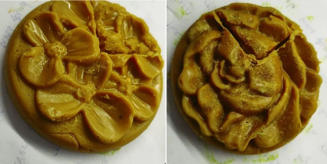
Figure 4: Visual Illustration of Quercetin Soap
Cytotoxicity evaluation: Cytotoxicity was seen on treated sample concentrations in 24 hours in the fibroblast cell line and it was further seen that an increase in the concentration of the drug increases the cytotoxicity effect on the fibroblast cell lines in comparison with the control sample. In MTT assay, formazan formation is directly proportional to viable cells.
Table 5: In-vitro Cytotoxicity Effect of Soap Formulation (F3) Against Fibroblast Cell Line (3T3-L1)
| Concentrations (µg/ml) | Absorbance | Average | Cell viability (%) | |
|---|---|---|---|---|
| I | II | |||
| Control | 0.912 | 0.922 | 0.917 | 100 |
| 25 | 0.898 | 0.893 | 0.8955 | 97.65539804 |
| 50 | 0.844 | 0.837 | 0.8405 | 91.65757906 |
| 75 | 0.796 | 0.802 | 0.799 | 87.13195202 |
| 100 | 0.757 | 0.763 | 0.76 | 82.87895311 |
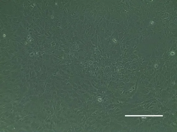
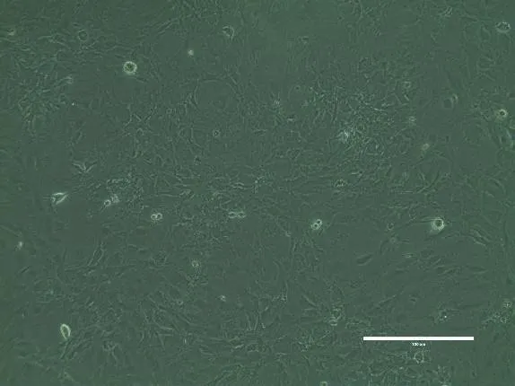
Figure 5: Control Fibroblast Cell Figure 6: Treated Cell-Dose 25Μgm
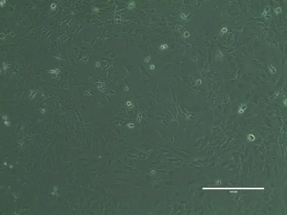
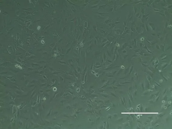
Figure 7: Treated Cell-Dose 50Μgm Figure 8: Treated Cell-Dose 75Μgm
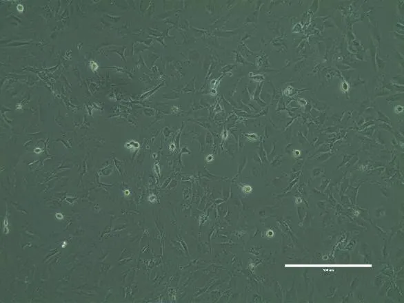
Figure 9: Treated Cell-Dose 100Μgm
Analysis of mitochondrial membrane potential: Two doses of quercetin soap formulation (F3) were used to analyze their effects on the mitochondrial membrane potential in fibroblast cell lines using rhodamine-123. The treatment caused reduced fluorochrome uptake, indicating mitochondrial membrane disruption and dysfunction, leading irreversibly to apoptosis.
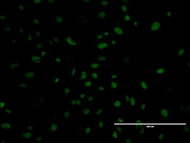
Figure 10: Control Fibroblast
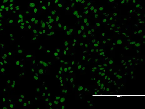
Figure 11: 250Μl of F-3
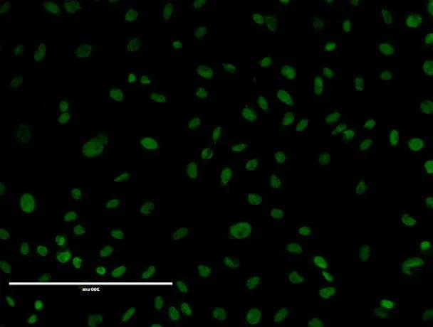
Figure 12: 500 Μl of F-3
The current study is devoted to investigating the effectiveness of quercetin-loaded soap formulation in stimulation of melanogenesis process; hence it may be considered as possible treatment of vitiligo. This study is carried out with the use of biomarkers and fibroblast cell culture technique for assessing the cellular and molecular responses to the quercetin using 3T3-l1 (normal fibroblast cell line). F3-induced mitochondrial dysfunction in 3T3-l1 cells was analyzed using lipophilic dye, Rhodamine-123. Increased generation of ROS induces alteration of the mitochondrial membrane permeability leading to mitochondria dysfunction and releasing of cytochrome c and induction of apoptosis. As one can see, obtained data demonstrate significant decrease in fluorochrome uptake in comparison to untreated 3T3-l1 cells. Decreased green fluorescence in F3-treated cells is indicative of damage of the mitochondrial membrane and thus of its dysfunction that can be considered as the first and irreversible step of apoptosis. Also, some previous researches have demonstrated that disruption of the mitochondrial membrane function by quercetin plays an important role in cell death/apoptosis. Thus, the results of this study indicate that quercetin-loaded soap formulation causes apoptosis due to depolarization and sensitization of mitochondrial membrane potential.
CONCLUSION
The main aim of this research is to synthesize the soap formulation of quercetin in order to activate melanocytes as a potential remedy for vitiligo. The experiment uses cd fibroblast cell lines and examines the influence of soap on melanogenesis. Four different soaps were formulated by using the trial-and-error approach in order to select the most efficient and stable soap. The results of cytotoxicity analysis by MTT test indicate that F3 does not exert cytotoxic effect on 3T3-l1 (normal fibroblast cell line) at 100 μg/ml. Further research will be directed towards conducting in vivo and clinical trials on humans as well as finding similar compounds from plants.
Acknowledgment
The authors are thankful to the management of Vels Institute of Science, Technology and Advanced Studies (VISTAS); Pallavaram; Chennai-600 117, Tamil Nadu, India for providing a research facility.
REFERENCES


R. Swetha, Manjuladevi Kasirajan, Formulation and Evaluation of Quercetin Loaded Soap by Invitro Study in Vitiligo Using Fibroblast Cell Line, Int. J. of Pharm. Sci., 2026, Vol 4, Issue 10, 240-248. https://doi.org/10.5281/zenodo.23110983
 10.5281/zenodo.23110983