

We use cookies to ensure our website works properly and to personalise your experience. Cookies policy
Dayanand Institute Of pharmacy, Latur.
Wound healing is a coordinated biological process involving haemostasis, inflammation, proliferation and tissue remodelling. Infection, excessive exudate, persistent inflammation and impaired tissue regeneration can delay this process, particularly in chronic wounds. Hydrogel wound dressings are three-dimensional, hydrophilic polymer networks capable of absorbing wound fluid, maintaining a moist environment and conforming to irregular wound surfaces. Natural and naturally derived biopolymers—including chitosan, alginate, gelatin, collagen, cellulose derivatives and hyaluronic acid—are widely investigated because of their biodegradability, tunable physicochemical properties and potential biological compatibility. Natural antimicrobial agents such as medical-grade honey and selected plant-derived compounds may be incorporated to provide additional activity; however, efficacy depends on composition, concentration, release, wound conditions and rigorous testing. This narrative review summarises the rationale for biopolymer-based hydrogel dressings, key polymer and bioactive-agent choices, common preparation approaches, quality evaluation, limitations and future directions. Preparation strategies discussed include physical blending and ionic gelation, covalent crosslinking, freeze–thaw processing and in situ gelation. Important evaluation parameters include swelling, gel fraction, degradation, mechanical strength, water-vapour transmission, pH, antimicrobial activity, cytocompatibility and in vitro release. The literature indicates strong potential for multifunctional dressings, but promising laboratory results should not be equated with proven clinical benefit. Batch variability of natural ingredients, sterilisation effects, residual crosslinkers, stability, manufacturing scale-up and regulatory requirements remain significant challenges. More standardised comparative studies and well-designed clinical investigations are needed before new formulations can be recommended for routine patient care.
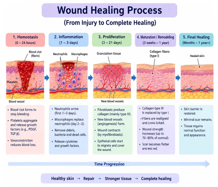
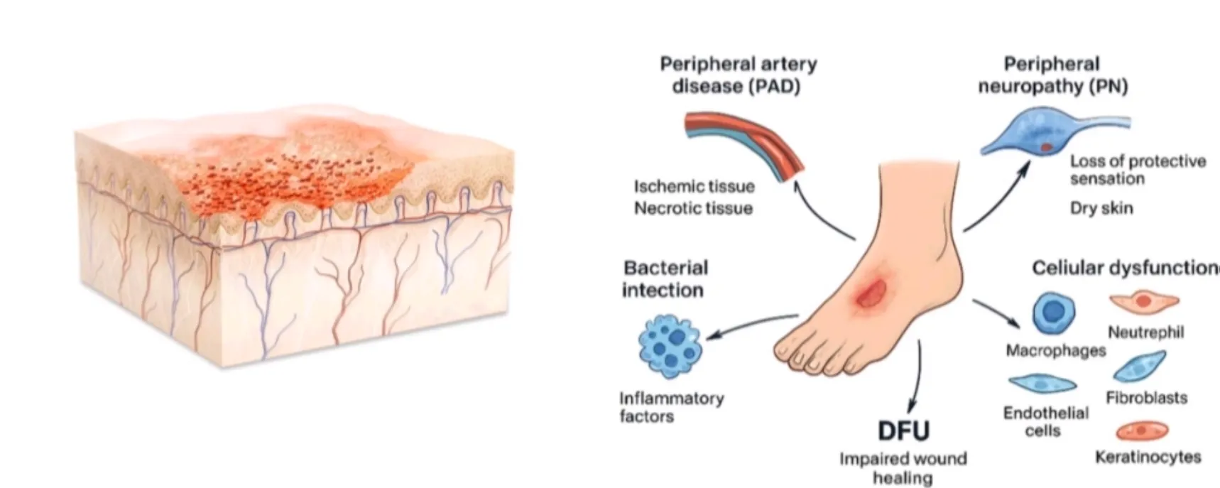
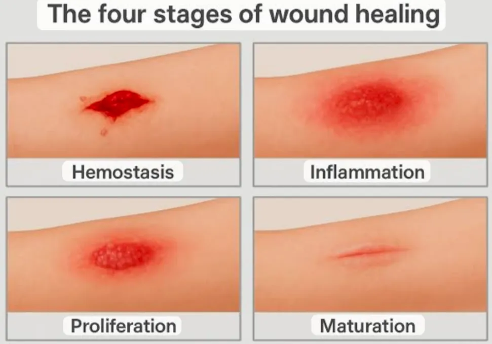
Skin acts as a protective barrier against mechanical injury, dehydration and microbial invasion. Following injury, wound repair proceeds through overlapping phases: haemostasis, inflammation, proliferation and remodelling. Platelet activation and clot formation initially limit blood loss; inflammatory cells remove microbes and damaged tissue; fibroblasts, keratinocytes and endothelial cells contribute to granulation tissue, re-epithelialisation and angiogenesis; and collagen remodelling strengthens the repaired tissue. Disruption of these stages can lead to delayed healing or chronic wounds. Wound dressings are intended to protect the wound, manage exudate, reduce trauma during removal and support an appropriate local environment. Traditional gauze can be useful for some purposes but may adhere to the wound and may not maintain ideal moisture conditions. Advanced dressings include films, foams, hydrocolloids, alginates and hydrogels. Hydrogels are particularly attractive where hydration and conformability are needed. Their water-rich networks can help maintain a moist wound interface and may serve as carriers for antimicrobial or healing-supportive substances. A hydrogel is a crosslinked hydrophilic polymer network that absorbs water without dissolving completely. The network may be formed by physical interactions—such as ionic association, hydrogen bonding or crystallite formation—or by covalent crosslinking. Hydrogel behaviour is controlled by polymer type, molecular weight, polymer concentration, crosslink density, pH, ionic strength and the nature of the incorporated agent. A formulation that is too weak may disintegrate; one that is too densely crosslinked may absorb insufficient fluid or release active compounds too slowly. Natural biopolymers are appealing because their structures can resemble components of the extracellular matrix or interact favourably with wound fluid. Nevertheless, ‘natural’ does not automatically mean safe, sterile, reproducible or clinically effective. Purity, source, processing, microbial burden, allergens and residual reagents must be assessed. This review therefore considers both the opportunities and the practical limitations of these materials.
2. Aim and Objectives
3. Review Methodology
This article is structured as a narrative review rather than a systematic review. Relevant literature was identified using keyword combinations related to biopolymer hydrogels, wound healing, chitosan, alginate, cellulose, hyaluronic acid, antimicrobial wound dressings and natural bioactive compounds. Sources included peer-reviewed review articles and selected research papers indexed in biomedical and multidisciplinary literature databases. Reference lists of relevant reviews were also considered to identify foundational publications. The discussion prioritises publications that describe polymer chemistry, hydrogel preparation, wound-care mechanisms, antimicrobial performance, evaluation methods or translational limitations. Because a fully reproducible systematic search and formal risk-of-bias assessment were not performed for this draft, it should not be described as a systematic review. Before academic submission, the author should conduct and document a date-stamped database search, record search strings and screening decisions, verify each reference against the publisher or PubMed record, and add any institutional requirements for review methodology.
4. Wound-Healing Requirements and Role of Hydrogels
4.1 Moisture management and exudate handling
A moist but not waterlogged wound environment can support cell migration and may reduce dressing adherence. Hydrogels can donate or retain water, while absorbent polymer combinations can take up exudate. The appropriate balance depends on the wound: a heavily exuding wound may require a more absorbent dressing or a secondary dressing, whereas a dry wound may benefit from a moisture-donating hydrogel.
4.2 Physical protection and conformability
Soft, flexible hydrogels can conform to irregular surfaces and provide a protective interface. Their mechanical strength must be sufficient for handling and removal without causing additional tissue trauma. Swelling and degradation should be considered together because a highly swollen gel may become fragile.
4.3 Antimicrobial action
Antimicrobial function may arise from the polymer itself, an incorporated natural agent, or a combination of mechanisms. Chitosan can interact with negatively charged microbial surfaces, although activity varies with molecular weight, degree of deacetylation, pH and organism. Honey can exert osmotic effects and, depending on type and processing, may contain additional antimicrobial factors. Plant-derived compounds such as curcumin and tannins have been investigated, but their solubility, stability, effective concentration and possible cytotoxicity require careful evaluation. Antimicrobial performance in a laboratory assay does not by itself prove prevention or treatment of clinical wound infection.
4.4 Support of tissue repair
A suitable hydrogel may support cell migration and tissue repair by maintaining hydration and providing a compatible interface. Collagen, gelatin and hyaluronic acid can interact with cells and biological molecules. Yet a dressing must avoid harming keratinocytes, fibroblasts and other relevant cells. The design goal is not simply maximum antibacterial activity: it is a balance between microbial control, biocompatibility, moisture management and physical performance.
5. Natural and Naturally Derived Biopolymers
5.1 Chitosan
Chitosan is produced by partial deacetylation of chitin, which is commonly obtained from crustacean shells or fungal sources. It contains amino and hydroxyl groups that support chemical modification and interactions with other polymers. Chitosan has been widely studied for wound dressings because it can form films, sponges, particles and hydrogels, and may contribute to haemostatic and antimicrobial functions. Its solubility is pH-dependent, and performance is influenced by molecular weight and degree of deacetylation. Source-related allergens, residual proteins and batch variability should be controlled.
5.2 Alginate
Alginate is an anionic polysaccharide commonly obtained from brown seaweed. In the presence of suitable divalent ions, especially calcium, alginate chains can form ionically crosslinked networks. Alginate materials can absorb exudate and are used in wound-care formats. Alginate itself is not a universal antimicrobial agent; it is often combined with chitosan or loaded with an antimicrobial compound. Ionic exchange in wound fluid can alter gel integrity and release.
5.3 Gelatin and collagen
Collagen is a major structural protein of the extracellular matrix, while gelatin is obtained through partial denaturation of collagen. These materials can provide cell-interactive motifs and can be formulated into hydrogels. Their limitations include weak mechanical properties in some formulations, enzymatic degradation and source-related variability. Crosslinking can improve stability, but the crosslinker and any residual reagent must be evaluated for toxicity.
5.4 Cellulose and cellulose derivatives
Cellulose is an abundant polysaccharide. Derivatives such as carboxymethyl cellulose and hydroxyethyl cellulose are used to tune viscosity, water uptake and gel formation. Cellulose-based materials can improve handling and fluid management, but typically need a separate antimicrobial component if a specific antimicrobial effect is required.
5.5 Hyaluronic acid
Hyaluronic acid is a glycosaminoglycan found in the extracellular matrix and is involved in tissue hydration and cell signalling. It can be formulated into hydrogels and combined with other polymers to adjust mechanical and biological properties. Its degradation and biological effects depend on molecular weight, modification and local context.
5.6 Other natural materials
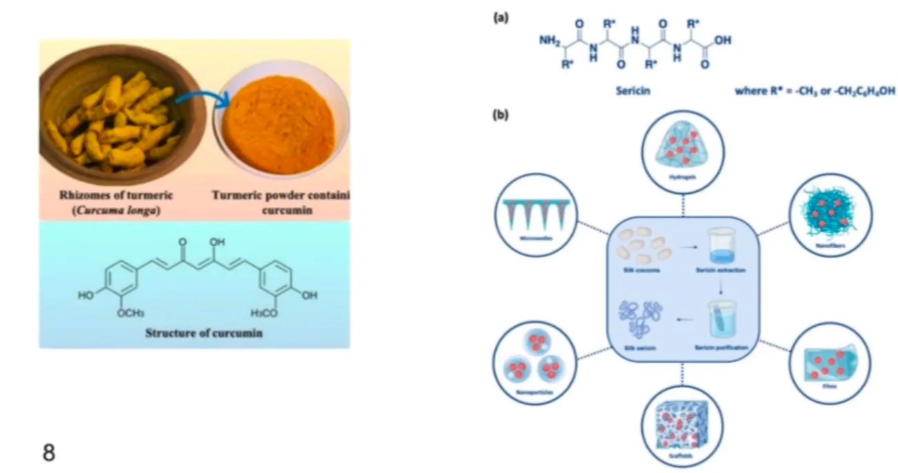
Silk fibroin, starch, pectin, dextran, carrageenan and plant-derived gums have also been explored in biomaterials. Selection should be based on defined quality attributes, reproducible supply, compatibility with the intended preparation method and evidence for the target wound-care use.
6. Natural Antimicrobial Agents for Hydrogel Incorporation
6.1 Medical-grade honey
Honey has been investigated for wound care because of its high sugar concentration, acidity and other composition-dependent factors. Medical-grade products are processed and quality-controlled for clinical use; ordinary household honey should not be assumed to be sterile or suitable for wounds. When incorporated into hydrogels, honey content, water activity, dilution, release and stability should be measured.
6.2 Curcumin
Curcumin, a polyphenolic compound from turmeric, has been studied for anti-inflammatory and antimicrobial-related effects. Its poor aqueous solubility and sensitivity to environmental conditions can limit direct incorporation. Encapsulation, dispersion systems or suitable polymer interactions may improve handling, but increased apparent solubility does not automatically establish bioavailability or clinical benefit.
6.3 Tannins and other plant polyphenols
Tannins and related polyphenols can interact with proteins and may contribute antioxidant or antimicrobial activity. Their chemical composition varies with plant species, plant part, extraction method and storage. Standardised extracts and quantitative marker assays are preferable to poorly characterised crude extracts.
6.4 Essential oils and plant extracts
Selected essential oils and plant extracts have been explored for antimicrobial dressings. Volatility, oxidation, odour, skin irritation and cytotoxicity can constrain use. Concentrations effective against microbes may also damage mammalian cells, so both antimicrobial and cytocompatibility testing are essential. A natural origin does not eliminate sensitisation risk.
6.5 Design considerations
The incorporated agent should be selected according to the target organism, desired release profile, chemical stability and compatibility with the polymer. A hydrogel may protect a labile compound or provide local delivery, but the active ingredient can also alter gelation, swelling, mechanical strength and degradation. Appropriate unloaded-polymer, free-agent and vehicle controls are required to interpret results.
7. Formulation and Methods of Preparation
There is no single universal recipe for antimicrobial wound-healing hydrogels. Polymer grades, intended wound type, active ingredient and crosslinking chemistry determine the process. The following are general formulation approaches for literature review and supervised research planning, not validated clinical manufacturing instructions.
7.1 Physical blending and polymer hydration
A common first step is to hydrate the selected polymer in a suitable solvent or buffer under controlled mixing until a uniform solution or dispersion is obtained. Hydration time, temperature, mixing speed and order of addition affect lump formation, viscosity and uniformity. The active ingredient may be dissolved, dispersed or separately encapsulated before incorporation, depending on its solubility and stability. Air bubbles may be reduced by allowing the mixture to stand or by a compatible degassing method.
7.2 Ionic gelation of alginate
Alginate can be crosslinked by contact with a solution containing suitable divalent cations, commonly calcium ions. The polymer solution may be shaped or cast before exposure to the crosslinking medium, or the components may be combined through a controlled mixing strategy. Gelation time, ion concentration, alginate composition and diffusion distance affect the final network. Excessive crosslinking can reduce swelling and slow release, while insufficient crosslinking can produce weak gels.
7.3 Chitosan-based gel formation
Chitosan may be dissolved in a compatible mildly acidic medium and then gelled through ionic interactions, polyelectrolyte complexation with an anionic polymer such as alginate, or a suitable crosslinking reaction. The pH must be adjusted carefully because it influences chitosan solubility, polymer charge and active-agent stability. Strongly acidic or alkaline conditions may damage sensitive ingredients or impair compatibility.
7.4 Blended chitosan–alginate hydrogels
Chitosan and alginate have opposite charge characteristics under appropriate conditions and can form polyelectrolyte complexes. Their ratio, concentration, pH, mixing sequence and ionic strength influence phase separation, pore structure, swelling and mechanical behaviour. A reproducible process should specify the polymer grades, mixing order, pH range, crosslinking conditions and final conditioning steps.
7.5 Gelatin/collagen and cellulose-based hydrogels
Gelatin and collagen may be combined with polysaccharides or crosslinked to improve stability. Cellulose derivatives may be used as gel-forming or viscosity-modifying components. Physical approaches, including cooling or freeze–thaw processing for suitable polymer systems, can create or strengthen networks without some chemical crosslinkers. The method must be chosen according to the polymer chemistry; freeze–thaw is not suitable for every natural polymer.
7.6 Incorporation of natural antimicrobial agents
The active agent can be added during polymer preparation if it remains stable under the process conditions, or introduced after network formation by soaking or loading. Adding it before gelation may provide more uniform distribution but may interfere with crosslinking. Post-loading can be simpler for sensitive agents but may result in surface-biased distribution or burst release. Content uniformity and release should be measured rather than assumed.
7.7 Drying, conditioning, packaging and sterilisation
Depending on the target product, the gel may be used in hydrated form or converted into a dried porous material and rehydrated before use. Conditioning should bring samples to a defined state before testing. Packaging must prevent contamination and maintain moisture and active-agent stability. Sterilisation is a major development issue: heat, radiation or chemical methods can change polymer molecular weight, crosslink density, colour, active-agent potency or gel strength. The chosen method must be validated for the specific formulation and package.
7.8 Illustrative development workflow
1. Define the intended use, target wound conditions and desired product properties.
2. Select polymer (s) and antimicrobial agent (s) using literature evidence and quality specifications.
3. Prepare small screening batches and document exact material grades, concentrations, pH, mixing order and processing conditions.
4. Optimise gel formation, homogeneity, handling, swelling and mechanical properties using a planned experimental design.
5. Evaluate antimicrobial activity against relevant test organisms with appropriate controls.
6. Assess cytocompatibility and other biological safety endpoints using validated methods.
7. Study release, degradation, packaging compatibility, stability and the effect of sterilisation.
8. Advance only formulations with reproducible results to more relevant preclinical and clinical evaluation.
8. Evaluation and Characterisation
8.1 Physical appearance and homogeneity
Record colour, odour, visible particles, phase separation, bubbles and dimensional uniformity. Natural extracts may impart colour or odour, but unexpected changes during storage can signal degradation.
8.2 pH
Measure pH using a defined method suitable for the hydrogel or its extract. pH affects polymer charge, antimicrobial-agent stability and tissue compatibility. A pH result alone cannot establish safety.
8.3 Swelling and fluid uptake
Swelling may be assessed by measuring the mass of a conditioned sample before and after exposure to a defined medium for a specified time. Report the medium, temperature, time points, blotting procedure and calculation. Wound-fluid-like media may provide more relevant information than pure water alone.
8.4 Gel fraction and degradation
Gel fraction or soluble fraction can help characterise network formation. Degradation studies should define the medium, temperature, agitation and sampling schedule. Mass loss can reflect dissolution, erosion or degradation and should not be interpreted as a direct measure of biological resorption without supporting evidence.
8.5 Mechanical properties and rheology
Tensile, compression or puncture testing can assess handling strength, while rheology may describe viscosity, viscoelasticity and gelation. Test geometry, strain rate, temperature and sample hydration should be reported because these factors affect results.
8.6 Moisture-vapour transmission and fluid management
Water-vapour transmission testing helps assess whether the dressing may retain excessive moisture or dry the wound. Methods and environmental conditions should be standardised. The ideal range depends on the wound and the complete dressing system, including any secondary layer.
8.7 Antimicrobial testing
Possible methods include agar diffusion screening, viable-count reduction and time-kill approaches. Agar diffusion can underestimate agents that diffuse poorly and may overemphasise highly diffusible compounds. Quantitative methods should include untreated, polymer-only, active-agent-only and positive controls where appropriate. Report strains, inoculum, contact time, replicates and statistical analysis. Laboratory antimicrobial activity is not equivalent to clinical infection control.
8.8 Cytocompatibility and biological performance
Relevant cell-based assays may examine viability and morphology in suitable cell models, such as fibroblasts or keratinocytes. More advanced studies may assess cell migration, inflammatory response, haemocompatibility or animal wound healing, subject to ethical approval and validated protocols. Cytotoxicity and antimicrobial effects should be interpreted together.
8.9 Drug or active-agent release
Release studies should use an appropriate medium and validated analytical method, with sampling conditions that preserve sink conditions where relevant. Report cumulative release, recovery, analytical specificity and replicate variability. Release data should be connected to biological activity where possible.
8.10 Stability and quality attributes
Stability assessment should monitor appearance, pH, polymer integrity, swelling, mechanical performance, active-agent content, microbial quality and package integrity under defined storage conditions. Critical quality attributes should be linked to product performance and established before scale-up.
9. Comparative Summary of Selected Materials
| Material | Potential contribution | Main formulation issue | Important evaluation |
|---|---|---|---|
| Chitosan | Potential inherent antimicrobial and haemostatic functions; film/gel formation | pH-dependent solubility; molecular-weight and source variability | pH, antimicrobial tests, cytocompatibility, swelling |
| Alginate | Fluid uptake; ionically crosslinked gel formation | Ion exchange and possible loss of integrity in wound fluid | Swelling, gel stability, mechanical properties, fluid handling |
| Gelatin/collagen | Cell-interactive protein-based matrix | Enzymatic degradation; source and crosslinker concerns | Degradation, cytocompatibility, mechanics |
| Cellulose derivatives | Viscosity control, water uptake and matrix formation | Often require an additional antimicrobial strategy | Swelling, rheology, moisture-vapour transmission |
| Hyaluronic acid | Hydration and extracellular-matrix-related interactions | Cost, degradation and molecular-weight effects | Rheology, degradation, cell response |
| Medical-grade honey | Potential multi-factor antimicrobial contribution | Composition, dilution, stability and quality control | Content uniformity, antimicrobial activity, cytocompatibility |
| Curcumin/polyphenols | Potential antioxidant and antimicrobial-related activity | Low solubility, instability and variable extracts | Content assay, release, antimicrobial tests, cytocompatibility |
10. Advantages and Limitations
10.1 Potential advantages
10.2 Limitations and risks
11. Future Perspectives
Future development should focus on reproducible formulations with clearly defined quality attributes rather than adding many ingredients without a clear rationale. Potential directions include responsive hydrogels that react to pH or enzymes, controlled delivery systems, antimicrobial combinations designed to reduce unnecessary exposure, and dressings that integrate sensing with wound assessment. Such systems must demonstrate that added complexity improves meaningful outcomes.
Design-of-experiments approaches can help identify interactions between polymer ratio, crosslinking and active-agent concentration. Better reporting of polymer grade, source, molecular weight, degree of deacetylation, extract composition and processing conditions would improve reproducibility. Standardised antimicrobial methods, clinically relevant wound models and comparative studies against existing dressings are essential. Manufacturing research should address sterilisation, packaging, long-term stability, batch release and cost-effectiveness.
For student projects, the most defensible scope is a focused comparison of a small number of polymer systems or a critical literature synthesis with a transparent search strategy. Claims should distinguish established wound-care use, preclinical evidence and speculative future applications.
CONCLUSION
Natural and naturally derived biopolymer hydrogels are promising platforms for wound dressings because their water-rich networks can support moisture management and can be engineered to carry antimicrobial agents. Chitosan, alginate, gelatin, collagen, cellulose derivatives and hyaluronic acid offer different strengths and limitations; no single polymer is optimal for every wound. Natural agents such as medical-grade honey and selected plant-derived compounds may add functionality, but their performance depends on standardisation, loading, release, stability and safety. Preparation method and crosslink density strongly influence the final hydrogel’s swelling, mechanics, degradation and active-agent release. A complete development programme must therefore assess physicochemical properties, antimicrobial performance, cytocompatibility, stability and sterilisation effects using reproducible methods. The field has substantial potential, but laboratory promise should not be overstated as clinical proof. Standardised testing, robust manufacturing controls and well-designed clinical studies remain necessary to establish which formulations provide meaningful benefits for patients.
SUMMARY
This review discusses natural antimicrobial wound-healing hydrogels made from biopolymer-based materials. The central concept is to combine a hydrophilic polymer network with appropriate wound-fluid management and, where justified, an antimicrobial agent. Chitosan may contribute antimicrobial activity; alginate is useful for fluid uptake and ionic gelation; gelatin and collagen provide protein-based matrices; cellulose derivatives support matrix and fluid-handling properties; and hyaluronic acid offers hydration and extracellular-matrix-related interactions. Natural antimicrobial candidates include medical-grade honey and selected plant-derived compounds, although natural origin does not guarantee safety or efficacy. Preparation may involve polymer hydration, blending, ionic or covalent crosslinking, active-agent loading, conditioning, packaging and validated sterilisation. The formulation should be evaluated for pH, swelling, degradation, mechanical strength, moisture-vapour transmission, antimicrobial activity, cytocompatibility, release and stability. The major barriers to translation are variability, potential cytotoxicity, weak mechanics, sterilisation effects, scale-up and limited clinical validation. Future work should prioritise standardisation, transparent reporting and evidence of clinically meaningful outcomes.
REFERENCES



Ganesh Todkar, Anvirappa Katte, Shredatta Shetkar, Ramprasad Khandare, Nikita Kale, Dnyaneshwari Marlapalle, Amarja Bhoge, Formulation of Natural Antimicrobial Wound-Healing Hydrogels Using Biopolymer-Based Materials: A Review, Int. J. of Pharm. Sci., 2026, Vol 4, Issue 11, 711-721. https://doi.org/10.5281/zenodo.23939652
 10.5281/zenodo.23939652