

We use cookies to ensure our website works properly and to personalise your experience. Cookies policy
KMCH College of Pharmacy, Coimbatore – 641048, Tamil Nadu, India
The vaginal route offers a mucosally accessible, first-pass-avoiding site for both local and systemic therapy, but conventional creams, tablets, and pessaries suffer from rapid clearance, leakage, and poor patient compliance. Gellan gum, an anionic exopolysaccharide produced by Sphingomonas elodea, has emerged as a leading excipient for mucoadhesive, ion-activated in situ gelling vaginal systems because it is instilled as a low-viscosity liquid that undergoes rapid sol-to-gel transition on contact with the monovalent and divalent cations present in vaginal fluid, yielding a mechanically robust, mucoadhesive depot at body temperature. This review synthesises current literature on the physicochemical basis of gellan gum gelation and mucoadhesion; formulation strategies including binary/composite polymer blends, thermosensitive gellan poloxamer hybrids, nanocarrier-embedded gels, and Quality-by-Design optimisation; and the standard panel of evaluation parameters (rheology, gel strength, mucoadhesive force, in vitro/ex vivo release, and stability) used to characterise these systems. Particular attention is given to the interaction between gellan gum-based delivery platforms and the vaginal microbiome, including delivery of Lactobacillus probiotics, antimicrobial and microbicide agents for bacterial vaginosis, vulvovaginal candidiasis, and trichomoniasis, and the broader concept of designing formulations that preserve or restore a Lactobacillus-dominant, eubiotic vaginal ecosystem rather than indiscriminately eliminating flora. Finally, the review surveys emerging smart technologies-stimuli-responsive nanocarriers, three-dimensional-printed vaginal rings and films, electrospun nanofibrous scaffolds, biosensor-integrated devices, and artificial-intelligence-guided formulation design-that are converging with gellan gum chemistry to enable personalised, long-acting, and microbiome-conscious vaginal therapeutics. Persisting challenges in scale-up, regulatory standardisation, and long-term safety evaluation are discussed alongside future research directions
Local and systemic delivery of drugs through the vaginal mucosa has been practised for millennia, yet it remains an underexploited route relative to oral and parenteral administration. The vaginal epithelium offers a large, richly vascularised surface area, avoidance of first-pass hepatic metabolism, ease of self-administration, and direct access to the site of many gynaecological pathologies, including bacterial vaginosis (BV), vulvovaginal candidiasis (VVC), trichomoniasis, and sexually transmitted infections such as HIV-1 [1,2]. Despite these advantages, conventional vaginal dosage forms-creams, pessaries, tablets, and simple aqueous gels-are limited by rapid mucus clearance, leakage, messiness, and the need for frequent redosing, all of which compromise patient adherence and therapeutic outcomes [2,6]. In situ gelling systems address these shortcomings by being administered as a low-viscosity liquid or semisolid that undergoes a rapid, environmentally triggered phase transition-in response to ionic strength, temperature, or pH-into a viscoelastic, mucoadhesive gel once in contact with vaginal fluid [1,2]. Among the polymers used for this purpose, gellan gum has attracted particular interest because of its unique ion activated gelation behaviour, its Generally-Recognised-As-Safe (GRAS) and pharmacopoeia status, and its capacity to form gels at very low polymer concentrations even in response to the modest monovalent cation content of physiological fluids [3,4]. Gellan gum-based systems have already been explored for ophthalmic, nasal, buccal, and rectal delivery, and this chemistry is now being adapted extensively for the vaginal compartment, frequently in combination with thermosensitive poloxamers, mucoadhesive cellulose derivatives, or nanocarrier systems to further tailor gelation kinetics, mechanical strength, and drug release [2,4,5]. A parallel and increasingly central consideration in vaginal formulation science is the vaginal microbiome. The healthy vagina is dominated by Lactobacillus species that maintain an acidic pH and inhibit pathogen overgrowth; disruption of this ecosystem (dysbiosis) is implicated in BV, increased susceptibility to sexually transmitted infections, and adverse reproductive outcomes [14,15,16]. Modern vaginal drug delivery design therefore increasingly aims not merely to deliver an antimicrobial payload, but to do so while preserving, or actively restoring, a Lactobacillus-dominant eubiotic state-for example, by co-delivering probiotics or by selecting excipients and gelation chemistries that are compatible with commensal flora [7,8,9]. Simultaneously, the field is being reshaped by "smart" technologies-stimuli-responsive nanocarriers, additive manufacturing, nanofibrous scaffolds, and digital/biosensor integration-that promise increasingly personalised, long-acting, and microbiome-conscious therapeutics [1,12,13]. This review provides an integrated, narrative synthesis of (i) the vaginal anatomical and physiological context relevant to mucoadhesive drug delivery; (ii) the physicochemical basis of gellan gum chemistry and gelation; (iii) formulation strategies and representative gellan gum-based vaginal systems reported in the literature; (iv) standard characterisation and evaluation parameters; (v) the interplay between formulation design and vaginal microbiome modulation; and (vi) emerging smart technologies that are converging with gellan gum-based platforms. The aim is to consolidate current knowledge into a single reference framework and to identify translational gaps that merit further research
The vaginal wall comprises four principal layers: an innermost stratified squamous epithelium (lacking goblet cells and therefore incapable of direct mucin secretion), an elastic lamina propria, a fibromuscular layer, and an outer adventitia [6]. Vaginal secretions arise instead from a combination of transudation across the epithelium, cervical mucus, exfoliated epithelial cells, and fluid from the endometrium and fallopian tubes, together forming cervicovaginal mucus (CVM), a complex, gel-forming mucin-based fluid that coats the epithelial surface [6,14]. This anatomical arrangement means that, unlike goblet-cell-rich mucosae, the vagina depends heavily on cervically derived mucus for its protective barrier, a distinction with direct implications for mucoadhesive formulation design.
CVM functions as a selective physical and biochemical barrier: its mucin glycoprotein network can immobilise pathogens (including viral particles) through low-affinity multivalent binding and steric obstruction, while permitting beneficial commensal organisms and appropriately engineered mucoadhesive or muco-penetrating particles to persist near the epithelial surface [14] Bulk rheological studies indicate that CVM microstructure is remarkably resistant to shifts in physiological pH, though it is markedly altered in dysbiotic states such as BV, where a thinner, more permeable, less mucoadhesive mucus layer has been observed and correlated with reduced protective capacity against pathogens including HIV-1 [14]. This has two direct implications for formulation science: first, an effective vaginal in situ gel must be able to interpenetrate and anchor within this mucin network (mucoadhesion) rather than simply resting on top of it; second, the barrier properties of CVM-and hence formulation performance-are not static but vary with the underlying microbiota composition, menstrual cycle stage, and pathological state [6,14].
Vaginal pH in reproductive-age women is normally acidic (approximately 3.8-4.5), maintained largely by lactic acid production from Lactobacillus fermentation of glycogen deposited in the oestrogen-primed epithelium [15,16]. This pH, together with epithelial thickness, is dynamically modulated by hormonal status (menstrual cycle, pregnancy, menopause), bacterial colonisation, and exposure to semen, all of which must be considered when designing an ion- or pH-triggered gelling system, since the local ionic strength and pH at the moment of administration determine gelation kinetics [6]. The resident microbiota itself has been classified into Community State Types (CSTs) based on the dominant Lactobacillus species (or, in CST IV, a more diverse anaerobic community), a framework that underpins much of the current understanding of eubiosis and dysbiosis [15].
Compared with oral therapy, vaginal administration offers localised high drug concentrations at the target tissue with reduced systemic exposure and side effects, avoidance of hepatic first-pass metabolism, and applicability to drugs with poor oral bioavailability or gastrointestinal instability [1,2]. However, conventional creams, ovules, tablets, and simple gels are typically cleared within a few hours by the vagina's continuous self-cleansing mucus flow, necessitating multiple daily doses, and are frequently associated with leakage and a sensation of messiness that undermines compliance, particularly for regimens intended to run for many days (as in standard BV or VVC therapy) or those intended for chronic prophylactic use (as in HIV pre-exposure prophylaxis) [2,5]. Sustained-release matrices such as vaginal rings mitigate the dosing frequency problem but require a rigid device to be inserted and later removed, which is not acceptable to all users and cannot be extemporaneously prepared [1]. In situ gelling systems occupy a favourable middle ground: they combine the ease of administration and accurate dosing of a liquid formulation with the prolonged retention and controlled-release profile of a solid or semisolid gel, because gelation occurs only after the correct dose has already been delivered to the target site [1,2,6].
Gellan gum is a linear, anionic, high-molecular-weight exopolysaccharide first described in 1978, produced industrially by aerobic fermentation of Sphingomonas elodea (formerly Pseudomonas/Sphingomonas paucimobilis) [3]. Its repeating tetrasaccharide unit consists of 1,3-linked beta-D-glucose, 1,4-linked beta-D glucuronic acid, 1,4-linked beta-D-glucose, and 1,4-linked alpha-L-rhamnose, with a single carboxyl side group per repeat unit that confers the polymer's anionic character and underlies its capacity for ionotropic (cation-mediated) cross-linking [3]. Native gellan gum is partially acetylated (bearing acetyl and glyceryl substituents), while the pharmaceutically dominant deacetylated form-marketed as Gelrite or Kelcogel-is produced by alkaline treatment that removes these substituents, producing a polymer capable of forming firmer, more brittle gels and, critically, one that gels in response to physiologically relevant concentrations of monovalent cations such as sodium, in addition to divalent cations such as calcium and magnesium [3]. This monovalent-cation sensitivity is the key property exploited in mucosal in situ gelling systems, since body fluids-including tear fluid, nasal secretions, and vaginal fluid-contain sufficient Na+, K+, Ca2+, and Mg2+ to trigger gelation of deacetylated gellan gum even at concentrations as low as 0.1-0.3% w/v [3].
Table 1. Comparative physicochemical properties of native (high-acyl) and deacetylated (low-acyl) Gellan gum relevant to mucoadhesive in situ gel formulation [3,4].
|
PROPERTY |
NATIVE (HIGH-ACYL) GELLAN GUM |
DEACETYLATED (LOW-ACYL) GELLAN GUM, Eg. GELRITE/KELCOGEL |
|
Source / production |
Direct fermentation product of Sphingomonas elodea (formerly Pseudomonas elodea) |
Obtained by alkali (KOH) treatment of native gellan to remove acetyl and glyceryl substituents |
|
Gel texture |
Soft, elastic, cohesive gel |
Hard, brittle, non-elastic gels with higher mechanical strength |
|
Gelation trigger |
Ionotropic (Ca2+, Mg2+ > K+, Na+) and thermal cooling |
Ionotropic gelation strongly promoted even by monovalent cations (Na+, K+) present in vaginal/lacrimal fluid; also thermoreversible |
|
Typical use concentration (mucosal in situ gels) |
Rarely used alone for mucosal in situ gel |
0.1-1.0% w/v; gels are formed even at 0.1 0.3% w/v on contact with physiological ionic strength |
|
Mucoadhesion |
Moderate, via H-bonding and chain entanglement with mucin |
Enhanced through interpenetration with mucin glycoprotein network and anionic cationic interactions; further increased by carboxymethylation |
|
Regulatory / safety status |
GRAS-listed food additive (E418 |
Approved by US FDA and EMA as gelling/stabilising/suspending agent; widely used in marketed ophthalmic in situ gel products |
|
Representative pharmaceutical role |
Viscosity modifier, tissue-engineering scaffolds, food-grade probiotic encapsulation |
Primary in situ gelling agent for ophthalmic, nasal, buccal, and vaginal mucoadhesive systems |
the molecular level, gelation proceeds through a coil-to-double-helix transition of gellan chains on cooling and/or cation exposure, followed by cation-mediated aggregation of these double helices into a three-dimensional network that entraps water and dissolved or dispersed drug [3,4]. The resulting gels are pseudoplastic and shear-thinning, which is pharmaceutically advantageous: the formulation flows readily during administration under the shear of a vaginal applicator, then rapidly rebuilds its gel structure once shear is removed and ionic triggering occurs at the mucosal surface [3]. Gellan gum's mucoadhesive character arises from a combination of physical entanglement with the mucin glycoprotein network, hydrogen bonding, and electrostatic/ionic interactions, and can be further enhanced by chemical modification (e.g., carboxymethylation), which increases mucoadhesive strength several-fold relative to unmodified gellan gum while maintaining a favourable ocular/mucosal tolerability profile [4]. Beyond its role as a gelling and mucoadhesive agent, gellan gum can also be electrospun (typically after blending with a carrier polymer such as polyvinyl alcohol to overcome its poor intrinsic spinnability) or fabricated into micro/nanoparticles, extending its utility into the nanofibrous and nanocarrier platforms discussed in Section 9 [4].
For vaginal application, ion-activated gelation is the principal and most extensively exploited mechanism for gellan gum systems. The formulation is prepared as a dilute aqueous sol of deacetylated gellan gum (typically 0.1-1.0% w/v); on contact with the Na+, K+, Ca2+, and Mg2+ ions present in vaginal fluid, the polymer undergoes rapid coil-to-helix transition and cation-bridged aggregation, forming a coherent gel in situ within seconds to minutes [3]. Because this transition does not require an external temperature stimulus, ion-activated gellan systems are comparatively simple to manufacture and are less prone to premature gelation during storage or in-process handling than purely thermosensitive systems, provided the formulation is protected from adventitious ionic contamination [3].
Gellan gum is frequently combined with thermosensitive block copolymers such as poloxamer 407 (and poloxamer 188) to create hybrid dual-trigger systems. Poloxamers self-assemble into micelles and then a cubic or hexagonal liquid-crystalline gel phase as temperature rises toward 37 Degree Celsius, but poloxamer-only gels are typically mechanically weak and poorly mucoadhesive, dissolving relatively quickly in physiological fluid [2,8]. Incorporating gellan gum (and sometimes additional mucoadhesive polysaccharides such as carrageenan or hyaluronic acid) into a poloxamer base combines the rapid, temperature-driven gelation and solubilising capacity of the poloxamer with the ionic cross-linking and superior mucoadhesive/mechanical reinforcement conferred by gellan gum, an approach used successfully for vaginal delivery of clindamycin (with prolonged vaginal residence reported up to approximately 9 hours), Lactobacillus gasseri probiotics, and combined metronidazole-curcumin formulations [2,6,8].
Although gellan gum itself is not strongly pH-triggered, it is frequently formulated alongside pH-sensitive polymers (e.g., polycarbophil, chitosan) to exploit the vagina's acidic resting pH and the transient pH elevation that occurs in the presence of semen or during BV, allowing formulations to be tuned so that gel strength, mucoadhesion, or drug release respond additionally to local pH shifts associated with the underlying pathology [2,6]. Composite systems combining gellan gum with hydroxypropyl methylcellulose (HPMC), sodium carboxymethylcellulose (NaCMC), or chitosan have also been used to fine-tune viscosity, reduce the total polymer load required for adequate gel strength, and further enhance bioadhesion, as demonstrated for clindamycin and secnidazole vaginal in situ gels [2,5].
Mucoadhesion of gellan gum-based gels is generally explained through a combination of classical mucoadhesion theories: the wetting/adsorption theory (spreading and interfacial contact between the low viscosity sol and the mucosal surface prior to gelation), the diffusion-interpenetration theory (entanglement of gellan chains with mucin glycoprotein chains once the sol is in contact with mucus), and the electronic/ionic-interaction theory (electrostatic and cation-bridging interactions between the anionic carboxyl groups of gellan and the mucin network, aided by the divalent cations that also drive gelation) [4,6]. In practice, these mechanisms act sequentially and synergistically: the sol first wets and spreads over the mucosal surface, ionic and thermal triggers then drive gelation and chain interpenetration with mucin, and the resulting entangled, cation-cross-linked network resists the shear forces of vaginal clearance, thereby prolonging residence time and localising drug release near the absorbing or infected tissue [4,6,14].
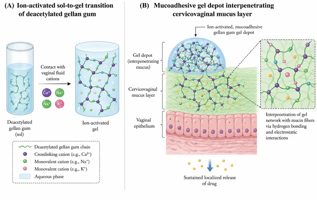
Figure 1: Schematic representation of (A) the ion-activated sol-to-gel transition of deacetylated gellan gum on contact with vaginal-fluid cations, and (B) the resulting mucoadhesive gel depot interpenetrating the cervicovaginal mucus layer overlying the vaginal epithelium, enabling sustained localized release [3,4,6]
The most common formulation approach combines gellan gum, as the primary ion-activated gelling agent, with one or more auxiliary polymers selected to reduce total polymer burden, augment mucoadhesion, or improve mechanical or optical properties. Reported combinations include gellan gum with HPMC (bioadhesive viscosity enhancer), sodium carboxymethylcellulose (which allows a reduction in gellan concentration without compromising gel strength while enhancing bioadhesion), chitosan (cationic mucoadhesive polymer providing complementary electrostatic interaction with anionic gellan and mucin), and carrageenan (additional gelling/mucoadhesive polysaccharide that has been shown to further prolong vaginal residence time when combined with gellan-poloxamer bases) [2,5]. Increasing the concentration of divalent cross-linker (e.g., calcium carbonate/calcium chloride) in gellan formulations has been shown to produce stronger gels through greater internal ionotropic cross-linking, while excessive polymer or cross linker concentration risks a formulation too viscous for comfortable administration [5].
Hybridising gellan gum with poloxamer 407/188 combines two independent, physiologically relevant triggers (temperature and ionic strength), producing formulations that gel rapidly and robustly across the range of conditions encountered in vivo. This strategy has been used for antiparasitic (metronidazole, metronidazole-curcumin), antifungal, antibacterial (clindamycin), and probiotic (Lactobacillus gasseri) vaginal formulations, the latter requiring the additional constraint that the gelation process, excipients, and any accompanying preservative system must not compromise bacterial viability [2,6,8].
To improve the solubility, stability, or targeting of poorly water-soluble or labile actives, gellan gum gels are increasingly used as a secondary mucoadhesive matrix that thickens and vaginally localises a primary nanocarrier dispersion (nanoparticles, nanocapsules, niosomes, or micelles). For example, indole-3 carbinol-loaded Eudragit RS100/rosehip-oil nanocapsules have been incorporated into a gellan gum hydrogel to create a vaginally applicable, mucoadhesive semisolid for trichomoniasis, addressing both the poor aqueous solubility of the phytochemical active and the need for prolonged mucosal contact [5]. More broadly, hydrogel-nanocomposite hybrid strategies-combining stimuli-responsive hydrogels with polymeric or lipid nanoparticles-are recognised across the vaginal drug delivery literature as a means of achieving both sustained matrix-level release and nanoscale control of drug pharmacokinetics and mucus penetration [1,2].
Modern formulation development of gellan/poloxamer vaginal in situ gels increasingly applies Quality-by Design (QbD) principles, using statistical experimental designs such as Box-Behnken response-surface methodology to systematically map the influence of critical material attributes (polymer ratios, cross-linker concentration) and process parameters on critical quality attributes such as sol-gel transition temperature, gel strength, mucoadhesion, and in vitro release, thereby identifying an optimised formulation space rather than relying on one-factor-at-a-time experimentation [6]. This approach has been applied, for example, to optimise combined poloxamer 407/188/polycarbophil gels co-delivering metronidazole and curcumin, in which the transition temperature, active-solubilising capacity, and release profile were all systematically tuned against formulation composition [6].
A distinct and rapidly growing formulation category uses gellan/poloxamer in situ gels as a delivery vehicle for live Lactobacillus organisms rather than a conventional small-molecule drug. Because probiotic viability is sensitive to shear, temperature, osmotic stress, and prolonged aqueous suspension, these formulations typically combine the in situ gelling matrix with a protective microencapsulation step (e.g., alginate encapsulation or co-extrusion with a prebiotic fructo-oligosaccharide), and are evaluated not only for standard physicochemical parameters but also for colony-forming-unit (CFU) viability over defined storage periods [8]. Complementary solid-dosage-form approaches-vaginal capsules, ointments, and gelatinous or waxy ovules-have also been compared for their ability to maintain Lactobacillus viability and support standardisation of magistral (compounded) probiotic formulations, underscoring that in situ gel and solid unit-dose strategies are complementary rather than mutually exclusive routes to intravaginal probiotic delivery [9].
Robust characterisation of gellan gum-based vaginal in situ gels follows a broadly standardised panel of physicochemical, mechanical, and biological evaluation parameters, These parameters collectively establish that a candidate formulation (i) is comfortable and non-irritant on administration (clarity, pH), (ii) reliably converts from a low-viscosity liquid to a robust gel on contact with vaginal fluid (gelling capacity, rheology, gel strength), (iii) will resist premature clearance (mucoadhesive strength, ex vivo retention), (iv) delivers the active in a controlled, reproducible manner (drug content, in vitro/ex vivo release), and (v) remains stable and safe over its intended shelf life and use period (stability, biocompatibility) [2,3,5,6]. Rheological characterisation deserves particular emphasis: rotational viscometry of the pre-gel sol establishes that the formulation is sufficiently fluid for comfortable vaginal application (a key patient acceptability parameter), while oscillatory rheometry of the resulting gel (storage modulus G' and loss modulus G'' as functions of frequency and temperature) confirms true gel-like viscoelastic behaviour and allows gel strength to be compared quantitatively across formulations [3]. Texture profile analysis, adapted from food science, is widely used as a practical surrogate for both gel strength (via hardness and cohesiveness) and mucoadhesive performance (via the work of adhesion required to separate a mucosal substrate from the gel), and has been applied specifically to gellan-based buccal and vaginal in situ gels [3,5].
8.Vaginal Microbiome Modulation
The healthy adult vaginal microbiome is typically dominated by one or a few Lactobacillus species (commonly L. crispatus, L. gasseri, L. iners, or L. jensenii), whose fermentation of glycogen to lactic acid maintains the protective acidic pH and whose production of hydrogen peroxide, bacteriocins, and biosurfactants inhibits colonisation by opportunistic pathogens; this composition has been formalised into Community State Types (CSTs) I-V based on the dominant taxon or, in CST IV, a more diverse anaerobic community associated with elevated risk of dysbiosis [15,16]. Depletion of protective Lactobacillus populations and overgrowth of anaerobic or opportunistic organisms (dysbiosis) underlies bacterial vaginosis and is associated with increased susceptibility to sexually transmitted infections including HIV-1, Mycoplasma genitalium, HPV, and HSV, as well as adverse pregnancy outcomes such as preterm birth [9,14]. Because CVM barrier properties are themselves altered in dysbiotic states-becoming thinner and more permeable-dysbiosis and impaired mucosal drug/pathogen barrier function are mutually reinforcing, which strengthens the rationale for formulation strategies that address the microbiome directly rather than only the presenting infection [14].
Direct intravaginal delivery of Lactobacillus probiotics has been proposed as a preventive and adjunctive strategy for restoring eubiosis, motivated partly by the recognition that antibiotic monotherapy for BV, while often initially effective, is associated with high recurrence rates because it does not itself repopulate the niche with protective organisms [8,9]. Gellan/poloxamer thermosensitive in situ gels have been specifically engineered for intravaginal delivery of Lactobacillus gasseri, with formulation optimisation targeting simultaneous achievement of appropriate gelation behaviour at body temperature, adequate mucoadhesion (aided by low-concentration hyaluronic acid), and preservation of bacterial viability (maintained at approximately 11 log CFU/mL over eight weeks of refrigerated storage using alginate or fructooligosaccharide co-encapsulation) [8]. Complementary work evaluating compounded vaginal capsules, ointments, and ovules containing certified Lactobacillus strains (including L. crispatus, L. johnsonii, L. gasseri, Limosilactobacillus reuteri, and Lacticaseibacillus rhamnosus) confirms that dosage-form selection materially affects microbial recovery and stability, reinforcing that in situ gel viability data must be interpreted alongside, and benchmarked against, solid-dosage-form alternatives [9]. A broader review of intravaginal probiotic delivery platforms for BV highlights emerging alternatives-including electrospun fibres and three-dimensional bioprinted scaffolds-as future directions for sustained, microbiome-compatible probiotic delivery, several of which intersect directly with the smart technologies [7].
Gellan gum-based in situ gels have been used to deliver a range of antimicrobial and antiparasitic agents targeted at the three most prevalent vaginal infections-bacterial vaginosis (metronidazole, tinidazole, clindamycin), vulvovaginal candidiasis (clotrimazole, miconazole), and trichomoniasis (metronidazole, indole-3-carbinol)-generally with the stated aim of improving local drug residence and efficacy relative to conventional gels or oral therapy while minimising the systemic drug exposure associated with adverse effects such as leukopenia [5,10]. Beyond individual anti-infective agents, the vaginal route has been explored for delivery of microbicide/pre-exposure-prophylaxis agents against HIV-1, such as tenofovir, where mucoadhesive formulation design must additionally contend with the barrier and immune-exclusion properties of cervicovaginal mucus and its interaction with resident microbiota [6,14]. An emerging and conceptually important theme is the recognition that antimicrobial vaginal formulations should ideally be selective for pathogenic organisms while sparing protective Lactobacillus populations, or should be paired with subsequent/concurrent probiotic administration, rather than achieving broad-spectrum microbial clearance that leaves the niche open to recolonisation by the same or different pathogens-an approach reflected in combination and sequential BV treatment regimens pairing antibiotics with Lactobacillus live biotherapeutic products [9,14].
Formulation excipients and antiseptic agents used around the vaginal compartment are not necessarily microbiome-neutral, and shifts in the relative abundance and diversity of Lactobacillus populations have been documented in association with a range of clinical and antiseptic exposures, reinforcing that any new gellan gum-based excipient system should itself be assessed for its net effect on the resident flora rather than being assumed inert [15,16]. This underscores the importance of evaluating not only the antimicrobial efficacy of a candidate gellan gum-based formulation against the target pathogen, but also its net effect on the broader vaginal microbial community, ideally using culture-independent methods such as 16S rRNA sequencing, as part of a comprehensive preclinical evaluation package [11,15].
9. Emerging Smart Technologies
9.1 Stimuli-Responsive Nanocarriers
Beyond the ion- and temperature-triggered gelation intrinsic to gellan gum itself, formulation scientists are increasingly embedding additional layers of stimuli-responsiveness within vaginal delivery platforms nanocarriers engineered to release their payload preferentially in response to pathological pH shifts, elevated local enzymatic activity (e.g., pathogen- or inflammation-associated proteases), or the modest temperature differential between ambient and vaginal conditions [1]. Because BV and other dysbiotic states are frequently associated with a measurable rise in vaginal pH above the healthy acidic baseline, pH responsive carriers principle be designed to remain relatively quiescent in a eubiotic environment and to release their antimicrobial payload preferentially once dysbiosis-associated pH elevation occurs, offering a route toward more targeted, lower-dose, microbiome-sparing therapy [1]. These stimuli-responsive nanoplatforms-spanning lipid-based carriers, polymeric nanoparticles, and hybrid nanocomposites-are reported to improve solubility, mucosal penetration, and controlled release relative to conventional nanocarriers, and are increasingly combined with gellan or related mucoadhesive matrices to add a macroscopic depot function on top of nanoscale targeting [1].
Additive manufacturing, particularly fused deposition modelling (FDM) and hot-melt extrusion-based 3D printing, has emerged as a route to personalised vaginal ring and film geometries that can be tailored the individual anatomy and dosing requirements far more readily than conventional industrial moulding [1,12]. In one proof-of-concept study, 3D-printed thermoplastic polyurethane vaginal ring shells were manually filled with jellified metronidazole or chloramphenicol (jellified using either gellan/agar-agar-type or chitosan/hydroxyethylcellulose gelling systems), directly marrying the geometric personalisation of 3D printing with the depot-forming, controlled-release properties of an in situ gelling matrix; the resulting devices showed distinguishable dissolution profiles depending on the jellifying agent used, and bactericidal activity against E. coli was confirmed for metronidazole- and chitosan-containing samples [12]. A related 3D printed clotrimazole intravaginal ring, manufactured by hot-melt extrusion of a drug-loaded filament, has been developed specifically for recurrent vulvovaginal candidiasis, illustrating a parallel, matrix-diffusion based (rather than gel-fill-based) route to personalised ring devices [13]. Beyond rings, 3D printing of short nanofibre/hydrogel composite vaginal films has been explored for anti-HIV microbicide delivery, combining the mechanical customisability of printing with the high surface area and controlled drug encapsulation of nanofibrous hydrogel matrices [1].
Electrospinning produces nanofibrous mats with very high surface-area-to-volume ratio, tunable porosity, and the capacity for one-step encapsulation of small-molecule drugs, nanoparticles, or, in principle, probiotic organisms, making it an attractive platform for vaginal films, inserts, and scaffold-type devices [4]. Gellan gum itself is intrinsically difficult to electrospin, owing to its complex gelling behaviour, comparatively low shear viscosity in solution, and limited chain entanglement in water, but this limitation can be overcome by blending gellan gum with a co-spinning polymer such as polyvinyl alcohol, which moderates electrostatic repulsion between gellan chains via hydrogen bonding and improves overall spinnability, yielding composite nanofibres that have been explored as extracellular-matrix-mimicking scaffolds and, by extension, as a platform adaptable to mucosal drug delivery [4]. Electrospun fibres are also specifically identified in the probiotic-delivery literature as an emerging alternative to gel- and capsule-based Lactobacillus delivery for BV [7].
The vaginal drug delivery devices with biosensing and digital health infrastructure: intravaginal rings or gel-based platforms integrated with microelectromechanical systems (MEMS) or embedded biosensors have been proposed to enable real-time monitoring of local physiological parameters (e.g., pH, temperature, or specific biomarkers of infection) and to trigger or modulate drug release accordingly, moving from a fixed-release to a feedback-controlled delivery paradigm [1]. Although this concept remains largely at the conceptual or early feasibility stage for the vaginal compartment specifically, it represents a logical extension of the stimuli-responsive nanocarrier and personalised 3D-printed device trends already underway, and is consistently identified in recent reviews as a key future direction for precision vaginal therapeutics [1].
Finally, artificial intelligence (AI) and machine-learning (ML) tools are increasingly being applied across pharmaceutical formulation science to predict drug-polymer interactions, optimise formulation parameters, and guide the design of 3D-printed dosage forms, complementing and potentially accelerating the statistical (Box-Behnken/QbD) optimisation approaches already used for gellan/poloxamer vaginal gels [1,6]. In the specific context of vaginal drug delivery, AI-guided design has been proposed both for optimising nanofibre/hydrogel composite formulations and, more broadly, for 3D-printing workflows, with AI-guided design and 4D printing (in which a printed structure is designed to change shape or function post-fabrication in response to a stimulus) highlighted as prospective future directions for vaginal ring technology in particular [1,12]. These computational approaches are not yet standard practice for gellan gum-based vaginal systems specifically, but represent a plausible and actively anticipated convergence point with the QbD methodologies already established in this field.
10. Marketed and Clinical-Stage Products: Translational Context
While gellan gum-based mucoadhesive vaginal in situ gels remain predominantly at the preclinical and early formulation-development stage in the literature reviewed here, they sit within a broader, partially translated landscape of vaginal drug delivery platforms. Conventional metronidazole vaginal gel (e.g., MetroGel-Vaginal and bioequivalent generics) remains the clinical benchmark for topical BV therapy and is frequently used as the comparator formulation against which novel gel-based candidates, including gellan based systems, are evaluated for bioequivalence and clinical non-inferiority [2]. Device-based platforms have achieved a higher level of clinical translation: the Dapivirine vaginal ring received a positive opinion from the European Medicines Agency in 2020 for HIV-1 pre-exposure prophylaxis and is frequently cited alongside dendrimer-based microbicide gels such as VivaGel as benchmark examples of translational nanomedicine in the vaginal space [1,12].
11. Safety, Toxicity, and Regulatory Considerations
Compounding-pharmacy stage; no standardised commercial product yet Gellan gum benefits from an established safety profile: it is GRAS-listed as a food additive (E418) and is approved by both the US FDA and the EMA for use as a gelling, stabilising, and suspending agent, underpinning its widespread use in marketed ophthalmic in situ gel products and supporting its extension to other mucosal routes, including vaginal delivery [3]. Ex vivo tolerability studies of gellan gum and its carboxymethylated derivative using the hen's-egg chorioallantoic membrane (HET-CAM) test and cytotoxicity screening on mammalian cell lines have generally reported non-irritant, biocompatible profiles, supporting continued mucosal application [4]. Nonetheless, formulation-specific safety and reproductive/developmental toxicity data specifically for the vaginal route remain comparatively limited relative to the ophthalmic and oral literature, and dedicated evaluation of gellan gum's reproductive and developmental safety profile in the context of vaginal drug delivery is an active area of investigation [3]. From a regulatory standpoint, there remains no harmonised, vaginal-route-specific guidance analogous to that available for ophthalmic in situ gels, and 3D-printed personalised vaginal devices in particular face design, manufacturing, and quality-control frameworks that are still being defined by regulatory agencies [12]. Robust, standardised preclinical packages-encompassing rheological/mechanical characterisation, ex vivo mucosal irritation and permeation testing, microbiological/CFU viability assays where probiotics are involved, and microbiome-impact assessment via culture-independent sequencing-will be essential to support clinical translation of gellan gum-based vaginal in situ gels [3,4,11,15].
12. Challenges and Future Perspectives
Several translational challenges remain. First, batch-to-batch variability in natural gellan gum (arising from fermentation-derived differences in molecular weight and degree of acetylation) can affect gelation kinetics and gel strength, necessitating tight raw-material specification and, potentially, greater reliance on chemically modified derivatives (e.g., carboxymethyl gellan gum) with more reproducible behaviour [3,4]. Second, the interplay between gelation triggers and the highly variable vaginal fluid volume and ionic composition across individuals, menstrual cycle phase, and disease states means that in vitro simulated vaginal-fluid testing, while necessary, may not fully predict in vivo gelation and residence behaviour, arguing for greater use of ex vivo tissue models and, ultimately, well-designed clinical pharmacokinetic/retention studies [6,14]. Third, formulations incorporating live Lactobacillus organisms face the additional complexity of balancing gelation chemistry, mucoadhesive strength, and manufacturing/storage conditions against the preservation of bacterial viability, an area where cold-chain requirements and shelf-life limitations remain significant practical barriers to commercialisation [8,9]. Fourth, while stimuli-responsive nanocarriers, 3D printed devices, and biosensor-integrated platforms are conceptually compelling, most remain at early feasibility stages for the vaginal compartment specifically, and will require dedicated scale-up, regulatory, and health-economic evaluation before clinical adoption [1,12]. Finally, there is a need for formulation science to more explicitly incorporate microbiome-impact assessment as a standard preclinical endpoint alongside conventional efficacy and safety testing, given the increasing recognition that vaginal therapeutics are acting within, and upon, a dynamic living ecosystem rather than a sterile anatomical compartment [11,14,15]. Addressing these challenges through interdisciplinary collaboration across pharmaceutics, microbiology, materials science, and clinical gynaecology will be central to translating the substantial preclinical promise of gellan gum-based mucoadhesive in situ gelling systems into clinically validated, patient-friendly vaginal therapeutics
CONCLUSION
Gellan gum's distinctive ion-activated, monovalent-cation-sensitive gelation behaviour, combined with its favourable safety profile and versatility for chemical and formulation modification, has established it as a leading platform for mucoadhesive, in situ gelling vaginal drug delivery. When combined with thermosensitive poloxamers, complementary mucoadhesive polymers, or nanocarrier systems, and optimised through systematic Quality-by-Design approaches, gellan gum-based systems can achieve prolonged mucosal residence and controlled, localised release of antimicrobial agents, antiparasitic agents, and, increasingly, live probiotic organisms intended to restore a eubiotic, Lactobacillus-dominant vaginal microbiome. The convergence of this well-established polymer chemistry with emerging smart technologies-stimuli-responsive nanocarriers, three-dimensional printing, electrospun nanofibres, and, prospectively, biosensor-integrated and AI-guided design-points toward a next generation of personalised, microbiome-conscious vaginal therapeutics. Realising this potential will require sustained investment in standardised characterisation, ex vivo and clinical validation, and microbiome-impact assessment, but the trajectory of the current literature strongly supports gellan gum-based platforms as a central pillar of future vaginal drug delivery innovation
REFERENCES


Jayalakshmi E, Gowtham P, Samiyuktha S, Gellan Gum-Based Mucoadhesive and In Situ Gelling Systems for Vaginal Drug Delivery: Formulation Strategies, Microbiome Modulation, and Emerging Smart Technologies, Int. J. of Pharm. Sci., 2026, Vol 4, Issue 9, 78-92, https://doi.org/10.5281/zenodo.22231065
 10.5281/zenodo.22231065