Topical drug delivery is a preferred approach for treating common skin infections because it targets the site directly and minimizes systemic side effects. However, the skin's outer layer, called the stratum corneum, often blocks drugs from reaching deeper where they're needed. Glycerosomes overcome this by blending phospholipids, cholesterol, water, and high levels of glycerol, which acts as a safe penetration enhancer and edge activator. This makes the vesicles more fluid, stable, and able to carry both water-loving and fat-loving drugs deeper into the skin, improving entrapment (often >80%), release, and efficacy compared to creams or basic liposomes. Studies show they boost permeation flux, reduce minimum inhibitory concentrations, and enhance zones of inhibition, making them ideal for conditions like otomycosis or cutaneous candidiasis, while offering improved safety profiles with reduced systemic toxicity.
Keywords
Topical drug delivery system, glycerosomes, skin infections, Glycerol, Penetration enhancer.
Introduction
×
Topical drug delivery system:
Topical drug delivery is one of the most convenient route of drug administration. Topical drug administration is a localized drug delivery system anywhere in the body through ophthalmic, rectal, vaginal and skin as topical routes. Topical medication delivery methods allow to minimize the entry of medicine to the systemic circulation. New drugs are being developed utilizing the transdermal approach in addition to the existing formulations due to the inherent benefits of delivery via this route. Although low skin permeability limits its applicability, it does provide a non-invasive route of medication administration1. Topical drugs are formulated by vehicle, or base which can be optimized for a particular site of the body or type of skin condition2. Topical drug delivery includes the better bioavailability, maintenance of plasma levels, longer duration of action so the dosing frequency can be reduced, reduction of side effects and enhanced and more consistent therapy by sustaining plasma levels through the entire dosing interval, unlike conventional oral dosage forms3. The main advantage of topical administration is that it bypasses first-pass metabolism, avoids gastrointestinal incompatibilities, and improves patient compliance. Another benefit is that topical formulation avoids the risks and drawbacks of intravenous therapy. Topical application may or may not require intracutaneous injection4.
The main limitations of the topical delivery are predominantly associated with the skin’s barrier function. The skin is a multi-laminate tissue; the outermost layer comprises the major barrier to drug permeation. A unique hierarchical structure of lipid-rich matrix with embedded corneocytes in the upper strata (15 μm) of skin, the stratum corneum (SC), is responsible for this barrier and severely constrains the absolute amount of a drug that is absorbed across a reasonable area of the skin during a dosing period.
The minimum requirements for a drug to penetrate the skin are as follows:
High potency (dose < 10 mg/day),
Small molar mass (molar mass < 500 g/mol),
Partition coefficient (moderate 1–5), and
Melting point (<250 °C).
Drugs administrated trough topical route is mainly for local actions like anti-septic, anti-inflammatory, anti-fungal, also as skin emollients for protection5.
Innovative research using penetration-enhancing technologies such as iontophoresis (electric current-driven delivery), electroporation (temporary membrane pores via pulses), microneedles (painless skin disruption for deeper access), sonophoresis (ultrasound waves to loosen tight junctions), and chemical permeation enhancers like alcohols or terpenes. These technologies promise broader clinical adoption of consumer-friendly transdermal dosage forms, from smart patches with biosensors to personalized microneedle arrays6.
As such Novel Drug Delivery Systems (NDDS) have transformed dermatological therapy by overcoming the limitations of traditional creams and ointments through sophisticated internal and external architectures7,8. And by utilizing phospholipids, the amphiphilic molecules that organize into supra-structures, these systems can be tailored in specific size, shape, and surface charge to meet specific clinical needs. Beyond foundational carriers like liposomes9,10, transferosomes, and niosomes11,12, the field has expanded to include high-stability options such as solid lipid nanoparticles (SLNs), nanostructured lipid carriers (NLCs) and nanoemulsions. Specialized vesicles like ethosomes (high ethanol content for deep penetration), liposomes, nanoparticles, invasomes (containing terpenes), and glycerosomes (utilizing glycerol to enhance stability and skin permeation) offer targeted action, while advanced platforms like dendrimers, emulsomes, bilosomes, and carbon nanotubes further broaden the scope of non-covalent drug entrapment for localized or systemic effects13. These are novel vesicular drug delivery systems and these have emerged to achieve targeted and controlled drug delivery.
Glycerosomes-
A new vesicular system with improved entrapment and penetration designed using lipids and glycerol, are glycerosomes. Glycerosomes are mainly used in topical preparation. Glycerosomes represent an innovative class of bilayer vesicles pioneered by Manca et al. specifically for enhanced dermal and transdermal delivery of diclofenac to the skin14. Unlike conventional liposomes, glycerosomes incorporate phospholipids combined with high concentrations of glycerol (typically 10-30% v/v), which imparts greater bilayer fluidity and distinguishes them fundamentally in structure and performance. Glycerosomes demonstrate superior stability and elevated fluidity compared to traditional liposomes, making them particularly suitable for topical drug delivery applications15.
The inclusion of glycerol plays a pivotal role by improving the deformability index of the liposomal bilayers, which in turn facilitates deeper skin penetration. Composed of phospholipids, water, and elevated levels of glycerol, these novel vesicular structures benefit from glycerol's inherently harmless, non-toxic, and non-irritating properties, ensuring safety for topical use. Glycerol enhances both the fluidity and overall stability of the vesicles, allowing glycerosomes to penetrate the skin surface more effectively than standard liposomes14.
Depending on the excipients and preparation technique, glycerosomes can yield unilamellar vesicles or multilamellar vesicles (MLVs). These nanostructures are surging in global interest due to their straightforward preparation, benign composition, and advantageous properties such as improved stability, fluidity, and penetration over conventional vesicular systems16.
Glycerosomes serve as versatile delivery systems capable of encapsulating both hydrophilic and hydrophobic therapeutic agents. In these formulations, poorly water-soluble (hydrophobic) drugs are typically embedded within the lipid bilayers composed of phospholipids and cholesterol, while hydrophilic compounds are sequestered within the aqueous core of the vesicles. Although thin-film hydration is a standard technique for loading water-soluble drugs, it often presents a trade-off, potentially reducing the overall encapsulation efficiency despite facilitating the initial entrapment. Ultimately, these vesicular networks are designed to protect the chemical entity from degradation, ensuring targeted delivery to the intended site of action17.
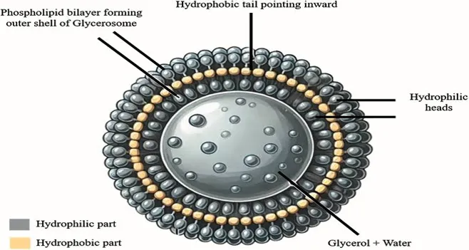
Structure of glycerosomes
Fig no:1 structure of glycerosome2
Composition of glycerosomes:
Glycerosomes are new vesicular systems composed of phospholipids, glycerol, water and cholesterol. Glycerosomes are analogous to conventional lipid-based vesicular system18. These contains the concentration of about 10,20,30,40, and 50% of water and glycerol. It is completely harmless and non-toxic method of drug delivery2.
Glycerol
phospholipids
cholesterol
a. Glycerol - Glycerol is a viscous liquid and an alcohol. It consists of three hydroxyl group which renders hydrophilic properties. It is used in pharmaceutical preparations as a lubricant, humectants, edge activator and emulsifier. And in glycerosomes it mainly acts as penetration enhancer19.
b. Phospholipids- Phospholipids are a class of amphipathic lipids that act as the primary structural component of cell membranes, forming a stable phospholipid bilayer. They consist of a hydrophilic phosphate head, a glycerol backbone, and two hydrophobic fatty acid tails. This structure allows them to regulate cellular transport and maintain membrane integrity20.
Glycerophospholipids and sphingomyelins are widely used in the glycerosomes preparation. These phospholipids are capable of including pharmaceutical ingredients in them and these are harmless. Phospholipids give rise to vesicles that are compatible with other excipients of the formulation.
c. Cholesterol- It is a vital lipid in animal cell membrane and synthetic vesicular systems like glycerosomes which acts as permeability barrier, structural regulator, retention efficiency and preventing the vesicle from collapsing.
Mechanism of action of Glycerosomes: In dermal and transdermal drug delivery, glycerosomes significantly enhance drug penetration through the skin. Glycerol, a key component, acts as a humectant, increasing the hydration of the stratum corneum. This hydration softens the skin and reduces its barrier function, facilitating drug penetration21. Additionally, the incorporation of glycerol into the lipid bilayer of glycerosomes increases membrane fluidity. This fluidization promotes the fusion of glycerosomes with the lipid matrix of the stratum corneum, allowing deeper drug penetration. Furthermore, glycerosomes can fuse with the lipid layers of the skin, releasing the drug directly into the deeper layers of the epidermis and dermis22. And these combination of phospholipids, glycerol, and cholesterol enhances permeability, solubility, entrapment efficiency and controlled release23.
Advantages of glycerosomes:
Enhanced Drug Solubility: By incorporating glycerol as a co-solvent, glycerosomes significantly increase the solubility of weakly water-soluble drugs.
Controlled Drug Release: These vesicles facilitate a sustained and prolonged release of their cargo, which enhances therapeutic efficacy and reduces the required frequency of administration25.
Potential for Targeted Delivery: Like other vesicular systems, glycerosomes can be engineered with surface ligands or modifications to enable site-specific drug delivery26.
Enhanced Skin Penetration: Glycerol acts as a humectant, increasing skin moisture and softening the stratum corneum. This allows glycerosomes to deliver bioactive compounds more effectively into and through the skin.
Improved Skin Permeability: The hydration of the stratum corneum provided by glycerol enhances drug permeation, making these systems highly efficacious for topical and transdermal administration.
Thermal and Osmotic Protection: Glycerosomes exhibit superior resistance to temperature fluctuations and osmotic stress relative to traditional liposomes, ensuring stability during storage and transport under varying conditions27.
Reduced Dehydration Risk: The presence of glycerol inhibits the dehydration of the vesicles, preserving their structural integrity and effectiveness over long durations, even in adverse environments.
Biocompatibility: Glycerol is a naturally occurring, non-toxic, and biocompatible substance. Consequently, glycerosomes are safe for medical and cosmetic use and are well-tolerated by tissues, minimizing the risk of irritation.
Versatility: These systems can encapsulate both hydrophilic and hydrophobic drugs, offering high flexibility. They are suitable for various delivery routes, including:
Topical: For localized treatments and hydration.
Transdermal: For systemic delivery via the skin.
Ocular: For treating eye-related conditions.
Oral: For improving absorption in the gastrointestinal tract27.
Improved Patient Compliance: By enhancing stability, enabling controlled release, and increasing permeability, glycerosomes simplify drug administration, particularly in chronic pain management and transdermal systems.
Stability Improvement: The addition of glycerol reinforces the lipid bilayer structure, which enhances the overall physical and chemical stability of the delivery system.
Enhanced Drug Loading Capacity: Glycerol reduces the rigidity of the bilayer membrane, which can significantly improve the encapsulation efficiency and loading capacity for specific drugs27.
Disadvantages of glycerosomes:
High glycerol concentrations can enlarge vesicles and slow down drug release rates.
While high viscosity enhances vesicle stability, it may simultaneously impede the migration speed of the vesicles to the skin surface21
Methods of preparation of Glycerosomes-
The various methods of preparations are used according to the vesicle lamellarity. The most commonly used method is thin film hydration method.
Mechanical methods
Lipid thin film hydration
Ultrasonic method
Organic solvent replacement methods
Reverse phase evaporation
solvent dispersion
Size transformation method
Freeze thaw extrusion method
the dehydration-rehydration method
All these methods follow basic four stages: Drying, hydrating, purifying and analysing29.
Mechanical methods
Lipid thin film hydration: The formulation of glycerosomes is a streamlined process that begins with dissolving phospholipids in an organic solvent to create a thin lipid film upon evaporation. This film is then hydrated using an aqueous phase consisting of a water and glycerol solution, followed by high-intensity ultrasonication to achieve a stable dispersion. This specific approach is preferred for its ability to produce vesicles with superior physical attributes namely a uniform spherical shape and smooth texture while yielding higher encapsulation efficiency than other techniques30. To ensure optimal drug loading, hydrophilic medications are incorporated via the aqueous hydration buffer, whereas lipophilic drugs are integrated directly into the initial lipid film30.
Ultrasonic Methods: The ultrasonic method is primarily utilized to produce small unilamellar vesicles, typically ranging from 15 to 25 nm in diameter. This process involves the ultrasonication of a glycerol-lipid dispersion through one of two distinct techniques:
Probe Sonication: In this high-energy approach, the sonicator tip is immersed directly into the dispersion, delivering intense energy to facilitate lipid breakdown. Because this process generates significant localized heat at the tip, the formulation must be kept in an ice/water bath to prevent thermal degradation. A notable drawback of this method is the potential for titanium particles to shed from the probe, contaminating the solution and necessitating a subsequent centrifugation step for purification31.
Bath Sonication: This method involves placing the container holding the glycerosomal dispersion into a sonication bath. The process is typically conducted for five to ten minutes at temperatures exceeding the lipid’s critical solution temperature (Tc). Unlike the probe method, bath sonication allows for easier temperature regulation and avoids the risk of metal contamination31.
Organic solvent replacement method:
This method lipid materials are co-solvated in organic solution which is then dispersed in glycerol solution containing drug that to be entrapped within the vesicles
Reverse phase evaporation: In the reverse-phase evaporation method, lipid materials are first co-solvated in an organic solution before being dispersed into a glycerol phase containing the substances targeted for entrapment. This process begins with the formation of a water-in-oil emulsion, created by sonicating a two-phase system of phospholipids and cholesterol in organic solvents such as ethanol, isopropyl ether, or a chloroform-ether mixture alongside an aqueous glycerol solution. As the organic solvent is removed under reduced pressure of 200rpm and 20-250C, the mixture transforms into a viscous gel; subsequent rotary evaporation then eliminates any residual solvent to yield the final glycerosomes. A primary advantage of this technique is its ability to produce both large unilamellar and multilamellar vesicles, which are particularly effective for encapsulating large macromolecules. However, the method is limited by the potential for material degradation, as the entrapped substances are subjected to both organic solvent exposure and mechanical stress during sonication32.
Solvent dispersion method: It is divided into two categories on the type of solvent used
Ether injection method: The ether injection method involves the gradual introduction of a lipid solution dissolved in diethyl ether or an ether-methanol blend into an aqueous or buffered glycerol phase containing the substances to be encapsulated. This procedure is typically executed at temperatures between 55°C and 65°C or under reduced pressure to facilitate solvent evaporation. While functional, the technique often yields a non-uniform glycerosome population, with vesicle sizes ranging widely from 70 to 190 nm. Furthermore, a significant limitation of this approach is the potential degradation of sensitive compounds due to their direct exposure to both organic solvents and elevated processing temperatures27.
Ethanol injection method: The ethanol injection method involves dissolving lipids in ethanol and rapidly forcing the solution through a small aperture, such as a syringe needle, into an excess of aqueous medium. Immediate and thorough mixing is critical to ensure that phospholipids disperse effectively as the ethanol is diluted into the hydration phase. A primary advantage of this technique is its ability to generate small, uniform liposomes typically under 100 nm without the need for secondary processes like sonication or extrusion. However, the method is constrained by the solubility of lipids in ethanol, which limits the total lipid concentration that can be incorporated. Additionally, while residual ethanol can be removed via dialysis, its presence during the initial formation remains a potential limitation for sensitive formulations30.
Size transformation method:
The following techniques utilize thermal and hydration cycles to manipulate vesicle size and structure:
Freeze-Thaw Extrusion Method: This technique relies on rapid cycles of freezing and thawing to transform small unilamellar vesicles (SUVs) into large unilamellar vesicles (LUVs). Primarily applicable to crude or charged phospholipids, the process causes the SUV bilayers to fuse during the thermal cycles, followed by sonication to stabilize the resulting LUVs. However, the efficiency of this method decreases with higher ionic strength or liposome concentrations. A notable limitation is its incompatibility with certain sensitive biological components, as the prolonged, temperature-sensitive nature of the process can lead to the loss or degradation of these materials28.
Dehydration-Rehydration Method: In this approach, pre-formed empty SUVs are mixed with an aqueous solution containing the target material and then dried. This drying process creates a finely subdivided lipid dispersion. Upon rehydration with the aqueous phase, the lipids reform into vesicles, typically yielding oligo-lamellar glycerosomes. This method is particularly useful for achieving a high degree of dispersion in the final formulation32.
Double emulsion evaporation: In this method W1/O/W2 is prepared it consists of inner and outer aqueous phase. Outer phase consists of dispersed individual oil globules and inner aqueous phase as small droplets in each oil globules of outer aqueous phase. This process is of double emulsion in which aqueous phase consisting of drug is dissolved in water and added to organic solvent contains lipids, this forms water in oil emulsion which is then homogenised to form primary emulsion and then combined with outer aqueous phase containing stabilizer gets converted into double emulsion.
Calcium-induced fusion method: In this method LUV’s (larger unilamellar vesicles) are formed in addition calcium to SUV’s (smaller unilamellar vesicles) and results in fusion, then give rise to large planar lamellae which transforms to cochleate cylinders. Then mixed with EDTA which re-establishes the negative charge and maintains the fluidity of the membrane and these further transforms to generate LUV’s27.
Characterization of glycerosomes:
The characterization of glycerosomes involves several critical analytical parameters to ensure their efficacy and stability as delivery vehicles. The following points detail the methodologies used for their evaluation:
a) Vesicle Size and Size Distribution
For parenteral administration, the particle size and distribution of glycerosomes are vital factors. Various techniques are employed for this assessment, including light microscopy, laser light scattering, photon correlation spectroscopy, gel permeation, and gel exclusion. However, transmission electron microscopy (TEM) is considered the most definitive method, as it facilitates the direct observation of individual vesicles to provide precise data on size distribution34.
b) Vesicle Shape and Lamellarity
The morphology and internal structure (lamellarity) of the vesicles are typically evaluated via advanced electron microscopy. Specifically, freeze-fracture electron microscopy and 31P nuclear magnetic resonance (NMR) studies are used to assess lamellarity, while freeze-etch techniques allow for the detailed analysis of surface morphology and overall shape35.
c) Percentage Encapsulation Efficiency
The quantity of drug successfully sequestered within the vesicles is often measured using the dialysis method.
The dialysis method purifies glycerosomes by removing unentrapped drug, excess glycerol, and free lipids using a cellulose-based semi-permeable membrane (MWCO ~12–14 kDa). After soaking the membrane, the crude glycerosomal dispersion is sealed in the dialysis bag and immersed in a large volume of release medium (e.g., buffer with 1% Tween 20) at 37 ± 0.5°C with stirring. Small molecules diffuse out through the membrane while glycerosome vesicles (with 20–40% glycerol) are retained, and the dialysate is refreshed periodically over 2–24 hours until free drug is negligible. The cleaned vesicles are used for further characterization and in vitro release studies, and encapsulation efficiency (%EE) is calculated from the drug obtained after dialysis relative to the initial drug content25,30.
%EE= (Total drug added – Free drug in dialysate/Total drug added) ×100
d) Drug Release Profile
The release kinetics of the encapsulated drug can be investigated using a calibrated Franz diffusion cell. These in vitro tests are essential for understanding the absorption characteristics and functional performance of the glycerosomal formulation prior to conducting in vivo (animal or human) studies.
The Franz diffusion method evaluates in vitro release and permeation of glycerosomes by applying the suspension to a membrane (e.g., cellophane or synthetic skin) placed between donor and receptor compartments. The receptor, filled with phosphate buffer (pH 6.8–7.4) to maintain sink conditions, is kept at 37 ± 0.5°C with magnetic stirring. Samples are withdrawn at set intervals, replaced with fresh buffer, and analyzed by UV or HPLC to quantify drug release and build cumulative release profiles38,39. Glycerosomes typically show biphasic or sustained release with high entrapment efficiency and enhanced permeability versus conventional liposomes, indicating controlled kinetics favorable for deeper topical absorption40.
e) Stability Studies
To determine the stability of glycerosomes over time, various dimensional analysis at regular intervals are performed. This involves measuring the Zeta potential, Dynamic Laser Light Scattering, and the polydispersity index (PDI) via Photon Correlation Spectroscopy to ensure the physical and chemical integrity of the system35.
f) Deformation Index Analysis
The flexibility or "deformability" of the vesicles is assessed by calculating the deformation index. This is determined by forcing the glycerosomal preparation through an extruder with a pore size significantly smaller than the vesicles' mean diameter, measuring their ability to adapt and pass through confined spaces without losing integrity32.
DI= J×(d0/p)×(d0/d0-d1)
where,
DI= Deformability index of glycerosome vesicle
J= The fraction of vesicle suspension recovered after extrusion
d0= The mean hydrodynamic diameter of the vesicles before extrusion.
d1= The mean hydrodynamic diameter of the vesicles after extrusion.
p= The nominal pore diameter of the polycarbonate membrane filter used30.
Therapeutic Applications of glycerosomes:
Recent research highlights the significant potential of glycerosomes in both cosmetic and dermatological applications due to their superior performance. Studies focusing on triptolide-loaded vesicles have demonstrated that glycerosomes, optimized through precise experimental designs, offer enhanced stability, biocompatibility, and transdermal permeability, making them ideal for deep dermal delivery. Furthermore, investigations into treating conditions like rosacea have compared various specialized systems including hexosomes, glycerosomes, and ethosomesutilizing ingredients such as soy phospholipids, tretinoin, and glycerol. While all these systems show promise, glycerosomes were particularly noted for their ability to boost skin penetration, suggesting that these advanced vesicular formulations could significantly improve the effectiveness of topical treatments for inflammatory skin disorders37.
Aerosolized glycerosomal transport: Glycerosomes are emerging as highly effective vehicles for pulmonary drug delivery, offering advantages like sustained release, improved stability, and reduced irritation compared to standard aerosols. Research into the delivery of rifampicin and curcumin shows that these vesicles, particularly when reinforced with polymers like sodium hyaluronate or trimethyl chitosan, significantly increase drug accumulation in the lungs while avoiding hepatic first-pass metabolism. These polymer glycerosomes form a unique hydrophilic mesh that stabilizes the unilamellar vesicles (typically 65–112 nm), enhancing their biocompatibility and ability to be efficiently aerosolized for deep lung penetration. By boosting the anti-inflammatory and antioxidant effects of the encapsulated drugs and ensuring high deposition rates, these advanced formulations represent a promising strategy for treating respiratory illnesses and improving patient adherence33.
Peroral delivery: These advanced delivery systems are specifically designed to treat diseases of the oral cavity by ensuring the effective dispersion and sustained release of therapeutic agents into the oral mucosa. Glycerosomes and penetration-enhancing vesicles significantly boost the biological performance of Citrus Lemon Extract, supporting keratinocyte viability in the oral epithelium when facing oxidative stress. Furthermore, these formulations provide robust antibacterial activity and potent antioxidant protection, making them highly effective tools for managing oral infections and promoting tissue repair within the mouth31.
Targeted Relief for Rheumatoid Arthritis: Managing rheumatoid arthritis effectively is often a struggle because traditional treatments like corticosteroids and NSAIDs fail to reach the synovial cavity in high enough concentrations. Glycerosomes solve this by acting as specialized carriers that can penetrate deep into the joint space. For example, when the immune-regulating drug papeiflorin is encapsulated in these vesicles, it accumulates much more effectively within the synovial cavity, overcoming its usual limitations and improving clinical outcomes41.
Reliable Sustained Drug Release: One of the standout benefits of using glycerosomes is their ability to maintain steady drug levels in the body over a set period. By providing a continuous release of medication, they minimize the effect of drug peaks and valleys, which also reduces the risk of side effects. This approach has been successfully used for medications like betamethasone and rifampicin, ensuring optimal therapeutic rates and better long-term stability in vivo33.
Precision Delivery to Hair Follicles: For skin and scalp conditions such as hair loss, getting the medication exactly where it’s needed like the sebaceous glands or hair follicles is crucial. Advanced glycerosomal formulations make it possible for substances that are normally hard to absorb, such as minoxidil, to be applied topically for localized action. This intrafollicular delivery ensures the treatment stays focused on the target area, boosting the effectiveness of hair restoration therapies16.
Boosting Anti-Inflammatory Power: While anti-inflammatory drugs are essential for many treatments, they often come with a laundry list of side effects. Encapsulating ingredients like diclofenac, celecoxib, or cupferron in glycerosomes significantly increases their therapeutic efficacy while maintaining high biocompatibility with human cells. This means the medicine works better where it's needed most without causing excessive irritation or damage to healthy keratinocytes42.
Stronger and Safer Antimicrobial Action: Glycerosomes improve antimicrobial therapy in two vital ways: they act as a protective shield against enzymatic breakdown and their lipid-based structure helps the medication slip into microbial cells more easily. By loading agents like resveratrol, gallic acid, and citrus lemon extract into these vesicles, researchers have seen a major boost in antibacterial performance against species like Streptococcus and Lactobacillus, all while lowering overall toxicity for the patient32.
Advancing Ocular Care: While primarily known for skin applications, glycerosomes are now making waves in eye care. Recent developments include eye drops formulated with the antifungal drug natamycin. These glycerosomal drops enhance the drug's entrapment and significantly improve its ability to penetrate ocular tissues, providing a more effective way to treat stubborn eye infections compared to traditional methods33.
Neurological disorders: Glycerosomes helps in delivery of drug to specific site of the brain by crossing blood-brain barrier, so makes possible to treat Alzheimer’s, Parkinson’s, and brain cancer. these are administered as neuroprotector drug or small interfering (RNA)31.
Example of Marketed Glycerosome:
Glycerosomal Technology is currently most prominent in the marketed dermo cosmetic sector rather than as a primary platform for mass-marketed prescription drugs.
The primary commercial application is found in the high-end skincare line Gen-Hyal, developed by the pharmaceutical company Prigen.
Marketed Products using Glycerosomes
Gen-Hyal Extreme: A high-performance serum designed with soothing and repairing active ingredients, utilizing glycerosomes to boost effectiveness by 3 to 5 times.
Gen-Hyal Urban Serum: A detoxifying and anti-aging serum that uses glycerosomes for deeper skin penetration of purifying active ingredients.
Gen-Hyal Eyes: An eye contour serum that uses the technology to deliver moisturizing and anti-aging agents more efficiently.
Gen-Hyal Plus / Premium / Elargan: Other facial care formulations within the Prigen line that leverage this patented delivery system for anti-aging results43.
CONCLUSION:
Glycerosomes represent an advanced vesicular drug delivery system that outperforms traditional liposomes in stability, flexibility, and penetration. They incorporate high glycerol concentrations (10-50% v/v) with phospholipids, enhancing bilayer fluidity for better drug entrapment and release control. Glycerosomes provide superior entrapment efficiency (up to 92%), smaller vesicle sizes (around 190 nm), and negative zeta potentials for stability. They enable controlled biphasic drug release, extended stability (up to 3 months at 2-8°C), and spherical morphology, making them ideal for topical, transdermal, and multi-route applications. Overall, they address limitations of conventional drug delivery by improving deformability, skin permeation via stratum corneum moisturization, and protection from harsh environments like the GI tract. Emerging Trends Recent 2025 advancements focus on hybrid variants like STO-glycerosomes (with essential oils), HY-glycerosomes (sodium hyaluronate), TMC-glycerosomes (trimethyl chitosan), glycethosomes, and glycerospanlastics for boosted stability and efficacy.
REFERENCES
Devi S, Jain A, Rathi G, Sharma R, Sharma R. Emugel for topical drug delivery: A novel approach. GSC Biol Pharm Sci. 2020;11(3):103-14.
Ahmed M, Ali M. Semisolid dosage form: topical gel formulation—a review. World J Pharm Res. 2016;5(12):1256–68.
Chourasia MK, Jain SK. Pharmaceutical approaches to colon targeted drug delivery systems. J Pharm Sci. 2012;6(1):33-66.
Kaur J, Singh G, Saini S. Aspects related to the solid lipid nanoparticles delivery through the topical route. J Drug Deliv Ther. 2012;2(6):111-16.
Katz MA, Cheng CH, Nacht S. Methods and compositions for topical delivery of benzoyl peroxide. US Patent 5879716. 1999 Mar 9.
Hadgraft J. Recent developments in topical and transdermal delivery. Eur J Drug Metab Pharmacokinet. 1996;21:165-73.
Wertz PW, Madison KC, Downing DT. Covalently bound lipids of human stratum corneum. J Invest Dermatol. 1989;92(1):109–11.
de Leeuw J, de Vijlder HC, Bjerring P, Neumann HA. Liposomes in dermatology today. J Eur Acad Dermatol Venereol. 2009;23:505-16.
Samad A, Sultana Y, Aqil M. Liposomal drug delivery systems: An update review. Curr Drug Deliv. 2007;4:297-305.
Rogerson A, Cummings J, Willmott N, Florence AT. The distribution of doxorubicin in mice following administration in niosomes. J Pharm Pharmacol. 1988;40:337-42.
Baillie AJ, Coombs GH, Dolan TF, Laurie J. Non-ionic surfactant vesicles, niosomes, as a delivery system for the anti-leishmanial drug sodium stibogluconate. J Pharm Pharmacol. 1986;38(7):502-05
Doktorovova S, Souto EB. Nanostructured lipid carrier-based hydrogel formulations for drug delivery: a comprehensive review. Expert Opin Drug Deliv. 2009; 6(2): 165-76.
Foster KW, Ghannoum MA, Elewski BE. Epidemiologic surveillance of cutaneous fungal infection in the United States from 1999 to 2002. J Am Acad Dermatol. 2004;50(5):748-52. doi:10.1016/S0190-9622(03)02117-20.
Ashtiani HRM, Bishe P, Lashgari NA, Nilforoushzadeh MA, Zare S. Liposomes in cosmetics. J Skin Stem Cell. 2016;3:1-6.
Zhang K, Zhang Y, Li Z, Li N, Feng N. Essential oil-mediated glycerosomes increase transdermal paeoniflorin delivery: optimization, characterization, and evaluation in vitro and in vivo. Int J Nanomedicine. 2017;12:3521-32.
Laura MI, Franco D, Cattel L. Stealth liposomes: review of the basic science, rationale, and clinical applications, existing and potential. Int J Nanomedicine. 2006;1:297-315.
Domenico L, Calandra P, Barreca D, Magazu S, Kiselev MA. Soft interaction in liposome nanocarriers for therapeutic drug delivery. Nanomaterials. 2016;6(125):1-26.
Manca ML, Peris JE, Melis V. Nanoincorporation of curcumin in polymer-glycerosomes and evaluation of their in vitro–in vivo suitability as pulmonary delivery systems. RSC Adv. 2015;127:1-28.
Caballero B, Trugo L, Finglas P. Encyclopedia of Food Sciences and Nutrition. 2nd ed. Vols 1–10. Amsterdam: Elsevier Science BV; 2003.
Manca ML, Manconi M, Zaru M. Glycerosomes: investigation of role of 1,2-dimyristoyl-sn-glycero-3-phosphatidylcholine (DMPC) on the assembling and skin delivery performances. Int J Pharm. 2017;532(1):401-07.
Zaki RM, Alfadhel MM, Alossaimi MA.Central composite optimization of glycerosomes for the enhanced oral bioavailability and brain delivery of quetiapine fumarate. Pharmaceuticals (Basel). 2022;15(8):940.
Melis V, Manca ML, Bullita E. Inhalable polymer-glycerosomes as safe and effective carriers for rifampicin delivery to the lungs. Colloids Surf B Biointerfaces. 2016;143:301–08.
Naguib MJ, Salah S, Halim SA, Badr-Eldin SM. Investigating the potential of utilizing glycerosomes as a novel vesicular platform for enhancing intranasal delivery of lacidipine. Int J Pharm. 2020; 582:119302.
AbouSamra MM, Farouk F, Abdelhamed FM, Emam KA, Abdeltawab NF, Salama AH. Synergistic approach for acne vulgaris treatment using glycerosomes loaded with lincomycin and lauric acid: formulation, in silico, in vitro, LC-MS/MS skin deposition assay and in vivo evaluation. Int J Pharm. 2023; 646:123487.
Kaur P, Verma S, Tomar B, Vyas M, Kakoty V, Saha P, et al. Exploring applications of flexible vesicular systems as transdermal drug delivery. Curr Drug Deliv. 2024;21(8):1062-72.
Jha A, Kumar M, Bharti K, Mishra B. Glycerosomes: a new tool for effective drug delivery. In: Systems of Nanovesicular Drug Delivery. London: Academic Press; 2022. p. 277-91.
Tan C, Wang J, Sun B. Biopolymer-liposome hybrid systems for controlled delivery of bioactive compounds: recent advances. Biotechnol Adv. 2021; 48:107727.
Jabin K, Husain Z, Ahmad M, Kushwaha P. Liposome: classification, preparation, and applications. World J Pharm Pharm Sci. 2018;7(9):1307-19.
Virden JW, Berg JC. Sodium chloride-induced aggregation of dipalmitoylphosphatidylglycerol small unilamellar vesicles with varying amounts of incorporated cholesterol. Langmuir. 1992;8(6):1532-37.
Gupta P, Mazumder R, Padhi S. Glycerosomes: advanced liposomal drug delivery system. Indian J Pharm Sci Rev Res. 2014;27(2):201-306.
Hamid MSS, Hatwar PR, Bakal RL, Kohale NB. A comprehensive review on liposomes: as a novel drug delivery system. GSC Biol Pharm Sci. 2024;27(1):199–210.
Rani D, Sharma V, Singh P, Singh R. Glycerosomes: a novel vesicular drug delivery system. Res J Pharm Technol. 2022;15(2):921-26.
Younes NF, Habib BA. Augmented local skin accumulation efficiency of sertaconazole nitrate via glycerosomal hydrogel: formulation, statistical optimization, ex vivo performance and in vivo penetration. J Drug Deliv Sci Technol. 2022;72:103364.
Traïkia M, Warschawski DE, Recouvreur M, Cartaud J, Devaux PF. Formation of unilamellar vesicles by repetitive freeze-thaw cycles: characterization by electron microscopy and 31P-nuclear magnetic resonance. Eur Biophys J. 2000;29:184-85.
Sharma D, Rani A, Singh VD, Shah P, Sharma S, Kumar S. Glycerosomes: novel nano-vesicles for efficient delivery of therapeutics. Recent Adv Drug Deliv Formul. 2023;17(3):173-82.
Kaddah S, Khreich N, Kaddah F, Charcosset C, Greige-Gerges H. Cholesterol modulates the liposome membrane fluidity and permeability for a hydrophilic molecule. Food Chem Toxicol. 2018; 113:40-48.
Gregoriadis G. The carrier potential of liposomes in biology and medicine (first of two parts). N Engl J Med. 1976; 295:704-10.
Salem HF, Kharshoum RM, Sayed OM, Hakim LFA Formulation design and optimization of novel soft glycerosomes for enhanced topical delivery of celecoxib and cupferron by box-behnken statistical design. Drug Development Ind Pharm 2018;44:1871-84.
Manca ML, Castangiaa I, Caddeoa C, Pando D, Escribano E, Valenti D, et al. Improvement of quercetin protective effect against oxidative stress skin damages by incorporation in nanovesicles. Colloids Surf B Biointerfaces 2014;123:566-74.
Manca ML, Zarub M, Manconi M, Lai F, Valenti D, Sinico C, et al. Glycerosomes: A new tool for effective dermal and transdermal drug delivery. Int J Pharm 2013,455:66-74.
Wang J, Guo F, Ma M, Lei M, Tan F, Li N. Nanovesicular system containing tretinoin for dermal targeting delivery and rosacea treatment: a comparison of hexosomes, glycerosomes and ethosomes. RSC Adv. 2014;4(85):45458-66.
Amiri A, Barreto G, Sathyapalan T, Sahebkar A. siRNA therapeutics: future promise for neurodegenerative diseases. Curr Neuropharmacol. 2021;19(11):1896.
Prigen Srl. Research and development: patented glycerosomal technology. Gen-Hyal Skincare.
Reference
Devi S, Jain A, Rathi G, Sharma R, Sharma R. Emugel for topical drug delivery: A novel approach. GSC Biol Pharm Sci. 2020;11(3):103-14.
Ahmed M, Ali M. Semisolid dosage form: topical gel formulation—a review. World J Pharm Res. 2016;5(12):1256–68.
Chourasia MK, Jain SK. Pharmaceutical approaches to colon targeted drug delivery systems. J Pharm Sci. 2012;6(1):33-66.
Kaur J, Singh G, Saini S. Aspects related to the solid lipid nanoparticles delivery through the topical route. J Drug Deliv Ther. 2012;2(6):111-16.
Katz MA, Cheng CH, Nacht S. Methods and compositions for topical delivery of benzoyl peroxide. US Patent 5879716. 1999 Mar 9.
Hadgraft J. Recent developments in topical and transdermal delivery. Eur J Drug Metab Pharmacokinet. 1996;21:165-73.
Wertz PW, Madison KC, Downing DT. Covalently bound lipids of human stratum corneum. J Invest Dermatol. 1989;92(1):109–11.
de Leeuw J, de Vijlder HC, Bjerring P, Neumann HA. Liposomes in dermatology today. J Eur Acad Dermatol Venereol. 2009;23:505-16.
Samad A, Sultana Y, Aqil M. Liposomal drug delivery systems: An update review. Curr Drug Deliv. 2007;4:297-305.
Rogerson A, Cummings J, Willmott N, Florence AT. The distribution of doxorubicin in mice following administration in niosomes. J Pharm Pharmacol. 1988;40:337-42.
Baillie AJ, Coombs GH, Dolan TF, Laurie J. Non-ionic surfactant vesicles, niosomes, as a delivery system for the anti-leishmanial drug sodium stibogluconate. J Pharm Pharmacol. 1986;38(7):502-05
Doktorovova S, Souto EB. Nanostructured lipid carrier-based hydrogel formulations for drug delivery: a comprehensive review. Expert Opin Drug Deliv. 2009; 6(2): 165-76.
Foster KW, Ghannoum MA, Elewski BE. Epidemiologic surveillance of cutaneous fungal infection in the United States from 1999 to 2002. J Am Acad Dermatol. 2004;50(5):748-52. doi:10.1016/S0190-9622(03)02117-20.
Ashtiani HRM, Bishe P, Lashgari NA, Nilforoushzadeh MA, Zare S. Liposomes in cosmetics. J Skin Stem Cell. 2016;3:1-6.
Zhang K, Zhang Y, Li Z, Li N, Feng N. Essential oil-mediated glycerosomes increase transdermal paeoniflorin delivery: optimization, characterization, and evaluation in vitro and in vivo. Int J Nanomedicine. 2017;12:3521-32.
Laura MI, Franco D, Cattel L. Stealth liposomes: review of the basic science, rationale, and clinical applications, existing and potential. Int J Nanomedicine. 2006;1:297-315.
Domenico L, Calandra P, Barreca D, Magazu S, Kiselev MA. Soft interaction in liposome nanocarriers for therapeutic drug delivery. Nanomaterials. 2016;6(125):1-26.
Manca ML, Peris JE, Melis V. Nanoincorporation of curcumin in polymer-glycerosomes and evaluation of their in vitro–in vivo suitability as pulmonary delivery systems. RSC Adv. 2015;127:1-28.
Caballero B, Trugo L, Finglas P. Encyclopedia of Food Sciences and Nutrition. 2nd ed. Vols 1–10. Amsterdam: Elsevier Science BV; 2003.
Manca ML, Manconi M, Zaru M. Glycerosomes: investigation of role of 1,2-dimyristoyl-sn-glycero-3-phosphatidylcholine (DMPC) on the assembling and skin delivery performances. Int J Pharm. 2017;532(1):401-07.
Zaki RM, Alfadhel MM, Alossaimi MA.Central composite optimization of glycerosomes for the enhanced oral bioavailability and brain delivery of quetiapine fumarate. Pharmaceuticals (Basel). 2022;15(8):940.
Melis V, Manca ML, Bullita E. Inhalable polymer-glycerosomes as safe and effective carriers for rifampicin delivery to the lungs. Colloids Surf B Biointerfaces. 2016;143:301–08.
Naguib MJ, Salah S, Halim SA, Badr-Eldin SM. Investigating the potential of utilizing glycerosomes as a novel vesicular platform for enhancing intranasal delivery of lacidipine. Int J Pharm. 2020; 582:119302.
AbouSamra MM, Farouk F, Abdelhamed FM, Emam KA, Abdeltawab NF, Salama AH. Synergistic approach for acne vulgaris treatment using glycerosomes loaded with lincomycin and lauric acid: formulation, in silico, in vitro, LC-MS/MS skin deposition assay and in vivo evaluation. Int J Pharm. 2023; 646:123487.
Kaur P, Verma S, Tomar B, Vyas M, Kakoty V, Saha P, et al. Exploring applications of flexible vesicular systems as transdermal drug delivery. Curr Drug Deliv. 2024;21(8):1062-72.
Jha A, Kumar M, Bharti K, Mishra B. Glycerosomes: a new tool for effective drug delivery. In: Systems of Nanovesicular Drug Delivery. London: Academic Press; 2022. p. 277-91.
Tan C, Wang J, Sun B. Biopolymer-liposome hybrid systems for controlled delivery of bioactive compounds: recent advances. Biotechnol Adv. 2021; 48:107727.
Jabin K, Husain Z, Ahmad M, Kushwaha P. Liposome: classification, preparation, and applications. World J Pharm Pharm Sci. 2018;7(9):1307-19.
Virden JW, Berg JC. Sodium chloride-induced aggregation of dipalmitoylphosphatidylglycerol small unilamellar vesicles with varying amounts of incorporated cholesterol. Langmuir. 1992;8(6):1532-37.
Gupta P, Mazumder R, Padhi S. Glycerosomes: advanced liposomal drug delivery system. Indian J Pharm Sci Rev Res. 2014;27(2):201-306.
Hamid MSS, Hatwar PR, Bakal RL, Kohale NB. A comprehensive review on liposomes: as a novel drug delivery system. GSC Biol Pharm Sci. 2024;27(1):199–210.
Rani D, Sharma V, Singh P, Singh R. Glycerosomes: a novel vesicular drug delivery system. Res J Pharm Technol. 2022;15(2):921-26.
Younes NF, Habib BA. Augmented local skin accumulation efficiency of sertaconazole nitrate via glycerosomal hydrogel: formulation, statistical optimization, ex vivo performance and in vivo penetration. J Drug Deliv Sci Technol. 2022;72:103364.
Traïkia M, Warschawski DE, Recouvreur M, Cartaud J, Devaux PF. Formation of unilamellar vesicles by repetitive freeze-thaw cycles: characterization by electron microscopy and 31P-nuclear magnetic resonance. Eur Biophys J. 2000;29:184-85.
Sharma D, Rani A, Singh VD, Shah P, Sharma S, Kumar S. Glycerosomes: novel nano-vesicles for efficient delivery of therapeutics. Recent Adv Drug Deliv Formul. 2023;17(3):173-82.
Kaddah S, Khreich N, Kaddah F, Charcosset C, Greige-Gerges H. Cholesterol modulates the liposome membrane fluidity and permeability for a hydrophilic molecule. Food Chem Toxicol. 2018; 113:40-48.
Gregoriadis G. The carrier potential of liposomes in biology and medicine (first of two parts). N Engl J Med. 1976; 295:704-10.
Salem HF, Kharshoum RM, Sayed OM, Hakim LFA Formulation design and optimization of novel soft glycerosomes for enhanced topical delivery of celecoxib and cupferron by box-behnken statistical design. Drug Development Ind Pharm 2018;44:1871-84.
Manca ML, Castangiaa I, Caddeoa C, Pando D, Escribano E, Valenti D, et al. Improvement of quercetin protective effect against oxidative stress skin damages by incorporation in nanovesicles. Colloids Surf B Biointerfaces 2014;123:566-74.
Manca ML, Zarub M, Manconi M, Lai F, Valenti D, Sinico C, et al. Glycerosomes: A new tool for effective dermal and transdermal drug delivery. Int J Pharm 2013,455:66-74.
Wang J, Guo F, Ma M, Lei M, Tan F, Li N. Nanovesicular system containing tretinoin for dermal targeting delivery and rosacea treatment: a comparison of hexosomes, glycerosomes and ethosomes. RSC Adv. 2014;4(85):45458-66.
Amiri A, Barreto G, Sathyapalan T, Sahebkar A. siRNA therapeutics: future promise for neurodegenerative diseases. Curr Neuropharmacol. 2021;19(11):1896.
Prigen Srl. Research and development: patented glycerosomal technology. Gen-Hyal Skincare.
Lekhana V
Corresponding author
Department of Industrial Pharmacy, Srinivas College of Pharmacy, Mangalore, Karnataka, India 574143
Bhavyashree T
Co-author
Department of Industrial Pharmacy, Srinivas College of Pharmacy, Mangalore, Karnataka, India 574143
Dr. Shripathy D
Co-author
Department of Industrial Pharmacy, Srinivas College of Pharmacy, Mangalore, Karnataka, India 574143
Lekhana V, Bhavyashree T, Dr. Shripathy D, Glycerosomes: Novel Vesicles Revolutionizing Topical Drug Delivery, Int. J. of Pharm. Sci., 2026, Vol 4, Issue 6, 4751-4764. https://doi.org/10.5281/zenodo.20752010


 10.5281/zenodo.20752010