

We use cookies to ensure our website works properly and to personalise your experience. Cookies policy
1 Student, M. Pharm, Department of Pharmacology, Ezhuthachan College of Pharmaceutical Sciences, Marayamuttom, Thiruvananthapuram.
2 Professor, Department of Pharmacology, Ezhuthachan College of Pharmaceutical Sciences, Marayamuttom, Thiruvananthapuram.
3 Professor/HOD, Department of Pharmacology Ezhuthachan College of Pharmaceutical Sciences, Marayamuttom, Thiruvananthapuram
4 Principal, Ezhuthachan College of Pharmaceutical Sciences, Marayamuttom, Thiruvananthapuram
Type 2 diabetes mellitus (T2DM) is a rapidly escalating metabolic disorder characterized by persistent hyperglycemia, insulin resistance, impaired insulin signaling, and chronic low-grade inflammation. Its progression is strongly associated with obesity, oxidative stress, altered lipid metabolism, and gut microbiota dysbiosis, which collectively contribute to metabolic endotoxemia and inflammatory activation through TLR4/NF-?B and PI3K/Akt dysregulation. Long-term hyperglycemia predisposes patients to cardiovascular disease, nephropathy, neuropathy, retinopathy, and hepatic dysfunction. Although current antidiabetic agents effectively control glycemia, most target a single pathway and are associated with limited long-term efficacy and adverse effects. Naturally derived bioactive compounds have gained attention as multi-target therapeutic strategies. Prenylated flavonoids exhibit improved lipophilicity, membrane permeability, and enhanced antioxidant and anti-inflammatory activity relative to parent compounds. Resistant starch (RS), a non-digestible carbohydrate, modulates gut microbiota and stimulates production of short-chain fatty acids (SCFAs)—butyrate, propionate, and acetate—that improve insulin sensitivity, reduce inflammation, and maintain intestinal barrier integrity. This review examines the therapeutic potential of prenylated flavonoids and RS as a synergistic multi-target strategy against T2DM, with emphasis on their combined effects on carbohydrate-digesting enzyme inhibition, oxidative stress reduction ,inflammatory pathway modulation, insulin signaling improvement, and gut microbiota regulation through SCFA production. The possible role of prenylated flavonoid–RS complexes in enhancing bioavailability and metabolic efficacy is also discussed. Overall, this integration represents a promising nutraceutical approach simultaneously targeting hyperglycemia, inflammation, insulin resistance, and gut dysbiosis.
Type 2 diabetes mellitus (T2DM) has emerged as one of the most pressing chronic metabolic disorders of the contemporary era, carrying substantial global morbidity and healthcare burden .1,2 Pathophysiologically, T2DM is characterized by progressive insulin resistance in peripheral tissues and deteriorating pancreatic β-cell function, resulting in sustained hyperglycemia and impaired glucose homeostasis.3
The rising prevalence of T2DM is driven by interacting factors including excess adiposity, reduced physical activity, calorie-rich dietary patterns, psychosocial stress, and genetic predisposition.4 Beyond hyperglycemia, T2DM is accompanied by oxidative stress, chronic low-grade inflammation, dyslipidemia, mitochondrial dysfunction, and vascular endothelial injury —all of which synergistically underlie serious long-term sequelae including cardiovascular events, kidney damage, peripheral neuropathy, retinal deterioration, and hepatic impairment.1,3,4
Accumulating evidence has established gut microbiota dysbiosis has a significant mechanistic role in T2DM pathogenesis by impairing intestinal barrier integrity, promoting metabolic endotoxemia, and driving chronic inflammation and insulin resistance.3,4 These observations have highlighted the gut–metabolic axis as an important therapeutic target in diabetes management.5,6 The detailed pathophysiology is discussed in Section 2.
Current pharmacological therapies—including metformin, sulfonylureas, thiazolidinediones, DPP-4 inhibitors, GLP-1 receptor agonists, and exogenous insulin—primarily achieve glycemic control through single or limited mechanistic targets.7 Prolonged use of these agents, however, is associated with gastrointestinal intolerance, hypoglycemic episodes, body weight changes, progressive attenuation of response, and insufficient modulation of the underlying inflammatory and microbial disturbances.7
Given the multifactorial nature of T2DM, there exists a compelling therapeutic imperative for interventions that can simultaneously address hyperglycemia, oxidative stress, inflammation, gut microbial imbalance, and impaired insulin signaling.8
In this setting, dietary polyphenols and flavonoids have gained considerable scientific attention for their capacity to restrain carbohydrate-digesting enzymes, enhance insulin signaling pathways, reduce oxidative damage, and dampen inflammatory cascades.9,10,11 A persistent limitation of many naturally occurring flavonoids is their inadequate aqueous solubility, restricted transmembrane passage, rapid biotransformation, and poor systemic bioavailability, constraining their clinical utility.12,13 Structural modification through prenylation—the enzymatic addition of dimethylallyl or geranyl moieties—substantially improves lipophilicity, cellular uptake, metabolic stability, and biological efficacy compared to parent compounds.14,15,16 Emerging evidence also indicates that prenylated polyphenols may support mitochondrial function and skeletal muscle energy metabolism, conferring supplementary metabolic advantages.17,18
Resistant starch (RS) is a dietary fiber-like non-digestible carbohydrate that bypasses small intestinal digestion and undergoes colonic fermentation by gut microbiota, yielding SCFAs that promote metabolic wellbeing, enhanced insulin responsiveness, and preserve intestinal mucosal integrity.19,20,21
Converging findings suggest that co-administration of polyphenols with RS may yield additive or synergistic benefits through complementary effects on gut microbial ecosystem, SCFA generation presents a comprehensive assessment of the therapeutic utility, inflammatory pathway regulation, and insulin signaling—providing a multi-target approach uniquely suited to the complex pathophysiology of T2DM.22,23
This review therefore presents a comprehensive assessment of the therapeutic utility of prenylated flavonoids and RS in T2DM, focusing on enzyme inhibition, gut microbiota modulation, SCFA-mediated metabolic regulation, HDAC inhibition, inflammatory pathway control, and improvement of insulin sensitivity as integrated multi-target natural interventions.
PATHOPHYSIOLOGY OF GUT DYSBIOSIS IN TYPE 2 DIABETES MELLITUS
T2DM is a multifactorial metabolic disorder defined by chronic hyperglycemia arising from progressive insulin resistance and pancreatic β-cell dysfunction. The gut microbiota—comprising approximately 10¹³–10¹⁴ microorganisms—maintains systemic metabolic homeostasis through coordinated regulation of nutrient metabolism, intestinal barrier integrity, immune function, and endocrine signaling.2 Gut dysbiosis—a qualitative and quantitative imbalance in microbial communities—has been recognized as an active mechanistic driver of T2DM through perturbations in microbial metabolite profiles, breakdown of mucosal barrier integrity, systemic endotoxin translocation, inflammatory activation, disrupted incretin and bile acid signaling, and eventual β-cell exhaustion.3,4
Shifts in Gut Microbial Composition and Ecological Diversity
A defining characteristic of T2DM-related dysbiosis is reduced alpha-diversity coupled with systematic compositional shifts, including an elevated Firmicutes-to-Bacteroidetes ratio and depletion of Verrucomicrobia and Actinobacteria populations.24 Beneficial commensals are substantially diminished: Akkermansia muciniphila (which fortifies the mucosal barrier and enhances insulin sensitization), Faecalibacterium prausnitzii (a key anti-inflammatory butyrate producer), Roseburia intestinalis and Eubacterium rectale (colonic butyrate synthesizers), and Bifidobacterium and Lactobacillus spp. (immunomodulators and barrier protectors).24,25 Concurrently, opportunistic taxa—Escherichia coli, Ruminococcus gnavus, and sulfate-reducing Desulfovibrio spp.—expand, augmenting LPS, BCAA, and trimethylamine (TMA) production while reducing beneficial SCFAs, secondary bile acids, and indole derivatives.26,27 Notably, hyperglycemia itself reshapes microbial ecology, establishing bidirectional reinforcing feedback loops.
Impaired Intestinal Barrier Function and Increased Permeability
The intestinal barrier comprises a mucus bilayer, polarized epithelial cells joined by tight junction (TJ) complexes—occludin, claudins-1/-3/-4/-5, and zonula occludens-1 (ZO-1)—and the gut-associated lymphoid tissue (GALT).2 In dysbiosis, depletion of SCFA-producing commensals reduces butyrate availability, impairing transcriptional upregulation of claudin-1, occludin, and ZO-1 via HDAC inhibition and PPARγ signaling.28,29 Reduced Akkermansia muciniphila abundance diminishes MUC2 production and mucus layer thickness. Expansion of proteolytic taxa promotes epithelial apoptosis through caspase-3/-9 activation and degrades TJ proteins via microbially derived proteases and hydrogen sulfide. The resulting increase in intestinal permeability permits translocation of luminal antigens and endotoxins, initiating and sustaining systemic inflammatory responses.
Metabolic Endotoxemia, TLR4 Activation, and Chronic Inflammation
Increased intestinal permeability enables systemic translocation of lipopolysaccharide (LPS), a glycolipid constituent of the outer membrane of Gram-negative bacteria.2,3 Elevated circulating LPS—referred to as metabolic endotoxemia—correlate with fasting insulin levels, insulin resistance indices, and inflammatory markers. LPS complexes with LPS-binding protein (LBP) and activates Toll-like receptor 4 (TLR4) via CD14 on macrophages, adipocytes, and hepatocytes.3 TLR4 engagement triggers the MyD88-dependent cascade (IRAK–TRAF6–IKK–NF-κB) and TRIF-dependent pathway (TBK1–IRF3), both activating MAPK (JNK, p38). NF-κB-driven transcription produces TNF-α, IL-1β, IL-6, and MCP-1, establishing chronic metaflammation. The NLRP3 inflammasome amplifies metabolic tissue injury by processing pro-IL-1β and pro-IL-18 into biologically active forms.8
Cellular and Molecular Mechanisms of Insulin Resistance
Under normal metabolic conditions, insulin receptor activation triggers IRS-1/IRS-2 tyrosine phosphorylation, PI3K–PIP₃–PDK1–Akt signaling, and GLUT4 translocation to the plasma membrane. TNF-α and IL-1β disrupt this cascade by activating JNK and IKKβ, which catalyze inhibitory serine phosphorylation of IRS-1 and promote its proteasomal degradation, attenuating PI3K-Akt signaling and impairing glucose uptake in skeletal muscle and adipose tissue.2,3 Within the liver, JNK and NF-κB activation upregulates gluconeogenic enzymes (PEPCK, G6Pase), augmenting hepatic glucose production. IL-6-induced SOCS3 further dampens insulin responsiveness via JAK-STAT3 signaling.5
Short-Chain Fatty Acid Deficiency and Epigenetic Consequences
SCFAs—butyrate, propionate, and acetate—are produced by microbial fermentation of dietary fiber at approximate molar ratios of 60:20:20. They signal through GPR41 (FFAR3) and GPR43 (FFAR2) on enteroendocrine cells and adipocytes to stimulate GLP-1/PYY secretion and suppress lipolysis.19 Butyrate exerts potent epigenetic effects through class I/II HDAC inhibition, suppressing NF-κB-driven inflammation and inducing Foxp3+ regulatory T cell (Treg) differentiation.28,29 Metabolically, butyrate activates AMPK and PGC-1α, promoting mitochondrial biogenesis and fatty acid β-oxidation.30,31 SCFA deficiency in dysbiosis simultaneously increases intestinal permeability, amplifies inflammation, impairs insulin signaling, and disrupts mucosal immune homeostasis.
Dysregulation of Incretin Hormones and Bile Acid Signaling
Incretin hormones—principally GLP-1 and GIP—account for up to 70% of postprandial insulin secretion. SCFA-mediated GPR41/43 activation on intestinal L-cells stimulates GLP-1 and PYY secretion; secondary bile acids further stimulate GLP-1 via TGR5; and indole derivatives from microbial tryptophan catabolism augment L-cell GLP-1 release through the aryl hydrocarbon receptor (AhR).32,33 In dysbiosis, reduced SCFA production and altered bile acid profiles impair L-cell stimulation, leading to GLP-1 deficiency, accelerated gastric emptying, reduced satiety signaling, and progressive β-cell secretory failure.31,33 Gut bacteria expressing bile salt hydrolase (BSH) and 7α-dehydroxylase convert primary bile acids into secondary forms (deoxycholic acid, lithocholic acid), activating FXR and TGR5. When dysbiosis shifts the bile acid pool toward conjugated primary forms, hepatic gluconeogenesisis augmented, attenuating GLP-1 secretion, and worsening insulin resistance.34
BCAA Dysmetabolism and TMAO-Mediated Dysfunction
Circulating BCAAs (leucine, isoleucine, valine) represent independent predictors of insulin resistance and T2DM risk. Dysbiotic species such as Prevotella copri and Bacteroides vulgatus augment BCAA biosynthesis while impairing catabolism through suppression of the BCKDH complex. Elevated BCAAs activate mTORC1–S6K1, inducing inhibitory serine phosphorylation of IRS-1 and attenuating PI3K-Akt signaling.35,36
Trimethylamine N-oxide (TMAO), generated from dietary choline, phosphatidylcholine, and L-carnitine via microbial CutC/D and hepatic FMO3, independently correlates with insulin resistance and T2DM risk. TMAO activates PERK-mediated ER stress, inhibiting Akt phosphorylation and amplifying inflammatory cytokine production. It additionally antagonizes FXR, promoting hepatic steatosis, and drives adipose macrophage M1 polarization.35,36
Immune Dysregulation, Oxidative Stress, and β-Cell Dysfunction
Gut dysbiosis disrupts the mucosal equilibrium between pro-inflammatory Th17 cells and immunosuppressive regulatory T cells (Tregs), shifting toward a pro-inflammatory state that amplifies intestinal permeability via IL-17A/F-mediated MMP activation and claudin-1 suppression.2,3 Chronic inflammatory signaling from gut-derived mediators drives pathological ROS generation through NOX2/NOX4 activation and mitochondrial electron transport chain disruption.4,5 Overwhelming ROS accumulation exceeds the capacity of endogenous antioxidant systems (SOD1/SOD2, catalase, Nrf2-Keap1), oxidatively modifying the insulin receptor and IRS proteins. In skeletal muscle, mitochondrial dysfunction reduces oxidative phosphorylation capacity; in the liver, ROS-mediated PP2A activation augments gluconeogenesis; and in endothelium, superoxide scavenging of nitric oxide impairs insulin-stimulated vasodilation.2,3,4
Pancreatic β-cells are particularly vulnerable to inflammatory and oxidative insults due to low antioxidant enzyme expression. IL-1β and IFN-γ activate NF-κB within β-cells, inducing iNOS-derived NO overproduction that impairs electron transport, depletes NAD+/ATP, and triggers apoptosis via caspase-3/-8. Sustained ER stress activates the unfolded protein response (UPR) through PERK, IRE1α, and ATF6 branches, ultimately inducing CHOP-driven apoptosis, suppressing key transcription factors (PDX-1, MafA, Nkx6.1), and promoting β-cell dedifferentiation. Concomitant incretin deficiency further removes the GLP-1 trophic and anti-apoptotic signal that normally supports β-cell survival.4,31
Adipose Tissue Inflammation and the Gut–Brain Axis
Gut-derived LPS, transported by postprandial chylomicrons, activates TLR4 on visceral adipose macrophages, polarizing them toward the M1 phenotype (TNF-α, IL-6, MCP-1 production) and stimulating hormone-sensitive lipase (HSL) and ATGL to drive lipolysis.2,3 Elevated circulating NEFAs propagate insulin resistance in muscle and liver through TLR4 activation and PKC-mediated IRS-1 serine phosphorylation. NF-κB-mediated suppression of adiponectin removes a key AMPK/PPARα-activating insulin-sensitizing signal, while chronic hyperleptinemia induces central leptin resistance.2,3,4
The bidirectional gut–brain axis, communicating via the vagus nerve, ENS, systemic microbial metabolites, and immune mediators, represents an additional dimension of dysbiosis-mediated metabolic dysfunction. Approximately 90–95% of peripheral serotonin is synthesized by enterochromaffin cells under microbial regulation; its dysregulation impairs gut motility, nutrient transit, and satiety signaling.3,4 Inflammatory cytokines and metabolites (LPS, TMAO, kynurenine) can traverse the blood–brain barrier to activate hypothalamic microglia, promoting central insulin and leptin resistance. SCFA-mediated GPR41/43 signaling in hypothalamic and brainstem nuclei, attenuated in dysbiotic states, further disrupts central appetite and energy homeostasis.8
Figure1 illustrates the mechanistic connection between gut dysbiosis and insulin resistance in T2DM. In the healthy metabolic state, a balanced dietary intake maintains tight junctions in the intestinal epithelium and supports beneficial bacteria such as Bifidobacteria, Faecalibacterium, and Akkermansia, along with robust SCFA production — particularly butyrate and acetate — that maintains gut barrier integrity. In contrast, an unbalanced diet leads to dysbiosis, characterized by expansion of pathogenic Gram-negative LPS-producing bacteria, reduced SCFA levels, and loosening of tight junctions, resulting in a leaky gut. This allows lipopolysaccharide (LPS) endotoxins to translocate into the bloodstream, where they activate TLR4 receptors on immune cells, triggering the release of inflammatory cytokines such as TNF-α and IL-6. These cytokines activate IKKβ and JNK signalling pathways, which induce serine phosphorylation of IRS-1 — acting as an "OFF switch" — thereby blocking the PI3K/Akt pathway, impairing GLUT-4 translocation, and ultimately reducing glucose uptake in muscle and fat cells, leading to insulin resistance and elevated blood sugar levels.
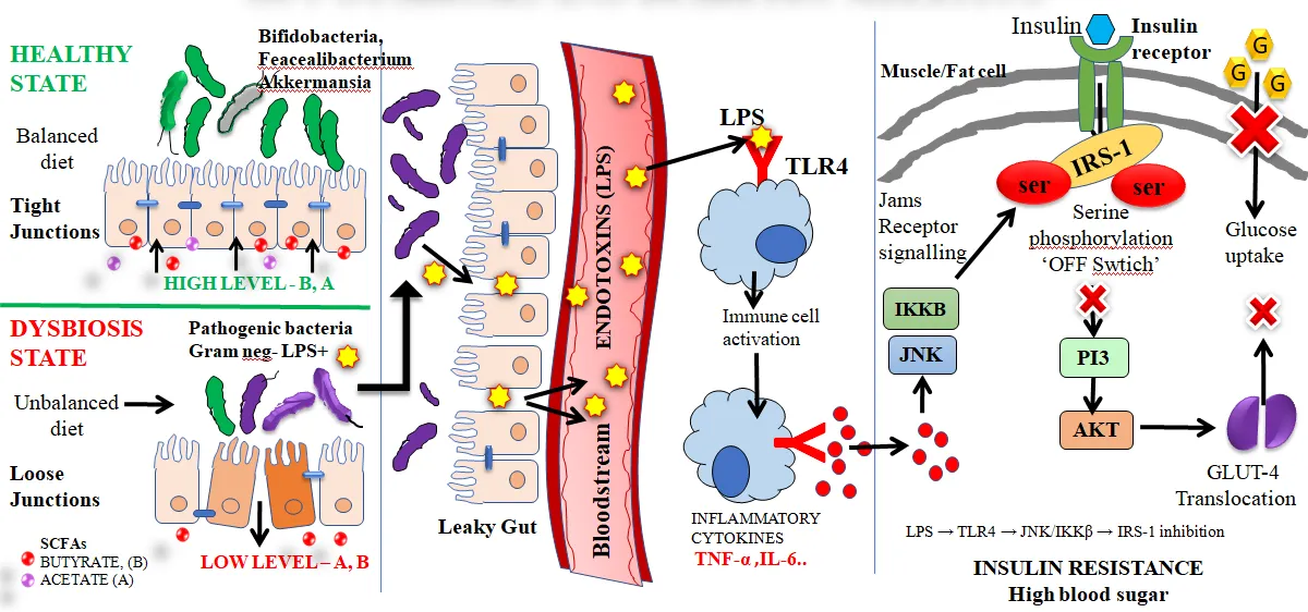
Figure 1; Pathophysiological mechanisms linking gut dysbiosis and showing the progression of Type 2 diabetes mellitus
PRENYLATED FLAVONOIDS IN TYPE 2 DIABETES MELLITUS
Flavonoids constitute a structurally diverse class of plant polyphenols found across fruits, vegetables, medicinal plants, tea, cocoa, and cereals, with documented beneficial effects in T2DM through inhibition of carbohydrate-digesting enzymes, improvement of insulin signaling, attenuation of oxidative stress, suppression of inflammatory pathways, protection of pancreatic β-cells, and modulation of gut microbiota.9,10,11 Despite their pharmacological promise, most flavonoids exhibit poor aqueous solubility, low intestinal absorption efficiency, rapid systemic clearence, limited membrane permeability, and inadequate bioavailability, which collectively impede clinical application.12,13
Among structural modification strategies, prenylation has emerged as particularly effective. Prenylated flavonoids contain one or more hydrophobic prenyl groups (dimethylallyl or geranyl moieties) attached to the flavonoid backbone by prenyltransferase enzymes in plants.14 This modification increases lipophilicity, facilitates membrane penetration, improves cellular uptake, enhances metabolic stability, and prolongs biological activity, generally conferring superior antioxidant, anti-inflammatory, antidiabetic, and neuroprotective activities relative to non-prenylated counterparts.15,16
Prenylation occurs through C-prenylation (direct carbon–carbon bond attachment, conferring enhanced chemical stability and resistance to metabolic degradation) or O-prenylation (ether linkage, improving membrane permeability but with greater susceptibility to hydrolysis).37,38,39 Accumulating evidence supports significant therapeutic potential of prenylated flavonoids in T2DM through multiple mechanisms: inhibition of α-amylase and α-glucosidase, enhancement of insulin sensitivity via PI3K/Akt and AMPK activation (increasing GLUT4 translocation and reducing hepatic gluconeogenesis), potent antioxidant activity through Nrf2 pathway activation, and suppression of NF-κB and NLRP3 inflammasome signaling. Recent investigations also indicate modulation of gut microbiota composition, with enhancement of SCFA production and improved intestinal barrier integrity providing additional metabolic benefits.40,41
Key Prenylated Flavonoids with Antidiabetic Activity
Several naturally occurring prenylated flavonoids have demonstrated promising pharmacological activities in metabolic disorders.15,38,39
Table 1: Prenylated flavonoids and their antidiabetic properties
|
Compound |
Natural source |
Antidiabetic mechanism |
Key activity |
|
Xanthohumol |
Hops |
AMPK activation, anti-inflammatory, antioxidant |
Antidiabetic, Antioxidant |
|
Isoxanthohumol |
Hops |
Regulation of glucose metabolism |
Anti-inflammatory |
|
8-Prenylnaringenin |
Hops |
Modulation of metabolic signaling pathways |
Antioxidant |
|
Glabridin |
Licorice root |
Improves insulin sensitivity; reduces oxidative stress |
Antioxidant, Insulin sensitizer |
|
Kuwanon G |
White mulberry |
α-Glucosidase inhibition |
Antihyperglycemic |
|
Sophoraflavanone G |
Ku Shen root |
Anti-inflammatory and antioxidant mechanisms |
Antioxidant |
|
Prenylated quercetin derivatives |
Multiple species |
Enhanced enzyme inhibition and glucose regulation |
Antidiabetic, Improved bioavailability |
Among these listed compounds, xanthohumol and glabridin are the most extensively investigated in metabolic disorders. Xanthohumol improves glucose homeostasis through AMPK activation and suppression of pro-inflammatory mediators, whereas glabridin improves insulin sensitivity and mitigates oxidative damage.42
Evidence from Experimental Diabetes Models
Prenylated flavonoids have demonstrated antidiabetic activity in experimental models through distinct molecular mechanisms. Xanthohumol ameliorates glucose homeostasis and improves insulin responsiveness through AMPK activation and attenuation of oxidative stress.42,43 Glabridin exerts insulin-sensitizing and antioxidant effects, while Kuwanon G exhibits potent α-glucosidase inhibitory activity contributing to reduced postprandial glucose levels. Prenylated quercetin derivatives demonstrate enhanced antioxidant capacity and stronger enzyme inhibitory activity compared with non-prenylated quercetin, attributable to increased lipophilicity and improved interaction with biological targets.37,44
Emerging evidence further suggests that prenylated flavonoids improve mitochondrial function, enhance skeletal muscle glucose utilization, and regulate cellular energy metabolism through AMPK-PGC-1α signaling, providing additional mechanisms supporting insulin sensitivity and reduced metabolic dysfunction in T2DM.44
RESISTANT STARCH AND GUT MICROBIOTA
Resistant starch (RS) refers to the portion of dietary starch that resists enzymatic hydrolysis in the small intestine and passes intact to the colon, where it serves as a fermentable substrate for resident gut microbiota.45 This digestive behavior renders RS functionally analogous to dietary fiber rather than conventional digestible starch, and it has been characterized as a bioactive food component with meaningful capacity to support gut health, regulate blood glucose, enhance insulin sensitivity, and suppress metabolic inflammation in T2DM.45,46
Dietary starch is classified into three fractions based on digestion rate:
Table 2: Classification of dietary starch types and their metabolic effects
|
Type |
Description |
Metabolic Effect |
|
Rapidly digestible starch (RDS) |
Digested within the first 20 min |
Rapid glucose release; elevated postprandial glycemia |
|
Slowly digestible starch (SDS) |
Digested between 20–120 min |
Gradual glucose release; improved glycemic control |
|
Resistant starch (RS) |
Escapes small intestinal digestion |
Colonic fermentation; SCFA production |
Quantification uses the equations: RDS = (G20 − FG) × 0.9; SDS = (G120 − G20) × 0.9; RS = TS − (RDS + SDS), where G20 and G120 are glucose released at 20 and 120 min, respectively, FG is free glucose, TS is total starch, and 0.9 is the glucose-to-starch conversion factor.21,45
Resistant Starch is mainly classified as five types:
Table 3: Classification of resistant starch types and their common sources
|
Type |
Description |
Common Sources |
|
RS1 |
Physically inaccessible starch enclosed within intact cell walls |
Whole grains, seeds, legumes |
|
RS2 |
Native ungelatinized granular starch |
Raw potato, green banana, high-amylose maize |
|
RS3 |
Retrograded starch formed after cooking and cooling |
Cooked-cooled rice, potato, breadfruit starch |
|
RS4 |
Chemically modified starch |
Cross-linked or esterified starches |
|
RS5 |
Amylose–lipid complex starch |
Starch–lipid complexes, processed foods |
Among these, RS3 is particularly important in functional food applications: formed during gelatinization–retrogradation, amylose chains reassociate into compact crystalline structures that are highly resistant to digestive enzymes and well-suited for colonic fermentation.47 RS5, formed through amylose–lipid complexation, represents an emerging class with distinct digestibility and fermentation characteristics.
Underutilized Sources of Resistant Starch
Many underutilized plant sources offer promising RS potential due to their local availability, nutritional value, and low cost.48,49
Table 4: Sources and significance of underutilized resistant starches
|
Source |
Significance |
|
Breadfruit |
Suitable for RS3 preparation by gelatinization and retrogradation |
|
Jackfruit seed |
Underutilized seed with high starch content |
|
Green banana |
Rich in RS2; suitable for functional food applications |
|
Yam and taro |
Tropical tubers with starch-rich composition |
|
Millets |
Traditional grains with slowly digestible starch and fiber content |
|
Cassava and sweet potato |
Abundant tuber sources for modified and retrograded starch preparation |
Fermentation of RS and SCFA Production
Upon reaching the large intestine, RS undergoes microbial fermentation with primary generation of acetate, propionate, and butyrate.30,48
Table 5: Short chain fatty acids primary site and metabolic roles
|
SCFAs |
Primary Site/Transport |
Key Metabolic Role |
|
Acetate |
Circulation; peripheral tissues |
Energy substrate; appetite regulation; lipid metabolism |
|
Propionate |
Liver |
Reduces gluconeogenesis; improves lipid metabolism |
|
Butyrate |
Colonocytes; immune cells |
Maintains gut barrier; reduces inflammation; HDAC inhibitor |
RS fermentation selectively promotes Bifidobacterium, Faecalibacterium prausnitzii, Roseburia, and environments favorable for Akkermansia muciniphila—bacteria associated with improved gut barrier function, lower endotoxemia, and better metabolic regulation.50,51
Metabolic Actions of SCFAs in T2DM
SCFAs act as both energy substrates and signaling molecules, improving T2DM-related metabolic imbalance through multiple mechanisms.20,31,32
Table 6: Actions and mechanisms of SCFAs
|
Action |
Mechanism |
|
Improves insulin sensitivity |
SCFAs enhance insulin signaling via AMPK activation, increasing glucose uptake and metabolic flexibility |
|
Reduces inflammation |
Butyrate and propionate suppress TNF-α, IL-6, and NF-κB through HDAC inhibition and receptor-mediated pathways |
|
Strengthens gut barrier |
Butyrate upregulates claudin-1, occludin, and ZO-1 expression, reducing LPS translocation |
|
Regulates gut microbiota |
RS selectively increases SCFA-producing beneficial microbial populations |
|
Improves lipid metabolism |
SCFAs regulate hepatic lipid synthesis and enhance fatty acid β-oxidation |
|
Supports β-cell function |
Butyrate protects β-cells from IL-1β-induced dysfunction through HDAC inhibition |
HDAC Inhibition by Butyrate
Among SCFAs, butyrate has particular epigenetic importance as a class I/II histone deacetylase (HDAC) inhibitor. HDAC inhibition modulates the expression of genes governing inflammation, glucose metabolism, mitochondrial biogenesis, and insulin signaling. By preventing IκB degradation and blocking NF-κB nuclear translocation, butyrate suppresses pro-inflammatory gene transcription.28,29 Through HDAC3 inhibition, butyrate promotes Foxp3+ Treg differentiation, restoring mucosal immune homeostasis. These epigenetic mechanisms collectively reduce metabolic inflammation and improve insulin sensitivity in T2DM.33
Figure 2, summarizes five key major metabolic actions of short-chain fatty acids (SCFAs) — butyrate, propionate, and acetate — in T2DM. First, through GPCR-mediated signalling, SCFAs activate GPR41 (FFAR3), GPR43 (FFAR2), and GPR109A (HCAR2) receptors, triggering downstream pathways that reduce lipolysis and inflammation, enhance glucose uptake via GLUT-4 translocation, and improve insulin sensitivity. Second, Through HDAC inhibition in the nucleus, butyrate drives epigenetic reprogramming, increasing histone acetylation and inducing transcriptional upregulation of PGC-1, IRS-2, and GLUT-4, promoting mitochondrial biogenesis, augmenting fatty acid oxidation, and improving insulin sensitivity. Third, SCFAs stimulate intestinal hormone modulation by activating L-cells and K-cells via GPR41/43, increasing GLP-1 and PYY secretion, which collectively enhance insulin secretion, suppress glucagon, slow gastric emptying, and promote satiety and weight control. Fourth, SCFAs contribute to improvement of intestinal barrier function by activating the AMPK pathway and upregulating tight junction proteins — ZO-1, occludin, and claudin-1 — thereby reducing intestinal permeability, endotoxemia, and metabolic inflammation. Finally, in hepatic tissue, SCFA-driven AMPK activation curtails gluconeogenesis, promotes fatty acid oxidation, inhibits de novo lipogenesis, and improves hepatic insulin responsiveness, collectively addressing the multifaceted metabolic dysfunctions characteristic of T2DM.
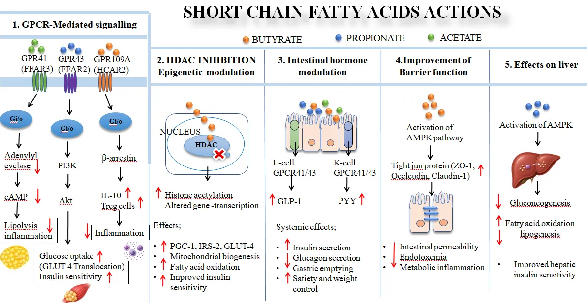
Figure 2; Metabolic and immunomodulatory actions of SCFAs in Type 2 diabetes mellitus.
SHORT-CHAIN FATTY ACIDS, SKELETAL MUSCLE METABOLISM, AND T2DM
Skeletal muscle constitutes the predominant site of insulin-driven glucose disposal, contributing approximately 80% of total postprandial glucose utilization.52 In T2DM, skeletal muscle displays impaired insulin signaling, diminished glucose uptake capacity, mitochondrial operational deficits, and altered energy substrate handling that collectively and substantially amplify whole-body insulin resistance and impair glycemic control.53 Emerging investigations indicate that gut microbiota-derived SCFAs modulate skeletal muscle metabolic function through several interconnected molecular mechanisms.54,55
Skeletal Muscle Dysfunction in T2DM
Under physiological conditions, insulin stimulates IRS-PI3K-Akt-dependent GLUT4 translocation to the sarcolemmal membrane, facilitating cellular glucose uptake. In T2DM, chronic inflammation, oxidative stress, elevated free fatty acids, and mitochondrial dysfunction impair this pathway.52 Progressive muscle atrophy and reduced muscle quality further exacerbate metabolic dysfunction, establishing a deteriorating cycle of worsening insulin resistance and declining glucose utilization capacity.52,53
Contributions to Skeletal Muscle Metabolic Function
SCFAs—particularly acetate, propionate, and butyrate—confer beneficial effects on skeletal muscle metabolism by enhancing insulin responsiveness, facilitating glucose uptake, and attenuating inflammatory signaling.55 SCFAs activate AMPK, increase fatty acid oxidation, and promote metabolic flexibility. Propionate may contribute to skeletal muscle glucose metabolism through gluconeogenic pathways and GPR41/43-mediated signaling. Butyrate-driven AMPK and PGC-1α activation stimulates mitochondrial biogenesis, enhances oxidative phosphorylation, and improves ATP production, addressing the mitochondrial dysfunction characteristic of insulin-resistant skeletal muscle.17,18,55
Prenylated Flavonoids and Muscle Metabolism
In addition to their antidiabetic and antioxidant activities, prenylated flavonoids may support skeletal muscle function through improvements in mitochondrial activity, reduction of oxidative burden, enhancement of insulin signal transduction, and promotion of myocellular glucose utilization.15,18 Prenylation may further augment these effects through improved membrane permeability and cellular uptake. Although direct evidence for prenylated flavonoids specifically in muscle metabolism remains limited, their capacity to activate AMPK-PGC-1α signaling and suppress inflammatory pathways represents a mechanistic basis for improved skeletal muscle metabolic health in T2DM.18
SYNERGISTIC APPROACH: PRENYLATED FLAVONOID–RESISTANT STARCH COMPLEX
The combination of prenylated flavonoids with RS represents a promising multi-target nutraceutical strategy for T2DM management. This synergistic approach integrates the direct antihyperglycemic and antioxidant effects of prenylated flavonoids with the gut-mediated metabolic benefits of RS.22,23 Relative to individual components used in isolation, flavonoid–RS complexes may offer improved physicochemical stability, enhanced oral bioavailability, controlled-release kinetics, improved colon delivery efficiency, and broader metabolic regulation through simultaneous engagement of multiple T2DM pathological pathways.56,57
Resistant Starch as a Natural Carrier System
RS can serve as a natural biocompatible carrier for bioactive phytoconstituents by virtue of its biodegradability, non-toxic profile, and fermentation-mediated colonic release properties.56,58 The compact retrograded structure of RS3, in particular, enables encapsulation or surface association with hydrophobic phytoconstituents such as prenylated flavonoids, protecting them from early degradation in the upper gastrointestinal tract.47,58 Because RS resists salivary and pancreatic enzyme digestion, associated bioactive compounds can remain protected until reaching the colon where microbial fermentation facilitates their controlled release. This makes RS an attractive platform for colon-targeted nutraceutical delivery.59
Molecular Interactions Underlying Complex Formation
Complex formation between prenylated flavonoids and RS occurs primarily through non-covalent molecular interactions. The increased hydrophobicity conferred by prenyl groups facilitates interaction with the starch matrix through several mechanisms23,60 like; Hydrogen bonding between flavonoid hydroxyl groups and starch polymer hydroxyls, hydrophobic interactions between prenyl groups and amylose helical regions, Van der Waals interactions, Electrostatic interactions and Inclusion complex formation within amylose helical structures
These non-covalent interactions may alter crystallinity, molecular arrangement, and physicochemical properties of the complex. Flavonoid–starch systems may form inclusion complexes (flavonoid entrapped within amylose helices), surface adsorption complexes (binding onto starch granule surfaces), or matrix entrapment systems (physical dispersion within retrograded starch matrices).60,61
It is important to acknowledge that dedicated experimental investigations into prenylated flavonoid–RS complexes specifically remain scarce. The proposed interactions discussed here are based on established knowledge of flavonoid–starch complexes and the physicochemical properties of prenylated flavonoids. Further targeted experimental work is needed to characterize these assemblies and confirm their in vivo metabolic benefits.23
Preparation: Solvent Dispersion Method
Among available fabrication techniques, solvent dispersion is regarded as a practical and accessible method for preparing flavonoid–RS complexes. The prenylated flavonoid is dissolved in a compatible organic solvent (e.g., ethanol), while RS is separately dispersed in distilled water. Both phases are combined under continuous mechanical agitation to promote flavonoid–starch molecular interaction, followed by controlled solvent evaporation and drying to produce a stable dry complex. This approach is valued for its operational simplicity, economical material requirements, mild processing conditions, and compatibility with thermosensitive phytoconstituents.62
Dual Mechanism of Antidiabetic Action
The prenylated flavonoid–RS complex may exert antidiabetic activity through complementary direct and indirect mechanisms:
Direct Mechanism: Enzyme Inhibition
Prenylated flavonoids inhibit α-amylase and α-glucosidase, delaying starch digestion and intestinal glucose absorption and thereby reducing postprandial hyperglycemia. Prenylation enhances lipophilicity and interaction with enzyme active sites, potentially improving inhibitory potency compared to native flavonoids.63,64,65
Indirect Mechanism: SCFA Production and Gut Modulation
RS undergoes microbial fermentation in the colon, yielding SCFAs that enhance insulin sensitivity, suppress systemic inflammation, regulate glucose metabolism, and maintain mitochondrial function in metabolically active tissues.19,20 Among SCFAs, butyrate plays a particularly important role through HDAC inhibition and transcriptional regulation of genes involved in glucose metabolism and inflammatory signaling.28,29
Concurrently the RS matrix functions as a prebiotic substrate promoting Akkermansia muciniphila, Bifidobacterium, and butyrate-producing bacteria, while flavonoids may help suppress pathogenic taxa and reduce intra-luminal oxidative stress. This combined ecological and biochemical effect strengthens intestinal barrier integrity, reduces metabolic endotoxemia, and enhances insulin sensitivity.51,66
CHALLENGES AND LIMITATIONS
Despite the promising therapeutic potential of prenylated flavonoid–RS systems in T2DM, several challenges limit clinical translation. Stability concerns are paramount: flavonoids are susceptible to oxidation, pH variation, light exposure, and enzymatic degradation during processing and gastrointestinal transit.12,13 Although prenylation and RS complexation may improve stability, maintaining long-term structural integrity and bioactivity remains challenging.22,23
Lack of standardization represents another significant limitation. Variability in plant source, extraction method, degree of prenylation, RS preparation, and complex formation techniques can substantially affect physicochemical properties and biological activity.14,38 Differences in amylose content, retrogradation conditions, and interaction efficiency further compromise reproducibility.22,23
Clinical validation of prenylated flavonoid–RS systems remains insufficient, with the preponderance of existing evidence originating from in vitro experiments, rodent disease models, and computational molecular docking analyses.46,50,51 Rigorously designed human clinical trials evaluating therapeutic efficacy remain scarce. Substantial interindividual variability in gut microbiota composition further complicates therapeutic outcomes, as SCFA production from RS fermentation is highly dependent on microbial diversity and abundance.67 These factors highlight the need for personalized, microbiome-informed nutritional approaches.67
Supporting mechanistic evidence for the individual components of this strategy remains fragmented across separate lines of investigation. Microbial biotransformation studies have characterized how gut microorganisms metabolize prenylated flavonoids such as prenylquercetin.68 Starch-based nano-microcapsule delivery platforms illustrate complementary encapsulation principles for bioactive compounds.69 Gut microbiota–metabolome alterations have been characterized in type 2 diabetic patients with peripheral neuropathy.70 Systems-level metabolic modelling has further revealed altered microbial community structure and metabolic autonomy within the diabetic gut microbiome.71 Starch–isoflavone complexation studies illustrate additional starch–polyphenol interaction principles relevant to complex formation.72 Functional-food interventions such as Bombyx batryticatus extract have demonstrated hypoglycemic and gut-microbiota-modulating effects in diabetic rodent models.73 Similarly, other gut-microbiota-targeted functional food ingredients have shown beneficial effects in rodent models of diabetes.74 Collectively, these findings support the broader feasibility of microbiota-directed nutraceutical strategies, although none directly evaluate a combined prenylated flavonoid–RS system.
Research specifically investigating prenylated flavonoid–RS complexes is still in early stages, representing an emerging and underexplored area in metabolic disease research.22,23 Further mechanistic, translational, and clinical studies are required to establish long-term efficacy, safety, bioavailability, and therapeutic relevance in diverse human populations.
FUTURE PERSPECTIVES
The combination of prenylated flavonoids and RS holds considerable potential for development of advanced nutraceuticals and functional foods targeting metabolic disorders. Their complementary mechanisms—enzyme inhibition, antioxidant activity, SCFA production, gut microbiota modulation, and colon-targeted delivery—make them attractive candidates for multi-target T2DM management.
Future research priorities include development of RS-based functional foods, encapsulated delivery systems, and synbiotic formulations optimized for bioavailability and metabolic efficacy. Exploration of underutilized starch sources and novel prenylated polyphenols may further expand the therapeutic scope. Large-scale clinical trials are essential to validate long-term efficacy, safety, and pharmacokinetics in diabetic patients.
Deepening insight into inter-individual gut microbial heterogeneity is anticipated to catalyze personalized nutritional medicine approaches in which dietary regimens are individualized according to the patient's microbial fingerprint, metabolic phenotype, and disease trajectory. Such precision nutrition strategies, combined with emerging omics technologies and microbiome-targeted formulations, may provide improved glycemic control and better management of metabolic complications in T2DM.
CONCLUSION
T2DM is a multifactorial metabolic condition encompassing chronic hyperglycemia, oxidative imbalance, persistent systemic inflammation, insulin resistance, and gut microbial dysbiosis. Conventional therapies primarily target individual metabolic pathways and may inadequately address the complex underlying pathophysiology. In this context, prenylated flavonoids and RS have emerged as promising nutraceutical components with antioxidant, anti-inflammatory, enzyme inhibitory, gut microbiota-modulating, and SCFA-mediated metabolic properties.
The strategic co-application or complex of prenylated flavonoids with RS creates a synergistic multi-target framework capable of improving oral bioavailability, enabling colon-targeted delivery, favorably remodeling gut microbial ecology, amplifying SCFA output, and enhancing whole-body insulin sensitivity. Through both direct enzyme inhibitory effects and indirect gut-mediated metabolic regulation, this combination may simultaneously addresses hyperglycemia, inflammation, insulin resistance, gut dysbiosis, and β-cell dysfunction—multiple pathophysiological axes of T2DM.
However, direct experimental studies investigating prenylated flavonoid–RS complexes remain limited, and current evidence is largely derived from preclinical models. Further mechanistic investigations, formulation studies, and well-designed clinical trials are required to validate efficacy, safety, bioavailability, and long-term therapeutic relevance in human subjects. Overall, simultaneous targeting of metabolic and gut-mediated pathways represents a valuable direction for future multi-target interventions against T2DM.
CONFLICT OF INTEREST
The authors have no conflicts of interest regarding this investigation.
REFERENCES


Akhila Crispin*, Dr. John Milton, Dr. Merlin N. J. , Dr. Shaiju Dharan, Harnessing The Gut–Metabolic Axis: Synergistic Potential Of Prenylated Flavonoids And Resistant Starch In Type 2 Diabetes Mellitus, Int. J. of Pharm. Sci., 2026, Vol 4, Issue 9, 2467-2486. https://doi.org/10.5281/zenodo.22874686
 10.5281/zenodo.22874686