

We use cookies to ensure our website works properly and to personalise your experience. Cookies policy
1,2,3,5 Rungta Institute of Pharmaceutical Sciences and Research
4,6 Rungta Institute of Pharmaceutical Sciences
Vitiligo is a skin condition that people have for a time. It is marked by patches on the skin that lose their color. This happens because the cells that give our skin its color, called melanocytes do not work properly1. The usual treatments for Vitiligo such as medicines like corticosteroids, light therapy and medicines that suppress the immune system do not always work well and can have bad side effects1,2. And within few years since people began to think about using herbal medicaments as the possible way to make the skin regain its color. Vitiligo and herbal medicines are being considered in tandem with each other as the herbal medicines can be used to help keep the skin healthy to enhance the system and to help the cells that give the skin its colour to work more efficiently. The concept of vitiligo and the use of medicines to cure the same is a matter of concern, to the majority of people today2.
Vitiligo is a skin condition that affects people over the world about one to two percent of them. So what happens with Vitiligo is that the cells that give our skin its color they get destroyed or they just stop working like they should. Vitiligo is a rare condition. It does happen to a lot of people. It is all because of these cells that are not doing their job the cells that make our skin look the way it does3.
People have lots of ideas about what causes Vitiligo. Some people think Vitiligo is caused by problems with the system while others think it is caused by stress on the cells or issues with the nerves that affect the skin and cause Vitiligo. When you have Vitiligo the skin loses its color4.
People try lots of things to get their natural skin color back. They use things that come from plants to help their skin make color again. Many plants that are used in medicine like Ayurveda, Siddha and Unani have been checked to see if they can help the skin make color again. The reason for people getting Vitiligo is not really known. It is thought that Vitiligo is a condition where the body's immune system gets attacked along with the bodys cells and it might run in some families5. Sometimes things like stress getting much sun or getting hurt can trigger Vitiligo. The symptoms of Vitiligo are patches on the skin these patches usually show up on the hands, face or feet first. Vitiligo can also affect the hair, eyes and the inside of the ear6. Vitiligo has these patches on the skin that're really easy to see. These Vitiligo patches come in sizes and shapes. You can get Vitiligo on your face on your hands on your feet and even around your eyes and mouth. The truth is, Vitiligo can show up anywhere on your body. These Vitiligo patches are usually pretty noticeable because they are a lot lighter, than the rest of your skin.
2. Vitiligo patches
Vitiligo is a persistent autoimmune disease which leads to loss of the natural color of the skin making the skin have characteristic smooth chalky-whites with clear boundaries. These spots are as a result of the immune system that the body uses to destroy the melanocytes which are the cells that the body uses to produce the pigment melanin7.
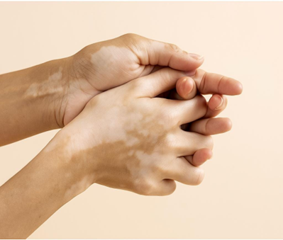
Figure 1 Vitiligo Patches
2.1 Historical background
Vitiligo is a skin ailment. It causes discolouration of the skin. This leads to distinct boundaries of white patches. These patches are created in case the body immune system attacks the cells which provide the skin color and it is known as melanocytes. Vitiligo takes its name after a word vitulus which means calf. This was the term used by a man by the name Aulus Cornelius Celsus in his book De Medicina some time ago. He compared the white patches of vitiligo to white parts of a calf8. The vitiligo is a very ancient disease, to an extent the disease is even mentioned in the Quran, Vedas or the bible. White spots were recorded in skin diseases in many years ago in India, Egypt, Greece and Rome in patients. People in India were aware of vitiligo in the period of time. It was called Shwitra or Kilasa in some books known as Charaka Samhita and Sushruta Samhita they called it. The individuals of the period held the view that vitiligo was as a result of something being amiss within the body which is referred to as Pitta. Therefore they made an effort to treat vitiligo with some of the herbs, including Bakuhi. They would also go out in the sun in a bid to better themselves. The ancient Egyptians also wrote the skin diseases that were marked with patches. These diseases were contained by the use of herbs and sunlight.
Vitiligo was not a well-known condition in Greece and Rome. They thought that it was on par with leprosy that disgraced individuals with vitiligo. The Chinese had a name of vitiligo which they called Bai Dian Feng translated as white patch wind. They believed it is because of the problem of something referred to as Qi and blood.
It has now become known that vitiligo is an illness in which the immune system of a human being attacks the cells that make the skin its colour. Nevertheless, even with the reality that today, individuals are aware of vitiligo and are left with some way of curing it such as through the use of herbs and sunlight particularly where Ayurvedic medicine and holistic medicine is practiced9,10.
2.2 Melanogenesis
Our skin has skin cells that are known to generate melanin that makes our skin colorful. This is referred to as melanogenesis. This group of cells is referred to as melanocytes and are enclosed by other cells referred to as keratinocytes. They are all involved in ensuring that the appropriate quantity of melanin is produced. It is the melanin that makes our skin, hair and eyes colored in the way that they are. It is also used in safeguarding us against the radiation of the suns.
The skin is variable and the physicians classify them under Fitzpatrick system. There are six categories under this system i.e., I- VI. It is pegged on the reaction of the skin to the sun. There are those individuals who are red and burnt under the sun and those who tan. The degree of melanin which we possess is governed by the genes and this makes us naturally coloured. The other influences that can influence the color of our skin are the hormones and the ultraviolet rays of the sun11.
Melanin production is a chronological process. When this is incorrect something will be out of place with our skin color. In other instances, individuals end up with lack of color on the skin which is called hypopigmentation. Sometimes there are instances of very pigmented people and this is known as hyperpigmentation. These skin issues may be at birth they may be at old age they may be temporary and permanent. They can just affect the skin. Get involved in a bigger health problem. There are even complications of pigmentation which can complicate the life of the individual. Normally these are issues that are not readily curable. This is what makes the researchers yet to know more about the melanin formation process. With the knowledge of melanogenesis mechanism we have a better position to elicit more information in regards to pathogenesis of skin ailments such as these. This would assist us in discovering a way of curing the pigmentation issues. This may be as an addition to the lives of people. Very essential are melanogenesis and skin and melanin. As we learn more of the skin and melanin and melanogenesis we are able to contrive some solution of these pigmentation problems. That is why it is so important to the research in the area of melanin and melanogenesis and the skin12,13.
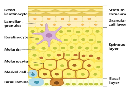
Figure 2 Melanocyte Cell
Costin, G. E. (n.d.). Decoding and modulating the color of human skin [Photograph]. http://www.thecosmeticchemist.com/education/skin_science/decoding_and_modulating_the_color_of_human_skin.html
Steps of melanogenesis :-
2.3 Etiology
Autoimmunity- because of common co-occurring autoimmune disorders like thyroiditis and type I diabetes, the anti-melanocyte antibodies are in the body, and the response to immunosuppressant therapy.
Cytotoxicity- the probability that compounds generated by the synthesis of melanin can cause the death of melanocytes.
Neural - chemical mediators that are secreted at nerve endings may kill melanocytes or prevent the production of melanin.
Free radicals- excessive free radicals could be toxic to melanocytes.
Genetic- It seems like there are a few genes (one of them being NALP1) that make a person prone to vitiligo development. The gene that is controlled by these remains to be known.
Triggering-- It seems that there must be an event that destroys the pigment cells. It has numerous suggested triggers, and they may not be universal (sunburn, trauma, pregnancy etc.)
Immune-Another aspect that may be involved is the one that leads to the destruction of the pigment cells by the immune system. That is why vitiligo is also referred to as an autoimmune disease. Vitiligo is not thought to have a definite cause. It is an autoimmune disease that is believed to be hereditary in nature. The theories are that stress, thyroid dysfunction and skin damage, excessive sunburns, chemicals and medications and the genetic predisposition of vitiligo are able to cause the condition. They are theories though that have not been proved yet16,17.
2.4 Epidemiology
Vitiligo is a polygenic and multifactorial disease which has a prevalence of 0.52 in the world. It mostly starts at childhood or young adulthood between the age of 10-30 years most of the time. It is found to be common to all ethnic groups as well as both sexes, but it is found that it is a little more common in women.
Another familiarity that is lacking towards vitiligo is in terms of its etiology but can be described as the depletion of melanocytes in the epidermis. Under normal conditions the functional unit of melanocyte growth and survival is the melanocytes and keratinocytes where the keratinocytes represent the melanocyte growth and survival by direct contact and growth factors. Destruction or apoptosis of the keratinocytes can also cause the loss of the growth factors like the stem cell factor, which triggers the death and depigmentation of the melanocytes in the vitiligo19.
There have been hypotheses that there exist several mechanisms which include autoimmune, oxidative stress, biochemical, neurological and viral. Autoimmunity and oxidative stress are two of them that are highly researched. Genetics also matters. Vitiligo has neither been mendelian in its inheritance, it possesses genetic heterogeneity, many susceptibility genes with incomplete penetrance20. The genes could cause the melanin production, immune regulation and oxidative stress response genes to make a person at risk. It is also justified by autoimmune correlation with certain human leukocyte antigen (HLA) haplotypes.
Vitiligo diagnosis may prove to be difficult in cases where the skin tone of a patient is very light. It must be distinguished with the other causes of depigmentation or hypopigmentation. Especially, Woods lamp test is beneficial to suggest the diagnosis among individuals with fair skin (Fitzpatrick skin type I and II)22.
2.5 Role of Melanogenesis in Skin Pigmentation
Melanogenesis is a physiological methodology of melanin production in Melanocytes. Their greatest one is the enzyme, tyrosinase, which changes the product, tyrosine, to dolaquinone and dopa which is further changed into the production of the melanin23.
Deficiencies of such path lead to hypopigmentation or depigmentation such as those seen in vitiligo. The agents may aid in the repigmentation either by activating the activity of the tyrosinase or by regaining melanocytes.
The latest finding of melanin re-pigmentation, plant extracts and natural cures of vitiligo will be the theme that will have to be targeted in this case. It will also find its way in the designing of therapeutic drugs that will be capable of pigmenting the skin with the vitiligo.stering pigmentation of the skin in vitiligo22.
3. Herbal Modulators and Their Mechanisms
They also have a few phytochemicals and herbs plants, which are known to cause melanogenesis or shielding of the melanocytes. These include the activation of an enzyme tyrosinase, scavenging of reactive oxygen species (ROS), immunomodulatory and regeneration of the melanocytes. Psoralea corylifolia (Bakuchi): It is a herb that is also composed of psoralen and bakuchiol that stimulates the melanin and skin sensitivity to UV-light. Ayurveda Ayurveda A popular Ayurvedic drug to cure Shwitra (vitiligo).
Acorus calamus (Sweet flag): this has b-asarone that acts as a melanocytic and tyrosinase inducer. Curcuma longa (Turmeric): Curcumin is an antioxidant, an anti-inflammatory; an inhibitor of the oxidative stress of melanocytes.
Glycyrrhizia glabra: This is a plant that comprises the glabridin that controls the activity of the tyrosinase and the depigmentation inhibition.
PIper nigrum ( Black pepper): Piperale is one which stimulates the growth of the melanocytes and repigmentation in the UV treatment.
Fig: Ficus carica (Fig): It is a furanocoumarin-based plant species, endowed with melanogenic property and antioxidant property.
Antioxidant (ferulic acid Polymyxa leucotomos)20.
3.1 Phytochemicals Acting as Melanogenesis Modulators
|
Phytochemical
|
Source Plant |
Mechanism |
Effect |
|
Psoralen |
Psoralea corylifolia |
UV sensitizer, stimulates tyrosinase |
Repigmentation |
|
Piperine |
Piper nigrum |
Melanocyte proliferation |
Pigment restoration |
|
Bakuchiol |
Psoralea corylifolia |
Antioxidant, melanogenic |
Melanin synthesis |
|
Glabridin |
Glycyrrhiza glabra |
Tyrosinase modulation |
Prevents depigmentation |
|
Ferulic acid |
Polypodium leucotomos |
Antioxidant |
Melanocyte protection |
4. Patient counseling
One of the primary reasons to successful implementation of dietary and plant-based interventions in the treatment of vitiligo is counseling of the patients. It has been recommended that the dietary therapies used in the real-life situations be taken as a supplement and not as a substitute of the general treatments. Perhaps, it is possible to prevent the use of inflammatory food and prescribe the intake of antioxidant rich food as a part of regular meals15. Examples of bioactive compounds that may potentially counter oxidative stress involve individual antioxidant-containing foods that have been suggested to contain apples, green tea, Indian gooseberry, onions and peppers.
It is also important that the method of preventing the foods that bring about the oxidative stress and including the anti-inflammatory foods should be corresponding. This would include ensuring that the patients are not advised to consume mangoes, some forms of nuts, and other forms of berries because they contain high phenol and may alternatively consume seafood because it contains high levels of omega-3 fatty acids8. The information shared between the dermatologists and dietitians/ nutritionists would occur on a regular basis to offer a cohesive solution and enhance adherence.
The clinicians have been encouraged to introduce the dietary and plantderived interventions as an adjuvant intervention to the growing yet inconclusive evidence. The patient expectations will be controlled through open dialogues on the purpose of the experiment, the potential benefits and limitations of such therapies in order to ensure trust24.
5. Benefits of Herbal Therapy.
Minimal side effects
Multi-targeted (antioxidant+ immune + melanogenic) mechanism.
Low cost and easily available.
Complementary to other modalities of treatment25.
6.Restrictions and Future outlook
Nevertheless, despite its promising results, there are still challenges in standardization of the herbal preparations, control of dosage and bioactivity even though promising results are achieved22. This kind of research should be conducted in the future by conducting clinical trials, molecular pathways and novel systems that can deliver their products in a better way such as nano-formulations to enhance their efficacies.
CONCLUSION
The herbal melanogenesis modulators are also safe and effective enough; hence making it a good alternative in the treatment of vitiligo. Phytochemicals like psoralen, piperine and bakuchiol are also capable of an enormous potential in pigmentation repairing as it has multi-pronged biological activities. The next step in natural therapies in the treatment of vitiligo will perhaps be the factor of incorporation of application of traditional herbal knowledge with scientific validation. Certain of herbal compounds would be discovered that they would be able to find viable therapies in treating vitiligo.
Specific Therapy: The spirit of research in the future will be more illuminated in the topic of vitiligo.
immunopathogenesis, which causes the emergence of new more therapeutics.
special treatment through special herbal substances.
Reduced Exacerbation: The first long term outcome is that, there could be considerable improvement of the severity and frequency of relapsing of the vitiligo therapy where some current therapies in case studies are already showing symptoms of the case relapse abating in a period of 9 months.
Improved Photoprotection: An improved intrinsic photoprotection and antioxidant activity of herbal extracts (e.g., Polypodium leucotomos, green tea, carotenoids, glabridin) will also be enhanced to achieve a superior adjuvant to traditional vitiligo cream. Increased Quality of Life: Once the formulations have worked, the quality of life of the patients will have improved significantly because they will have access to safe, effective and well-tolerated therapeutic choices with minimal side effects that accompaniment of the same will have vitiligo. Safety and Stability: It has been observed that good physical stability is usually observed in the developed herbal creams.
It is expected to produce (appearance, consistency, pH and no phase separation) at a temperature range. They are mostly discovered to be non-irritant and harmless when they are applied on the skin, which was not so in the investigation of the irritancy. Mechanism of Action: The existence of natural compounds such as flavonoid, tannin and phenolic compounds that have a strong antioxidant, anti inflammatory as well as UV absorbing properties expounds greatly on the mechanism of action.
Specific Ingredients: Aloe vera, Curcuma longa (turmeric), Ginkgo biloba and Polypodium leucotomos (Fernblock(r)) extract of plants have been specifically tested and found to be effective in repigmenting and ameliorating the effect of vitiligo.
Synergistic Effects: When these herbal extracts are used in combination with one another they are likely to have a synergistic effect.
increasing general effectiveness of the treatment compared to single extracts.
Patient Compliance: The majority of the formulations are oil in water (O/W) and therefore not greasy, therefore can easily be washed, and does not cause pain to an individual using it, thus increasing his or her patient compliance25.
DISCUSSION
Vitiligo is a chronic depigmentary disorder characterized by the loss of melanocytes, leading to the appearance of white patches on the skin. Conventional therapies such as corticosteroids, immunomodulators, and phototherapy are commonly used for treatment; however, prolonged use of these therapies may cause adverse effects. Therefore, there is growing interest in herbal formulations as safer and more effective alternatives27. Herbal modulator creams formulated with plant-based ingredients may provide antioxidant, anti-inflammatory, and immunomodulatory effects that support melanocyte protection and repigmentation. One promising herbal ingredient is Polypodium leucotomos, a tropical fern known for its strong antioxidant and photoprotective properties. Extracts of this plant have been shown to reduce oxidative stress, protect melanocytes from UV-induced damage, and improve skin tolerance to phototherapy, which may contribute to repigmentation in vitiligo patients29. Another important herbal component is Glycyrrhiza glabra (licorice), which contains bioactive compounds such as glabridin and liquiritin that possess anti-inflammatory, antioxidant, and skin-healing properties. Liquiritin has been reported to promote melanin dispersion in depigmented areas and support the melanogenesis process. The combination of Polypodium and licorice in a topical herbal modulator cream may produce synergistic effects by protecting melanocytes from oxidative damage, regulating immune responses, and promoting melanin production. Such herbal formulations offer advantages including better skin compatibility, lower risk of side effects, and suitability for long-term use. However, further scientific studies, including standardized formulations and clinical trials, are necessary to confirm the efficacy, safety, and optimal concentrations of these herbal extracts in the management of vitiligo30.
REFERENCES

V. Ramya Sri, Nishika Tamrakar, Praveen Kumar Sahu, Suchita Wamankar, Dr. Gyanesh Kumar Sahu, Dr. Chanchal Deep Kaur, Herbal Modulators Cream for Melanogenesis: A Review on Phytochemicals in Vitiligo Management, Int. J. of Pharm. Sci., 2026, Vol 4, Issue 3, 1675-1684. https://doi.org/10.5281/zenodo.19046843
 10.5281/zenodo.19046843