

We use cookies to ensure our website works properly and to personalise your experience. Cookies policy
Lokmanya Tilak Institute of Pharmacy, Navi Mumbai, Maharashtra, India.
Hydrogels are three-dimensional, hydrophilic polymeric networks capable of absorbing and retaining large quantities of water while maintaining their structural integrity. Due to their unique physicochemical properties, including swelling capacity, biocompatibility, flexibility and tunable mechanical strength, hydrogels have gained considerable attention in pharmaceutical and biomedical applications. This review provides an overview of hydrogel formulation techniques, focusing on the selection of natural and synthetic polymers, crosslinking methods, preparation approaches and various factors influencing hydrogel characteristics. Different formulation strategies, including physical and chemical crosslinking, freeze–thaw methods, free-radical polymerization and other advanced techniques, are discussed. Furthermore, important characterization parameters such as swelling behaviour, gel fraction, mechanical strength, rheological properties, drug content and drug-release profiles are highlighted. The pharmaceutical applications of hydrogels in controlled drug delivery, topical formulations, wound management, tissue engineering and other biomedical fields are also explored. A comparative discussion of selected hydrogel formulations demonstrates how polymer composition and preparation conditions influence their performance. Despite their promising potential, challenges such as limited mechanical strength, formulation instability, difficulties in characterization, reproducibility and biocompatibility require further investigation. Overall, advancements in polymer science and formulation optimization are expected to support the development of more effective, stable and application-specific hydrogel systems for future pharmaceutical and biomedical use.
A hydrogel is a three-dimensional (3D) network of hydrophilic polymers that, through chemical or physical cross-linking of individual polymers in the chain, can swell in water and absorb a significant amount of water while maintaining its shape. Wichterle and Lím (1960) described the first report of hydrogels. Compared to any other class of synthetic biomaterials, these are most similar to natural living tissue in terms of high water content, porosity, and softness. Hydrogels can be resorbed and dissociate over time or be resistant to chemical breakdown. If molecular entanglements and secondary interactions like ionic, hydrogen bonding, or hydrophobic interactions are responsible for the formation of the linkage, hydrogels are also called “reversible” or “physical” gels. Physical gels are usually recovered by simple washing with an appropriate solvent and reground by altering the temperature, pH, or ionic strength of the medium. Networks created by cross-linking polymers in a dry state or in solution can also be formed by covalent bonds that hold the macromolecular chains together in “permanent” or “chemical” gels. Almost all water-soluble polymers with different bulk physical properties and chemical composition can be used to make hydrogels. They can also be produced in different physical forms such as slabs, coatings, films, microparticles, and nanoparticles.[1]Hydrogels are also referred to as super absorbent hydrogels due to their tendency to take in considerable quantities of water, amounting to approximately 10–20 times their original molecular weight once fully swollen.[2]
1.1Properties of hydrogel
Application of hydrophilic gels (also called hydrogels) in pharmaceutical and biomedical engineering have attracted much attention.
Swelling characteristics
Hydrogel can be quickly and reversibly transformed due to minor changes in the environment. The physical structure of the hydrogel can change due to different environmental parameters such as temperature, pH, electric signal, presence of enzymes or other ionic species.
Mechanical properties
The extent of cross-linking can be adjusted to tune the hydrogel’s mechanical properties; that is, increasing the degree of cross-linking leads to higher mechanical strength of the hydrogel network, but hydrogels become more fragile at the same time.[1]
1.2Advantages of hydrogels
1.3 Disadvantages of hydrogel
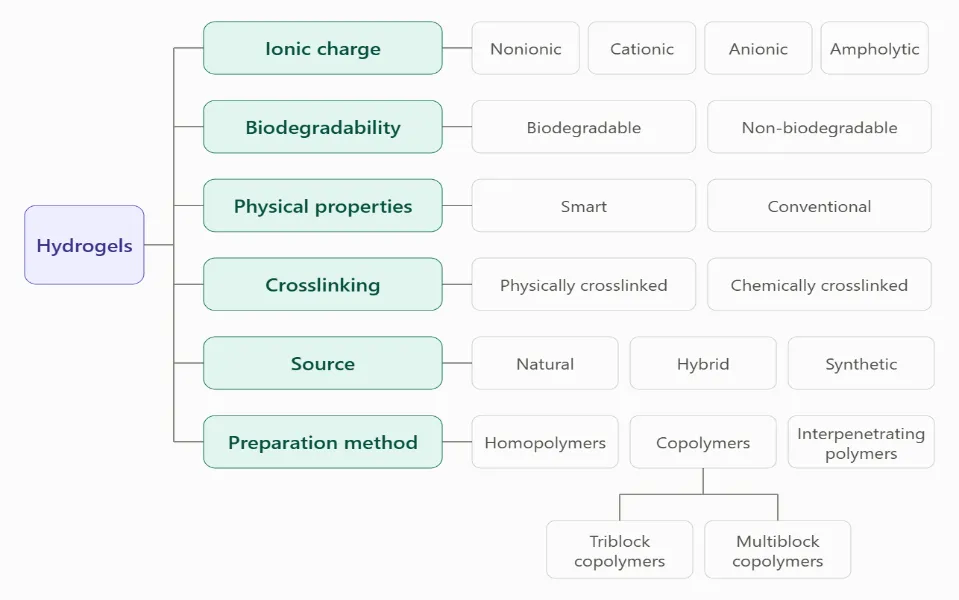
Figure 1: Classification of Hydrogel
2.CLASSIFICATION:
Hydrogels are classified primarily by the presence of side functional groups in polymer chains, which can be neutral or ionic. The swelling properties of neutral hydrogels are mostly due to the overall unbound energy provided by water-polymer thermodynamic mixing and expandable polymer contribution.[2] Hydrogels can be categorized according to their network structure's physical properties, including amorphous, hydrogen-bonded, semi-crystalline, super-molecular, and hydro colloidal aggregates.[2]
3.MATERIALS USED FOR PREPARATION:
Both synthetic and natural polymers are used to create hydrogel. Chitosan, gelatin, alginate, hyaluronic acid, and fibrin are examples of common natural polymers. To improve their mechanical strength, stability, and functional qualities, these materials are frequently mixed with synthetic monomers or polymers.[3]
MONOMERS USED FOR PREPARATION:
Table 1: Monomers for Preparation of Hydrogels .[4]
| Monomer | Name | Abbreviation | Chemical structure |
|---|---|---|---|
| I | Hydroxyethyl methacrylate | HEMA | CH2=C(CH3)COOCH2CH2OH |
| II | Hydroxyethoxyethyl methacrylate | HEEMA | CH2=C(CH3)COOCH2CH2OCH2CH2OH |
| III | Hydroxydiethoxyethyl methacrylate | HDEEMA | CH2=C(CH3)COOCH2CH2OCH2CH2OCH2CH2OH |
| IV | Methoxyethyl methacrylate | MEMA | CH2=C(CH3)COOCH2CH2OCH3 |
| V | Methoxyethoxyethyl methacrylate | MEEMA | CH2=C(CH3)COOCH2CH2OCH2CH2OCH3 |
| VI | Methoxydiethoxyethyl methacrylate | MDEEMA | CH2=C(CH3)COOCH2CH2OCH2CH2OCH2CH2OCH3 |
| VII | Ethylene glycol dimethacrylate | EGDMA | CH2=C(CH3)COOCH2CH2OOCC(CH3)=CH2 |
| VIII | N-vinyl-2-pyrrolidone | NVP | CH2=CHNCOCH2CH2CH2 |
| IX | Methacrylic acid | MA | CH2=C(CH3)COOH |
| X | Vinyl acetate | VAc | CH2=CHCOOCH3 |
4. PREPARATION TECHNIQUES OF HYDROGEL
4.1 Polymerisation via free radicals
Formed a chemically crosslinked network by free-radical polymerisation, copolymerising acrylic acid with ethyl cellulose and employing benzoyl peroxide as the initiator and N,N'-methylenebisacrylamide (MBA) as the crosslinker. The initiator and MBA were dissolved in acrylic acid (50 to 85 g per 100 g), the solution was mixed with an ethanolic ethyl cellulose solution (5 to 10 g per 100 g), and nitrogen was passed through the mixture for 30 minutes. Polymerisation was then carried out in a water bath with the temperature raised stepwise from 45 °C to 65 °C, after which the cylinders were cut into 6 mm discs, rinsed with 50% ethanol and dried under vacuum at 45 °C. Higher amounts of acrylic acid and MBA produced a denser network, which reduced both swelling and drug release. A new infrared band at 1701 cm⁻¹ was regarded as evidence that graft copolymer had formed.[5] Gonsalves et al. used a redox-initiated approach in which a citric acid-based polymer, PEGMC, was synthesised by heating PEG 200, maleic acid and citric acid (6:4:1 molar ratio) at 160 °C and subsequently at 140 °C, and it was crosslinked with PEG diacrylate (PEGDA of 3.3, 6 or 8 kDa) using ammonium persulfate and TEMED. A half-factorial design across PEGMC, PEGDA and TEMED concentrations yielded six formulations that cured within the desired 3 to 5 minutes, and the 8kDa-H1 gel, which set in 313 s, was selected. To produce a nanocomposite, 10 mg of drug-loaded PLGA and carboxymethyl chitosan nanoparticles, prepared earlier by double emulsion solvent evaporation, was incorporated into the precursor solution before gelation.[6]
4.2 Physical crosslinking through freezing and thawing
Paranhos et al. produced poly (vinyl alcohol) (PVA) gels without using any chemical crosslinker. A 10% w/v PVA solution (Mw 120,000, 99% hydrolysed) was combined with a 5% sulfonated polyester in 90/10, 75/25 and 50/50 ratios, poured into Petri dishes and put through five freezing and thawing cycles. The first cycle kept the sample at -22 °C for 17 h, and every subsequent cycle consisted of 1 h at -22 °C followed by 30 minutes at room temperature. As the authors explain, this process generates PVA crystallites that serve as physical crosslinks, making the gel insoluble without any added chemicals, and repeated cycles are required to achieve a well-defined network. The polyester was included because neutral PVA gels respond poorly to pH and ionic strength.[7] A review of PVA cryogels points out that a higher polymer concentration gives a stiffer gel with less porosity, and raising the solution from 10% to 15% and 20% increased the tangent modulus by 69% and 137% respectively. The same review reports that crystallisation dominates the first three cycles, that phase separation continues for at least six, and that stiffness often levels off at about six cycles. Slower thawing was found to give stiffer gels, and thawing rates above 10 °C/min were described as unsuitable. Holding the frozen sample for long periods, up to 10 days in one cited study, gave the strongest gels, whereas holds of 1 or 6 hours had no measurable effect on tensile properties.[8]
4.3 Photocrosslinking
Nguyen et al. used lithium phenyl-2,4,6-trimethylbenzoylphosphinate (LAP) at 0.1, 0.25, 0.5 and 1% w/w to crosslink 10% w/w gelatin methacryloyl (GelMA, 50% methacrylation). A single millilitre of each solution was irradiated with UV-A light at 86, 342 or 855 mJ/cm², which yielded twelve formulations in total. LAP was chosen over Irgacure 2959 because its absorbance peaks at 370 nm and it still retains 14.1% at 405 nm, while Irgacure 2959 peaks at 280 nm. The elastic modulus ceased to rise beyond 342 mJ/cm² (120 s), so a longer exposure was not needed.[9] According to a review on GelMA, the polymer is made by dissolving gelatin in phosphate-buffered saline at pH 7.4, adding methacrylic anhydride, stopping the reaction by dilution, dialysing the product and freeze-drying it. The degree of substitution rises with the amount of methacrylic anhydride added. Using dimethyl sulfoxide as the solvent or replacing PBS with carbonate-bicarbonate buffer raises the degree of substitution further.
Visible-light systems based on eosin Y with triethanolamine and N-vinyl-2-pyrrolidinone are an alternative to Irgacure 2959 and are described as less toxic. Both UV and visible light, however, penetrate tissue poorly.[10]
4.4 Schiff base reaction and natural crosslinker, genipin
Hu et al. produced a hydrogel without any separate crosslinker by allowing the aldehyde groups of oxidised hyaluronic acid (OHA) to react with the amino groups of carboxymethyl chitosan (CMC). OHA was synthesised by oxidising hyaluronic acid with sodium periodate for 2 h in the dark, which gave degrees of oxidation ranging from 27.9 to 56.0% depending on the periodate ratio. CMC was produced from chitosan using chloroacetic acid under alkaline conditions. Equal volumes of OHA (1 to 3% w/v) and CMC (1 to 4% w/v) in PBS at pH 7.4 were mixed and held at 37 °C until gelation occurred.[11] Matcham and Novakovic used genipin to crosslink chitosan and poly (vinyl pyrrolidone) (PVP). Chitosan (1.5% w/v in 1% acetic acid), PVP (5% w/v) and genipin (0.5% w/v) were prepared separately, and 0.15 mL chitosan, 0.75 mL PVP and 0.1 mL genipin were combined under stirring. Genipin was selected because it is reported to be 5000 to 10,000 times less cytotoxic than glutaraldehyde. The reaction is pH-dependent, since in basic conditions genipin molecules first polymerise among themselves and fewer remain available to react with chitosan. [12] A review of genipin-crosslinked chitosan notes that genipin crosslinks chitosan spontaneously at a small molar ratio and gives blue, fluorescent gels. Nanogels prepared by reverse microemulsion had hydrodynamic diameters of 270 to 390 nm. The same review mentions that a small shift of pH within 4.00 to 5.50 noticeably changes the properties of the gel and that the reaction can take up to four days. It also points out that genipin is expensive to produce.[13]
4.5 Variables controlling the network
In the ethyl cellulose system, acrylic acid and MBA increased the gel fraction and reduced porosity. More acrylic acid and MBA also lowered swelling and drug release because of the higher degree of crosslinking. [5] In the PEGMC system, short PEGDA chains produced compact networks with little void space whereas long chains permitted more diffusion. The polymer and PEGDA concentrations were the formulation factors that significantly affected swelling, while the nanoparticles had a negligible effect.[6] In the OHA-CMC system, gelation became faster as the precursor concentration rose, and gels made from OHA with a higher degree of oxidation formed more quickly.[11] The energy dose is also a variable, as the GelMA modulus rose between 86 and 342 mJ/cm² and then reached a plateau.[9]
For PVA, the number of freezing and thawing cycles and the long first freezing step are part of the recipe, because the network is built from crystallites formed over repeated cycles.[7] Medium conditions such as pH and temperature influenced the genipin reaction, and fluorescence was lower at pH 10 than at pH 4.[12]
5. FACTORS AFFECTING HYDROGEL FORMULATION
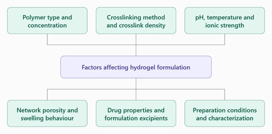
Figure 2: Factors Affecting Hydrogel Formation
Hydrogels are three-dimensional networks of hydrophilic polymers capable of absorbing and retaining substantial quantities of water or aqueous fluids without dissolving. Their properties depend on polymer composition, network architecture, crosslinking conditions and interactions with the surrounding environment. These characteristics determine the suitability of a hydrogel for applications such as drug delivery, topical formulations, wound dressings and tissue.[14,15]
5.1 Polymer type and concentration
The selection of polymer is a major determinant of hydrogel performance. Natural polymers, including chitosan, cellulose, alginate, gelatin and collagen, are widely investigated because of their potential biocompatibility and biodegradability. Synthetic polymers, including poly (vinyl alcohol), poly (ethylene glycol) and poly (2-hydroxyethyl methacrylate), provide opportunities to adjust mechanical, chemical and swelling properties. Combining natural and synthetic polymers may improve characteristics that are inadequate in an individual polymer system, particularly mechanical strength and stability[15,16].
Polymer concentration influences the density of the polymer network, consistency, viscosity, swelling behaviour and transport of incorporated substances. A change in polymer concentration may therefore affect spreadability and drug release simultaneously. However, the direction and magnitude of these effects depend on polymer chemistry, crosslinking and formulation conditions; a higher concentration does not necessarily produce a formulation with better overall performance.[15,17]
5.2 Crosslinking method and crosslink density
Crosslinking establishes the three-dimensional structure responsible for the hydrogel’s integrity. Physical crosslinking involves interactions such as hydrogen bonding, ionic interactions, hydrophobic associations or polymer-chain entanglement. Chemical crosslinking forms covalent connections between polymer chains. Radiation-induced crosslinking and grafting polymerization are additional approaches used to prepare or modify hydrogel networks.[15,16]
Crosslink density affects the mobility of polymer chains, pore structure, swelling and diffusion through the network. Increasing crosslinking can restrict swelling and the movement of incorporated molecules, while also changing the stiffness and resistance of the gel to deformation. These relationships are formulation-dependent and must be evaluated experimentally rather than assumed to be linear.[15]
Lambrecht et al. (2024) demonstrated the importance of crosslinker selection in carbohydrate-based hydrogels. Their formulations differed in gelation behaviour and mechanical response depending on the crosslinker used and, for polyethylene glycol diacrylate (PEGDA), its molecular mass and concentration. Several formulations displayed high stiffness but limited stability against shear deformation, illustrating that a mechanically rigid gel is not automatically a mechanically suitable gel.[18]
5.3 pH, temperature and ionic strength
Environmental conditions influence polymer ionisation, intermolecular interactions and the swelling response of hydrogels. Changes in pH may alter the charge of ionisable groups within a polymer network, while temperature can affect chain mobility and temperature-responsive interactions. Ionic strength can modify electrostatic interactions and swelling, particularly in charged hydrogels. These variables may also influence the loading and release of drugs or other bioactive molecules.[15]
For pharmaceutical applications, formulation behaviour should be examined under conditions relevant to the intended administration site. A hydrogel designed for topical use, for example, should be evaluated for appropriate pH and application characteristics. A stimulus-responsive hydrogel intended for controlled delivery requires additional assessment of its response to the specific environmental trigger.
5.4 Network porosity and swelling behaviour
Pore size, pore-size distribution, network connectivity and the amount of water retained within the hydrogel influence molecular diffusion and the transport of incorporated agents. These properties depend on polymer composition, crosslinking and preparation conditions. The size and chemical characteristics of the drug, as well as its interactions with the polymer, also affect release behaviour.[15]
Jayawardena et al. (2023) investigated agarose hydrogels containing different agarose concentrations and compared methods for determining pore-size distributions. Their findings demonstrate that measured pore characteristics depend not only on the hydrogel itself but also on the analytical technique and sample preparation. Consequently, porosity measurements should be interpreted alongside the method used to obtain them.
5.5 Drug properties and formulation excipients
Drug loading and release depend on factors such as molecular size, charge, solubility and interactions with the polymer network. The pH and ionic strength of the loading medium may also affect the amount of drug incorporated. Excipients and processing conditions can further influence homogeneity, consistency, stability and release behaviour.[15]
In topical formulations, additional ingredients may be included to achieve the desired application properties. Gehalot et al. (2023), for example, prepared neomycin sulfate hydrogels using Carbopol-940 and guar gum, with variations in the amounts of the formulation components. Their findings show that changing the composition can produce measurable differences in viscosity, spreadability, drug content and in-vitro release.[19]
5.6 Preparation conditions and characterization
Preparation variables include the order of ingredient addition, mixing conditions, polymer hydration time, temperature, crosslinker concentration and curing or polymerization conditions. These factors can affect network formation and batch-to-batch consistency. Gehalot et al. (2023) allowed the Carbopol-940 and guar gum dispersions to swell for 24 hours and used magnetic stirring during formulation preparation, illustrating the importance of controlled processing conditions in a topical hydrogel system.[19]
Characterization should be selected according to the intended use of the formulation. Common assessments include appearance, pH, viscosity, spreadability, drug content, swelling, mechanical properties, in-vitro release and stability. More advanced investigations may include rheological measurements, microscopy, infrared spectroscopy, Raman spectroscopy and thermal analysis. Lambrecht et al. (2024) showed that combining rheology and Raman spectroscopy can provide information about gelation and polymerization as they occur. No single method, however, provides a complete assessment of all chemical, structural and mechanical properties.[18]
6. COMPARISON OF HYDROGEL FORMULATIONS AND CHARACTERIZATION APPROACHES
Comparing hydrogel formulations requires attention to the polymer system, preparation method, intended application and endpoints measured. The studies considered here are not a single head-to-head comparison: they investigate different materials and purposes. Their results are therefore best interpreted as complementary examples of how formulation composition and characterization methods affect measured performance.
Table 2: Comparison of Formulation and Material Studies
| Hydrogel system | What was varied | Key findings |
|---|---|---|
| Neomycin sulfate topical hydrogel (Carbopol-940 and guar gum) | Proportions of the two polymers across 5 formulations | Viscosity 918–987 cP; spreadability 12.24–14.54 g·cm/s; drug content 89.1–96.4. Composition changed several properties, and no single formulation was best at everything. |
| Chemically modified chitosan (thioglycolic acid, preactivated) | Chitosan modified vs unmodified | 7.46-fold higher skin adhesiveness and 1.9-fold more controlled release of the model compound Rhodamine 123. Modification improved adhesion and altered release. |
| Carbohydrate-based hydrogels | Crosslinker type, and PEGDA molecular mass and concentration | Gelation, stiffness and shear resistance all varied with the formulation. Several gels were stiff but unstable under shear, so crosslinker choice affects mechanical suitability. |
6.1 Interpreting the neomycin hydrogel results
In the study by Gehalot et al. (2023), formulation F2 had the highest reported viscosity (987 ± 2.1 cP), whereas F5 had the highest reported spreadability (14.54 ± 0.01 g·cm/s). F1 had the highest reported drug content (96.4 ± 0.2). These differences demonstrate that one formulation may perform better for one measured attribute without necessarily being superior in all attributes.[19]
The paper also reports cumulative in-vitro drug-release values that vary among formulations and over time. However, the release table labels the time points in minutes, while the methods describe sampling at hourly intervals over eight hours. This inconsistency limits confident interpretation of the release results. The data should therefore be quoted cautiously, and the study should not be used alone to establish a definitive optimal formulation or a general rule for hydrogel drug release.
6.2 Comparison of pore-characterization methods
Jayawardena et al. (2023) compared stimulated emission depletion microscopy (STED), atomic force microscopy (AFM) and cryogenic scanning electron microscopy (Cryo-SEM) for assessing agarose hydrogel morphology and pore-size distributions. For a 1.0% agarose gel, the average pore sizes reported using STED and Cryo-SEM were 240 nm and 230 nm, respectively. For 1.5% and 2.0% gels, AFM-derived average pore-size values were 10–15% smaller than those obtained from Cryo-SEM, consistent with a potential AFM tip artefact when measuring concave features.
These results show why comparisons should account for analytical method and sample preparation. Apparent differences in hydrogel structure may partly reflect how the structure was measured, rather than a true difference in the material alone.[21]
6.3 Overall comparison
Taken together, the studies demonstrate that hydrogel performance is multidimensional. Polymer composition can affect application properties and drug release; chemical modification can alter adhesion; crosslinker selection can change gelation and mechanical behaviour; and characterization methods can influence the structural parameters reported. Formulation selection should therefore be based on the intended application and a predefined set of performance criteria rather than on a single result such as viscosity, swelling or stiffness.
7. CHARACTERIZATION AND ASSESSMENT OF HYDROGELS
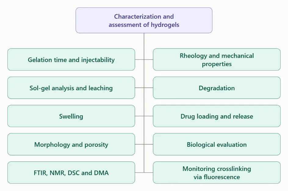
Figure 3: Characterization and Assessment of Hydrogels
7.1 Gelation time and injectability
Using the vial inversion method at 37 °C, Hu et al. observed that gels with greater oxidation and higher precursor concentration formed more quickly, with three compositions gelling within 45 s.[11] Gonsalves et al. defined curing time as the point when a stirring bar first moved, targeted roughly 5 minutes, and verified injectability by extruding the precursors through a 21G needle. [6] A review reports that the stiffness of genipin-crosslinked chitosan gels increased with genipin content, reaching about 502 Pa at 0.5% w/v, while the gelling time became shorter.[13]
7.2 Sol-gel analysis and leaching
Ranjha et al. extracted discs with water in a Soxhlet apparatus for 4 h and determined the sol and gel fractions from the change in weight.[5] For freeze-thaw gels, Paranhos et al. stored samples in water for 15 days and identified released PVA through its green complex with iodine and boric acid at 670 nm, detecting no more than 1.2% dissolution.[7]
7.3 Swelling
The ethyl cellulose and acrylic acid gels swelled only slightly at pH 1.2 (coefficient 3.45 for O1) but markedly at pH 7.5 (40.04), and the gel containing the most MBA swelled less (20.69 at pH 7.5). [1]The OHA-CMC gels attained equilibrium swelling ratios of 48 to 58 g/g within 24 h, and the weaker gel showed the greatest swelling.[11] The PEGMC gel 8kDa-H1 swelled by roughly 283% in 2 h, and loading with nanoparticles altered this only marginally.[5] For PVA with polyester, water uptake increased above pH 4 and declined as ionic strength rose, dropping 150% for the 50/50 blend between ionic strengths of 10⁻⁴ and 1.[7] A GelMA review gives the swelling ratio as (wet weight - dry weight) / dry weight × 100% and reports that it falls as the degree of substitution or the gel concentration increases. [10] Another review notes that the swelling capacity and zeta potential of chitosan microspheres decreased as the extent of genipin crosslinking increased.[13]
7.4 Morphology and porosity
Porosity of the ethyl cellulose gels was assessed by solvent replacement in ethanol and fell as acrylic acid and MBA content increased.[5] Scanning electron microscopy of freeze-dried OHA-CMC gels revealed interconnected pores averaging 61.1 to 77.3 μm in size, with the denser gels showing smaller pores.[11] Cryo-SEM of the PEGMC gel indicated that around 60% of pores measured 0.25 to 0.5 μm, with no change after nanoparticle addition.[6] In GelMA hydrogels, SEM images are usually taken after freeze-drying and gold coating, and a higher degree of substitution or gel concentration gives smaller pores and lower porosity. [10] In chitosan-protein scaffolds, porosity increased from 25.75% without genipin to 44.75% with 2.0% genipin.[13] Small-angle neutron scattering of PVA cryogels showed crystallites of about 3 nm spaced about 19 nm apart, together with water-filled macropores of micrometre size.[22]
7.5 FTIR, NMR, DSC and DMA
FTIR verified the chemistry in the ethyl cellulose gel, where a band at 1701 cm⁻¹ pointed to grafting. [1]The FTIR band at 1726 cm⁻¹ indicated aldehyde groups in OHA, and a band at 858 cm⁻¹ signified the Schiff base in the gel. NMR of OHA showed two new proton peaks at 4.8 and 4.9 ppm.[11] Bands at 1639, 1400 and 1093 cm⁻¹ demonstrated that every component was present in the PEGMC nanocomposite. The PEGMC gels exhibited a thermal transition near 46 °C irrespective of the nanoparticles.[6] For the PVA and polyester gels, differential scanning calorimetry revealed reduced crystallinity as polyester content increased, and dynamic mechanical analysis showed a lower glass transition temperature, pointing to strong interaction between the polymers.[7] For GelMA, the degree of substitution is measured by ¹H NMR on samples dissolved at 10% w/v in D₂O or by the TNBS method read at 335 nm. FTIR of GelMA, run on samples mixed with KBr at 1:100, shows that methacrylation moves the amide bands to higher frequencies. [10]
7.6 Rheology and mechanical properties
For the OHA-CMC gels, the storage modulus was roughly ten times the loss modulus, at approximately 240 Pa for the stronger gels and 146 Pa for the weaker one. [11) The PEGMC gels likewise displayed solid-like behavior, with G′ around seven times G″. Under compression, the PEGMC gel exhibited a modulus of 5.34 MPa, which increased to 6.66 MPa with nanoparticles.[6] Nanoindentation of GelMA yielded plateau moduli of approximately 20 to 25 kPa, somewhat lower at higher LAP content.[9] In the GelMA review, cylinders of 8 mm diameter were exposed to UV for 60 s and compressed after 24 h in buffer, and the compressive modulus rose with gel concentration, exposure time and degree of methacrylation.[10] Crosslinking electrospun chitosan membranes with 10 mM genipin was reported to raise the ultimate tensile strength by 165% to 32 MPa.[13] For PVA cryogels, reported compressive moduli span roughly 2 to 20 MPa depending on the composition and the number of cycles. The tensile curves of PVA cryogels are nonlinear, and a 15 wt% gel after four cycles had a modulus of about 350 kPa, close to that of porcine aortic root. A 10 wt% PVA cryogel relaxed to about 45% of its initial stress after one hour.[8]
7.7 Degradation
After 10 days in PBS, the two stronger OHA-CMC gels retained 12.2% and 7.3% of their weight, whereas the weaker gel had disappeared by day 9, and hyaluronidase only slightly accelerated this process.[11] The PEGMC nanocomposite dropped to 54% of its weight within 3 days and then remained constant for 21 days, since PEGDA does not degrade.[6] GelMA degradation is tested in buffer containing 2 U/mL collagenase type II at 37 °C, and it becomes slower as the degree of functionalisation increases. Electrospun chitosan mats crosslinked with genipin lost about 22% of their mass after four months in vitro, compared with 34% without genipin.[13]
7.8 Drug loading and release
Release from the ethyl cellulose gels decreased as acrylic acid content rose, for instance from 84.91 to 75.85% at pH 7.5, and the data followed first-order kinetics at pH 1.2 and zero-order kinetics at pH 6.5 and 7.5.[5] From the OHA-CMC gel, curcumin displayed a burst release of 55.3% on day 1, while EGF released 88.1% over 9 days, more slowly owing to its size and covalent binding to the network. [11] In the PEGMC nanoparticles, 5-fluorouracil release on day 3 reached 72% at pH 5.5 versus 51% at pH 7.4.[6] In PVA cryogels, release of bovine serum albumin was mainly diffusion-controlled, with about 95% released within 30 hours. Water diffusion in the same gels fell by 62% between the second and the fifth freeze-thaw cycle.[8]
7.9 Biological evaluation
Extracts of the GelMA-LAP gels produced 92.5, 85.4, 78.9 and 67.1% viability in the CyQuant assay at 0.1 to 1% LAP, so only the highest concentration fell below the 70% toxicity threshold, even though the Alamar Blue assay revealed no effect.[9] OHA-CMC extracts produced 100% viability in a CCK-8 test, and hemolysis ranged between 0.41 and 2.36%. S. aureus was inhibited by 71.4 to 98.7% by the OHA-CMC gels, and in diabetic mice the gel loaded with curcumin and EGF closed 88.3% of the wound by day 10.[11] The PEGMC nanocomposite maintained about 80% fibroblast viability up to 500 μg/mL and generated inhibition zones of roughly 2.2 cm² for E. coli and 3.8 cm² for S. aureus.[6] A GelMA review reports that mesenchymal stem cells showed higher viability at a low GelMA concentration and a low UV dose, and that 5% GelMA supported osteogenic differentiation better than 10% GelMA.[10] Fibroblast viability on genipin-crosslinked chitosan gels was 88.4 to 90.9%, compared with 93.7% on blank plates. In tendon explants, about 50% of the cells remained viable after 72 hours at 6.2 mM genipin, and a concentration of 5 mM or lower was suggested for in vivo use.[13]
7.10 Monitoring crosslinking via fluorescence
Matcham and Novakovic tracked genipin crosslinking using fluorescence at 485/520 nm over 24 h and observed lower fluorescence at pH 10 compared with pH 4, with decreases of 63% at 24 °C and 35% at 37 °C. At 37 °C the signal declined following an initial peak, which they interpreted as degradation of the crosslinks. Surface scans revealed that crosslinking was non-uniform.[12] A review also notes that genipin-crosslinked chitosan gels are blue and fluorescent, which is what makes such monitoring possible.[13]
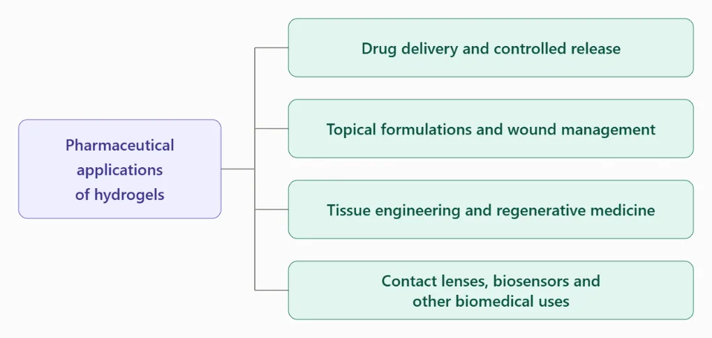
Figure 4: Pharmaceutical Applications of Hydrogels
8. PHARMACEUTICAL APPLICATIONS OF HYDROGELS
8.1 Drug delivery and controlled release
Hydrogels are investigated as drug-delivery matrices because their hydrated polymer networks can incorporate therapeutic agents and regulate their movement through the material. Drug release is influenced by network structure, swelling, diffusion, polymer–drug interactions and environmental conditions. Depending on the formulation, hydrogels may be designed for sustained or stimulus-responsive delivery.[14,15]
Stimuli-responsive hydrogels can alter their swelling or other properties in response to changes such as pH or temperature. This behaviour offers opportunities for designing delivery systems that respond to conditions at a target site. Nevertheless, the actual release profile and suitability for a particular medicine must be established experimentally.
8.2 Topical formulations and wound management
Hydrogels are suitable candidates for topical applications because their high water content can help maintain a moist environment. Their consistency, spreadability and ability to incorporate active ingredients make them useful for investigating formulations intended for wound management and other skin applications. Important formulation requirements include acceptable pH, ease of application, retention at the application site, appropriate drug release and physical stability.[16,19]
Alven and Aderibigbe [2020] reviewed chitosan- and cellulose-based hydrogels for wound management. They discussed the use of these biopolymers in wound dressings and their potential contribution to moisture management, wound protection and re-epithelialization. They also noted that combining biopolymers with selected synthetic polymers can improve mechanical and physicochemical properties.[16]
Gehalot et al. (2023) investigated a neomycin sulfate hydrogel prepared with Carbopol-940 and guar gum. The study evaluated properties relevant to a topical formulation, including pH, spreadability, viscosity, drug content, in-vitro release and accelerated stability. The results provide a formulation-specific example of how polymer selection and composition can be assessed during the development of a medicated hydrogel.[19]
8.3 Tissue engineering and regenerative medicine
Hydrogels have been investigated as scaffolds and matrices for tissue engineering because their hydrated structures can provide an environment that resembles aspects of the extracellular matrix. Their geometry, functional properties and degradation behaviour can be adjusted through material selection and formulation design. Depending on the application, hydrogels may also support cell incorporation or be delivered through minimally invasive approaches.[23]
Karami et al. (2023) reviewed the preclinical evaluation of hydrogel-based devices for cartilage repair. They highlighted the importance of demonstrating suitable biological compatibility, degradation behaviour, cellular support where relevant and integration with surrounding tissue. Their review also emphasizes that promising laboratory results do not, by themselves, establish clinical effectiveness.
8.4 Contact lenses, biosensors and other biomedical uses
Hydrogels are used or investigated in contact lenses, biosensors, tissue-engineering matrices and other biomedical systems. Their water retention, polymer-network structure and capacity for chemical modification support a range of material designs.[14,15,18]
Hydrogels can also serve as matrices for immobilizing biological or catalytic components. Their suitability depends on the specific mechanical, chemical and transport properties required by the application. Consequently, a formulation developed for a topical product should not be assumed to have the properties needed for a contact lens, biosensor or tissue-engineering scaffold.
9. CHALLENGES AND FUTURE PERSPECTIVES
9.1 Mechanical weakness and stability
A key challenge for some hydrogel systems is limited mechanical strength. A formulation may absorb substantial quantities of water yet lack sufficient resistance to deformation, handling or loading. In topical applications, inadequate adhesion or structural integrity may reduce residence at the intended site. In tissue engineering, insufficient mechanical properties and poor integration with surrounding tissue can restrict performance.[16,23]
Future work should focus on matching polymer composition and crosslinking strategy to the mechanical requirements of each application. Polymer blending, chemical modification and optimized network architectures are possible approaches, but improvements in stiffness should be evaluated alongside flexibility, deformation tolerance, swelling and biological compatibility.
9.2 Balancing swelling, drug release and network integrity
Hydrogel properties are interdependent. Changes intended to improve swelling or drug loading may also alter mechanical strength, release rate or stability. Similarly, increasing crosslinking may strengthen the network while restricting swelling or molecular diffusion. This creates a need for systematic optimization rather than adjustment of individual variables in isolation.[15,18]
Future formulation studies should define target performance criteria before optimization and evaluate multiple responses together. Experimental designs that investigate the effects of polymer concentration, crosslinker content and processing conditions can help identify useful combinations while reducing unnecessary experimental runs.
9.3 Characterization limitations and reproducibility
Hydrogel characterization remains challenging because these materials contain substantial amounts of water and may be difficult to analyze using techniques developed for dry or soluble samples. For example, water can interfere with infrared spectral bands, while drying before microscopy may alter pore morphology. Rheological testing and complementary spectroscopic or imaging approaches can provide a more complete understanding of the material.[18,21]
Future studies should clearly report sample preparation, test conditions, replicate numbers and analytical procedures. Using complementary methods can improve confidence in conclusions about chemical structure, pore distribution, gelation and mechanical properties. Standardized reporting would also make results from different laboratories easier to interpret.
9.4 Biocompatibility, safety and preclinical validation
For pharmaceutical and biomedical use, promising physical properties are not sufficient to establish suitability. Formulations must also be evaluated for biocompatibility and other safety considerations relevant to the intended route and duration of exposure. For tissue-engineering devices, additional requirements may include degradation behaviour, tissue integration, reproducibility and appropriate preclinical evaluation.[23]
The choice of raw materials and crosslinking agents should account for potential biological risks, including the effects of residual reagents where chemical crosslinking is used. Safety assessment should be integrated into development rather than postponed until after optimization of the physical formulation.
CONCLUSION:
Hydrogels have emerged as promising polymeric systems with significant potential in pharmaceutical and biomedical applications due to their high water absorption capacity, swelling behaviour, flexibility, biocompatibility and ability to incorporate and release therapeutic agents. This review highlights the importance of polymer selection, polymer concentration, crosslinking techniques and preparation conditions in determining the physicochemical and functional properties of hydrogel formulations. Different preparation approaches and crosslinking strategies offer opportunities to modify the structure, stability, mechanical strength and drug-release characteristics according to specific application requirements.The reviewed studies demonstrate that hydrogel performance depends on the combined influence of formulation composition, network structure and processing parameters. Their applications in controlled drug delivery, topical formulations, wound management and tissue engineering indicate their versatility and importance in modern pharmaceutical research. However, limitations such as inadequate mechanical strength, stability concerns, difficulties in characterization, reproducibility and the need for comprehensive biocompatibility evaluation remain important challenges.Future research should focus on developing optimized polymer combinations, improving mechanical properties, establishing reliable characterization methods and conducting systematic safety and performance evaluations. The use of advanced formulation techniques and stimulus-responsive polymer systems may further expand the applications of hydrogels. In conclusion, continued research and technological advancements in hydrogel formulation can contribute to the development of innovative, efficient and application-specific pharmaceutical and biomedical systems.
REFERENCES

Shubhangi Kasture, Jitendra Prajapati, Shivanjali Bhosale, Priya Singh, Shrutika Patil, Hydrogel Formulation and Development: A Comprehensive Review of Preparation Methods, Characterization and Pharmaceutical Applications, Int. J. of Pharm. Sci., 2026, Vol 4, Issue 11, 931-945. https://doi.org/10.5281/zenodo.23972009
 10.5281/zenodo.23972009