

We use cookies to ensure our website works properly and to personalise your experience. Cookies policy
1,2,3MBBS 5th year, Department of Pharmacology, Fergana Medical Institute of Public Health, Uzbekistan.
4MBBS 6th year, Department of Pharmacology, Fergana Medical Institute of Public Health, Uzbekistan.
5Assistant Professor Department of Pharmacology, Fergana Medical Institute of Public Health, Uzbekistan.
Background: Cardiovascular-Kidney-Metabolic (CKM) syndrome represents a novel, integrative clinical framework endorsed by the American Heart Association in 2023, encompassing the pathophysiological convergence of metabolic dysfunction, chronic kidney disease (CKD), and cardiovascular disease (CVD). Emerging evidence demonstrates that systemic inflammation, insulin resistance, and endothelial dysfunction are not merely comorbidities in this triad but represent mechanistically interlinked drivers that perpetuate organ damage through shared molecular circuits. Objective: This comprehensive review systematically examines the molecular and cellular mechanisms underlying the inflammatory–metabolic–endothelial crosstalk in CKM syndrome, with the objective of identifying shared pathophysiological nodes that can serve as diagnostic biomarkers and pharmacological targets. Methods: A narrative review of peer-reviewed literature was conducted using PubMed, Scopus, and Web of Science databases, with search terms including 'CKM syndrome', 'cardiovascular-kidney-metabolic', 'endothelial dysfunction', 'insulin resistance', 'NF-?B signaling', 'RAAS', 'AMPK', 'SGLT2 inhibitors', and 'GLP-1 receptor agonists'. Publications from 2000 to 2026 were included, with preference for randomised controlled trials, meta-analyses, and mechanistic studies published in Q1 journals. Results: The pathogenesis of CKM syndrome involves three intersecting axes of dysfunction: (i) chronic low-grade inflammation driven by NF-?B and JAK/STAT activation with resultant cytokine dysregulation (TNF-?, IL-1?, IL-6); (ii) insulin resistance mediated through impaired IRS-1/PI3K-Akt signalling, mitochondrial ROS generation, and adipokine imbalance;
The global burden of non-communicable diseases has undergone a dramatic epidemiological transformation over the past three decades, with cardiovascular disease (CVD), chronic kidney disease (CKD), and metabolic disorders — principally type 2 diabetes mellitus (T2DM) and obesity — emerging as the dominant causes of premature mortality worldwide [1,2]. Historically, these conditions were studied in relative isolation, each commanding dedicated clinical subspecialties and therapeutic frameworks. However, the growing recognition of shared pathophysiological mechanisms has catalysed a paradigm shift in our understanding of chronic non-communicable disease, culminating in the formal definition of Cardiovascular-Kidney-Metabolic (CKM) syndrome by the American Heart Association in 2023 [1].
CKM syndrome is defined as a systemic health disorder arising from pathophysiological interactions between metabolic risk factors, CKD, and the cardiovascular system, in which the interplay of these conditions predisposes individuals to a disproportionately elevated risk of cardiovascular events, CKD progression, and all-cause mortality [1]. The syndrome spans a continuum — Stage 0 representing no risk factors, through Stages 1–2 (metabolic risk factors and early CKD), to Stage 3 (high cardiovascular risk) and Stage 4 (established CVD with metabolic and/or kidney dysfunction) — and is estimated to affect more than a quarter of the global adult population [1,2].
At the molecular level, the pathogenesis of CKM syndrome cannot be adequately explained by any single organ system or pathway. Rather, it emerges from a complex web of bidirectional interactions between the heart, kidneys, vasculature, and metabolic tissues — particularly adipose tissue, liver, and skeletal muscle — mediated by circulating hormones, inflammatory cytokines, oxidative stress signals, metabolic substrates, and neurohumoral factors [3,4]. Central to this network are three cardinal pathophysiological processes: chronic systemic inflammation, insulin resistance, and endothelial dysfunction — each reinforcing the others through multiple crosstalk mechanisms.
Chronic low-grade inflammation, termed 'metaflammation' by Hotamisligil [16], is now recognised as a foundational driver of insulin resistance, endothelial injury, renal fibrosis, and myocardial dysfunction.
The activation of nuclear factor kappa-B (NF-κB) and Janus kinase/Signal Transducer and Activator of Transcription (JAK/STAT) pathways in immune and parenchymal cells orchestrates the production of pro-inflammatory cytokines — including tumour necrosis factor-alpha (TNF-α), interleukin-1 beta (IL-1β), and interleukin-6 (IL-6) — that directly impair insulin receptor substrate (IRS) signalling and nitric oxide (NO) bioavailability [17,18]. Insulin resistance, in turn, shifts vascular endothelial signalling towards the mitogen-activated protein kinase (MAPK) pathway, favouring vasoconstriction, endothelial adhesion molecule expression, and pro-atherogenic responses, while simultaneously activating the renin-angiotensin-aldosterone system (RAAS) and driving glomerular hyperfiltration [19,20].
Endothelial dysfunction serves as both a consequence of, and a contributor to, the inflammatory–metabolic axis. Impaired endothelial nitric oxide synthase (eNOS) activity reduces NO-mediated vasodilation, promotes platelet aggregation, and increases endothelial permeability, thereby facilitating the transmigration of inflammatory monocytes into the subendothelial space [21,22]. The resultant vicious cycle — where inflammation drives insulin resistance, which drives endothelial dysfunction, which amplifies inflammation — creates a self-sustaining pathobiological network that accelerates the progression of atherosclerosis, heart failure, glomerulosclerosis, and renal tubular damage simultaneously [23,24].
The molecular pathways mediating these interactions are diverse and include the RAAS, AMP-activated protein kinase (AMPK), mechanistic target of rapamycin (mTOR), Sirtuin-1 (SIRT1), and the hypoxia-inducible factor-1 alpha (HIF-1α) network, all of which converge on common effector mechanisms involving oxidative stress, mitochondrial dysfunction, epigenetic reprogramming, and impaired autophagy [35,36,37]. Understanding the integration of these pathways is not merely of academic interest; it has profound clinical implications for therapeutic targeting, as several currently approved agents — most notably sodium-glucose cotransporter 2 (SGLT2) inhibitors, glucagon-like peptide-1 receptor agonists (GLP-1RAs), and the non-steroidal mineralocorticoid receptor antagonist (MRA) finerenone — exert multi-organ protective effects precisely because they modulate multiple nodes of this crosstalk network [8,10,42,43].
Despite significant advances in individual drug development, the integrated molecular landscape of CKM syndrome remains incompletely characterised, particularly with respect to the mechanistic crosstalk between inflammation, insulin resistance, and endothelial function at the organ-interface level. Current clinical guidelines, while acknowledging CKM as a unified syndrome, still tend to treat individual risk factors in relative isolation, potentially missing opportunities for synergistic therapeutic benefit [74,75].
This review aims to: (i) comprehensively map the molecular crosstalk between inflammation, insulin resistance, and endothelial dysfunction in CKM syndrome; (ii) elucidate the key signalling pathways
— RAAS, AMPK, mTOR, SIRT1 — that integrate these three pathological axes; (iii) critically appraise traditional and emerging biomarkers for CKM syndrome; (iv) evaluate the mechanistic basis of current and emerging pharmacological therapies; and (v) identify future directions in precision medicine, multi-omics, and digital health that will shape the next generation of CKM management.
2. CKM AXIS OVERVIEW
The CKM axis represents a bidirectional network of physiological and pathophysiological interactions among the cardiovascular system, kidneys, and metabolic organs — particularly adipose tissue, liver, skeletal muscle, and the pancreas. Understanding the systems biology of this network is essential to appreciating why single-organ therapeutic strategies are inherently insufficient to fully address CKM syndrome.
2.1 Heart–Kidney Interactions
The cardiorenal axis is one of the most extensively characterised organ-organ interactions in medicine. Cardiorenal syndrome (CRS), as formally classified by Ronco and colleagues, encompasses five distinct subtypes reflecting the direction and acuity of the cross-organ insult [48,49]. In the context of CKM syndrome, the most clinically relevant interactions are chronic: reduced cardiac output in heart failure leads to renal venous congestion, decreased renal perfusion pressure, and neurohormonal activation that promotes sodium and water retention, aggravating pulmonary congestion and further impairing cardiac function [50]. At the molecular level, the failing heart generates haemodynamic and neurohumoral signals — including
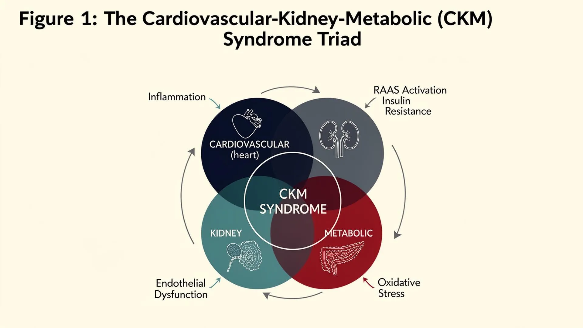
Figure 1: The Cardiovascular-Kidney-Metabolic (CKM) Syndrome Triad
Caption. CKM syndrome arises from the convergence of cardiovascular disease, chronic kidney disease, and metabolic dysfunction. Five shared molecular drivers — chronic inflammation, insulin resistance, endothelial dysfunction, RAAS over-activation, and oxidative stress — interconnect the three organ systems and underpin the unified pathobiology of the syndrome.
.
elevated angiotensin II, aldosterone, sympathetic catecholamines, and arginine vasopressin — that promote renal tubular sodium avidity, mesangial expansion, and interstitial fibrosis, ultimately driving CKD progression [48,50].
Conversely, the diseased kidney actively drives cardiac pathology through multiple mechanisms. Reduced eGFR is associated with accumulation of uraemic toxins (e.g., indoxyl sulphate, p-cresyl sulphate), phosphate retention, FGF-23 elevation, and reduced erythropoietin production, each of which independently promotes myocardial inflammation, hypertrophy, and arrhythmogenesis [72,73]. FGF-23, in particular, acts directly on cardiac myocytes — despite the absence of the obligate co-receptor Klotho in the heart — via FGFR4-dependent signalling to promote left ventricular hypertrophy, a finding that identifies FGF-23 as a critical molecular bridge in the cardiorenal continuum [72].
2.2 Kidney–Metabolic Interactions
The kidney is not a passive victim of metabolic disease but an active participant in metabolic regulation. The proximal tubule is a major site of glucose reabsorption (via SGLT2 and SGLT1) and gluconeogenesis, and its dysfunction under hyperglycaemic conditions — characterised by increased tubuloglomerular feedback disruption, hyperfiltration, and tubular hypertrophy — initiates a cascade of inflammation and fibrosis that accelerates diabetic nephropathy [40,100]. Furthermore, the kidney plays a central role in adipokine clearance, with reductions in GFR leading to elevated circulating leptin and resistin levels that perpetuate systemic insulin resistance [3].
Metabolic factors, in turn, exert profound effects on renal haemodynamics and tubular function. Obesity-related expansion of visceral and renal adipose tissue promotes perinephric inflammation, mechanical compression of the renal medulla, and the secretion of pro-fibrotic adipokines including leptin, visfatin, and resistin [67,68]. Adiponectin — the primary anti-inflammatory adipokine — is paradoxically reduced in obesity despite its protective effects on glomerular podocytes and mesangial cells, creating a condition of 'adipokinopathy' that accelerates both renal and cardiovascular disease progression [67].
2.3 Heart–Metabolic Interactions
The heart is an extraordinarily energy-demanding organ, consuming approximately 6 kg of ATP per day in the resting adult, and is highly sensitive to alterations in metabolic substrate availability and signalling [46,47]. Under physiological conditions, the healthy heart derives approximately 60–70% of its energy from fatty acid oxidation and the remainder primarily from glucose and lactate oxidation [46]. In the setting of insulin resistance and T2DM, however, this metabolic flexibility is markedly impaired: the heart shifts toward excessive fatty acid oxidation with simultaneous impairment of glucose uptake, generating lipotoxic intermediates (ceramide, diacylglycerol) and reactive oxygen species (ROS) that promote cardiomyocyte apoptosis, mitochondrial dysfunction, and interstitial fibrosis — the hallmarks of diabetic cardiomyopathy [88].
Epicardial adipose tissue (EAT) — a metabolically active visceral fat depot that shares a microcirculation with the myocardium — plays a particularly important role in heart–metabolic crosstalk [67,68]. In patients with metabolic syndrome and T2DM, EAT undergoes phenotypic transformation from a cardioprotective depot to an active source of pro-inflammatory cytokines, free fatty acids, and lipopolysaccharide-binding proteins that diffuse directly into the myocardial interstitium, promoting coronary microvascular dysfunction, pericardial inflammation, and atrial fibrosis [68].
2.4 Systems Biology Perspective
When viewed through a systems biology lens, CKM syndrome exemplifies the concept of 'disease network medicine' — where perturbation of one node (e.g., adipose tissue inflammation) generates ripple effects across the entire biological network via shared molecular pathways and circulating mediators [37]. The concept of 'organokines' — bioactive peptides secreted by individual organs that act on distal tissues
→ heart/kidney), and adipokines (adipose → all organs) [36,67,68]. These organ-specific secretory
products, when dysregulated, collectively disrupt the homeostatic balance that normally maintains cardiovascular, renal, and metabolic health.
3. INFLAMMATION IN CKM SYNDROME
Chronic, low-grade systemic inflammation — distinct from the acute inflammatory response — is a unifying pathobiological feature of all three components of CKM syndrome. This 'metaflammation', characterised by sub-pyrexial cytokine elevations and immune cell activation without classical signs of infection, is now recognised as both a driver and a consequence of the metabolic and cardiovascular dysfunction that defines CKM syndrome [16,17].
3.1 NF-κB Signalling Pathway
Nuclear factor kappa-B (NF-κB) is the master transcriptional regulator of the inflammatory response and occupies a pivotal position in CKM pathogenesis [26]. In the canonical NF-κB pathway, inflammatory stimuli — including TNF-α, IL-1β, Toll-like receptor (TLR) ligands such as lipopolysaccharide (LPS) and saturated fatty acids, as well as advanced glycation end-products (AGEs) — activate IκB kinase (IKK) complex, leading to phosphorylation and proteasomal degradation of the inhibitory protein IκBα, thus liberating the p65/p50 NF-κB heterodimer to translocate to the nucleus [26]. Once activated, NF-κB drives transcription of a broad array of inflammatory genes including those encoding TNF-α, IL-1β, IL-6, IL-8, MCP-1, VCAM-1, ICAM-1, cyclooxygenase-2 (COX-2), and inducible nitric oxide synthase (iNOS) [26,27].
Critically, NF-κB activation also directly inhibits insulin signalling: IKK-β phosphorylates insulin receptor substrate-1 (IRS-1) on serine residues (Ser307 in rodents; Ser312 in humans), converting it from an activating phosphorylation to an inhibitory one, thereby disrupting the PI3K-Akt insulin signalling cascade [18]. This mechanistic link between inflammation and insulin resistance — now well validated in both in vitro and in vivo models — explains why anti-inflammatory interventions consistently improve insulin sensitivity in preclinical and clinical studies [18,19].
3.2 JAK/STAT Signalling
The Janus kinase/Signal Transducer and Activator of Transcription (JAK/STAT) pathway constitutes a second major inflammatory signalling axis in CKM syndrome, particularly relevant to cytokine-driven cardiovascular and renal pathology [28,29]. IL-6, the most clinically significant JAK/STAT activator in CKM, acts through its soluble receptor (sIL-6R) to activate JAK1 and JAK2, driving STAT3 phosphorylation and nuclear translocation [29]. STAT3 activation promotes the transcription of acute-phase reactants (C-reactive protein, fibrinogen, serum amyloid A), pro-fibrotic mediators (TGF-β, CTGF), and anti-apoptotic proteins that collectively drive myocardial remodelling, glomerulosclerosis, and hepatic insulin resistance [29].
In the context of obesity and adipose tissue expansion, IL-6 is produced in large quantities by both adipocytes and resident macrophages, creating a systemic cytokine milieu that chronically activates JAK/STAT signalling in the liver, heart, and kidney. Notably, chronic hepatic STAT3 activation by IL-6 promotes gluconeogenesis and inhibits hepatic insulin signalling, contributing to fasting hyperglycaemia and systemic insulin resistance independently of the classical IRS-1/PI3K pathway [29,56].
3.3 NLRP3 Inflammasome
The NLRP3 (nucleotide-binding oligomerisation domain-like receptor family pyrin domain-containing 3) inflammasome is a multiprotein intracellular complex that serves as a critical amplifier of metabolic inflammation in CKM syndrome [28]. NLRP3 is activated by a diverse range of 'danger signals' that are elevated in CKM, including cholesterol crystals, uric acid, saturated fatty acids, glucose, and mitochondrial ROS [28]. Upon activation, NLRP3 recruits the adaptor protein ASC and caspase-1, forming a functional complex that cleaves pro-IL-1β and pro-IL-18 into their bioactive forms, and initiates pyroptosis — a form of inflammatory cell death that amplifies local inflammation through the release of damage-associated molecular patterns (DAMPs) [28].
NLRP3-derived IL-1β has been specifically implicated in the pathogenesis of all three components of CKM syndrome: in the pancreatic islets, it promotes beta-cell apoptosis and impairs insulin secretion; in the vasculature, it activates endothelial cells and smooth muscle cells to express adhesion molecules and produce vasoconstricting prostanoids; and in the kidney, it promotes mesangial cell proliferation and tubular injury [28,65]. The clinical validation of NLRP3 targeting in cardiovascular disease came from the CANTOS trial, in which the anti-IL-1β monoclonal antibody canakinumab significantly reduced major adverse cardiovascular events in patients with prior myocardial infarction and elevated high-sensitivity CRP, independently of lipid-lowering effects [13].
3.4 Innate and Adaptive Immune Contributions
Beyond the cytokine signalling axes, cellular components of both the innate and adaptive immune system contribute significantly to CKM pathogenesis. Macrophage polarisation plays a central role: classically activated (M1) macrophages, induced by IFN-γ and LPS, produce IL-1β, TNF-α, and IL-12, and are enriched in the adipose tissue, kidneys, and atherosclerotic plaques of individuals with CKM syndrome, where they perpetuate organ inflammation and fibrosis [17]. Conversely, alternatively activated (M2) macrophages, which are anti-inflammatory and pro-resolving, are relatively depleted in the metabolically diseased state due to the dominance of pro-inflammatory cytokines [17].
CD4+ T helper cells, particularly Th1 and Th17 subsets, contribute to vascular inflammation and promote autoimmune-like endothelial injury in hypertension and atherosclerosis, while regulatory T cells (Tregs) — which normally suppress vascular inflammation — are functionally impaired in obesity and T2DM [17,21]. Mast cells, positioned at the perivascular and perirenal adipose interfaces, release histamine, tryptase, and IL-6 upon activation by FFA and DAMPs, adding another dimension to the inflammatory milieu of CKM syndrome [17].
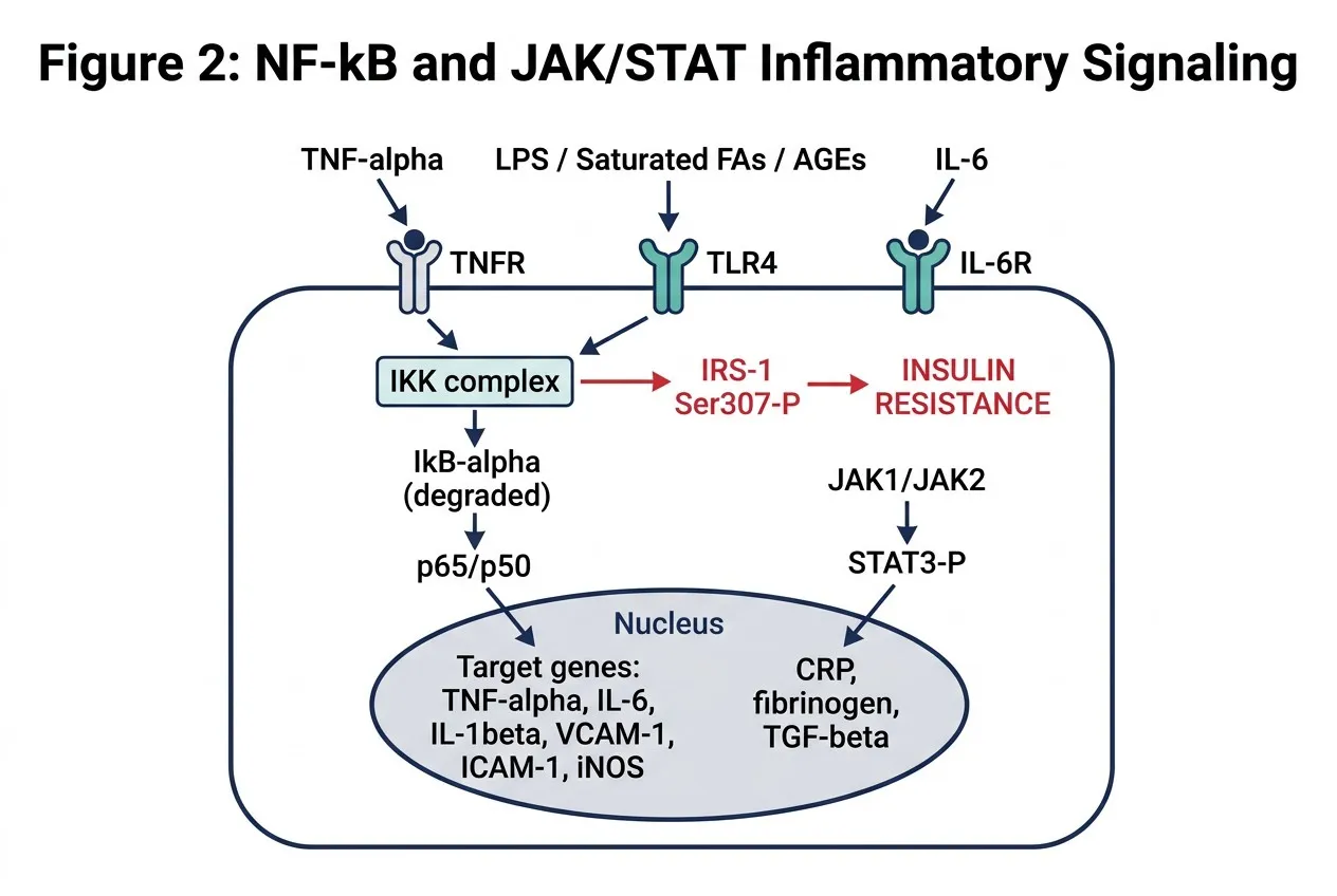
Figure 2: NF-B and JAK/STAT Inflammatory Signaling in CKM
Caption. TNF-, IL-1, IL-6, LPS, saturated fatty acids and AGEs activate IKK, leading to IB degradation and nuclear translocation of p65/p50, driving transcription of TNF-, IL-6, VCAM-1, ICAM-1 and iNOS. IL-6 also activates JAK1/2–STAT3, producing CRP, fibrinogen and TGF-. IKK- concurrently phosphorylates IRS-1 at Ser307, mechanistically coupling inflammation to insulin resistance.
4. INSULIN RESISTANCE: MECHANISMS AND METABOLIC CONSEQUENCES
Insulin resistance — defined as a diminished biological response to insulin at target tissues, principally skeletal muscle, liver, and adipose tissue — is the pathophysiological cornerstone of CKM syndrome and the primary metabolic driver that links obesity and T2DM to cardiovascular and renal disease [56,57]. Far from being a uniform defect, insulin resistance in CKM syndrome involves multiple, hierarchically organised molecular mechanisms that converge on impaired PI3K-Akt signalling and mitochondrial bioenergetic dysfunction.
4.1 Normal Insulin Signalling
Under physiological conditions, insulin binds to its receptor (IR) — a heterotetramer composed of two α and two β subunits — on target cell surfaces, triggering receptor autophosphorylation on tyrosine residues within the β-subunit activation loop [31]. This autophosphorylation creates docking sites for insulin receptor substrate proteins (IRS-1 and IRS-2), which, upon tyrosine phosphorylation by the IR, recruit and activate phosphatidylinositol 3-kinase (PI3K) [32]. PI3K phosphorylates phosphatidylinositol 4,5-bisphosphate (PIP2) to generate phosphatidylinositol 3,4,5-trisphosphate (PIP3), which recruits phosphoinositide-dependent kinase-1 (PDK1) and AKT/PKB to the plasma membrane [33]. AKT activation subsequently mediates the canonical metabolic effects of insulin: GLUT4 translocation to the plasma membrane for glucose uptake, inhibition of glycogen synthase kinase-3 (GSK-3) for glycogen synthesis, and inhibition of forkhead box protein O1 (FOXO1) to suppress gluconeogenesis [31,33].
4.2 IRS-1/PI3K-Akt Disruption in CKM
In CKM syndrome, this signalling cascade is disrupted at multiple levels. The most widely characterised defect involves serine/threonine phosphorylation of IRS-1, which competitively inhibits insulin-stimulated tyrosine phosphorylation and promotes proteasomal degradation of IRS-1, reducing its cellular abundance [18,31]. Multiple kinases catalyse inhibitory IRS-1 serine phosphorylation in the setting
of CKM, including IKK-β (activated by NF-κB), c-Jun N-terminal kinase (JNK, activated by ceramide and ER stress), S6 kinase 1 (S6K1, activated by mTORC1 in nutrient excess), and protein kinase C (PKC), the latter activated by diacylglycerol accumulation from lipid overflow [18,31,34].
JNK activation, in particular, represents a critical node in the inflammation–insulin resistance crosstalk. JNK1 is activated by saturated fatty acids — particularly palmitate — through TLR4-dependent signalling, and by ceramide, a sphingolipid metabolite generated from excess fatty acid flux that is markedly elevated in obese adipose tissue, liver, and skeletal muscle in CKM syndrome [18]. Ceramide simultaneously inhibits AKT through protein phosphatase 2A (PP2A)-mediated dephosphorylation and through the activation of atypical PKCζ, creating a lipid-driven insulin resistance that is particularly relevant in the context of the ectopic fat deposition that characterises CKM [18].
4.3 Mitochondrial Dysfunction
Mitochondrial dysfunction is both a cause and consequence of insulin resistance in CKM syndrome, creating a self-amplifying bioenergetic and oxidative stress cycle [5,30]. In healthy individuals, mitochondrial oxidative phosphorylation efficiently processes fatty acid and glucose-derived acetyl-CoA to generate ATP, with minimal 'electron leakage' to molecular oxygen. In CKM syndrome, however, the combination of nutrient excess, elevated circulating free fatty acids, and inflammatory cytokine-driven inhibition of mitochondrial biogenesis (via suppression of PGC-1α/SIRT1/AMPK signalling) leads to mitochondrial dysfunction characterised by reduced electron transport chain (ETC) activity, mitochondrial membrane potential dissipation, and generation of excess superoxide [30,35].
This mitochondrial superoxide production initiates a cascade of downstream oxidative stress: superoxide dismutase-mediated conversion to hydrogen peroxide (H₂O₂), Fenton reaction-generated hydroxyl radical, and the non-enzymatic reaction of superoxide with nitric oxide to form peroxynitrite (ONOO⁻) — a highly reactive nitrogen species that nitrosylates and inactivates multiple insulin signalling kinases including AKT and eNOS, thereby linking mitochondrial dysfunction directly to endothelial
dysfunction and insulin resistance [69,70]. Peroxynitrite-mediated nitration of insulin receptor substrate proteins represents a particularly important mechanism of persistent insulin resistance in conditions of chronic metabolic stress [70].
4.4 Adipokine Dysregulation
Adipose tissue in CKM syndrome transitions from a metabolically neutral energy depot to a pathologically active endocrine organ, secreting an altered adipokine profile that systemic promotes insulin resistance [67,78]. The two most clinically significant adipokines in this context are leptin and adiponectin. Leptin, normally a satiety signal, is elevated in obesity — producing 'leptin resistance' in the hypothalamus
5. ENDOTHELIAL DYSFUNCTION IN CKM SYNDROME
The vascular endothelium — a 700 m² monolayer of approximately 60 trillion cells lining the entire circulatory system — functions not merely as a passive conduit barrier, but as a highly sophisticated metabolic and signalling organ that continuously integrates local and systemic inputs to maintain vascular homeostasis [21,71]. Endothelial dysfunction, defined as impaired endothelium-dependent vasodilation and acquisition of a pro-inflammatory, pro-thrombotic, and pro-atherogenic endothelial phenotype, is a hallmark of CKM syndrome that precedes the clinical emergence of CVD, CKD, and T2DM and independently predicts adverse cardiovascular outcomes [21,22].
5.1 Normal Endothelial Function and eNOS
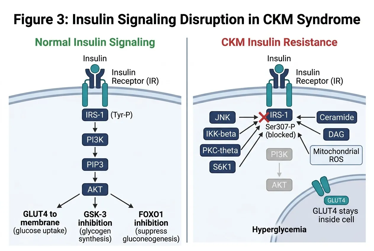
Figure 3: Insulin Signaling and Its Disruption in CKM Syndrome
Caption. Normally, insulin engages the insulin receptor IRS-1 (Tyr-P) PI3K PIP3 AKT, promoting GLUT4 translocation, glycogen synthesis and suppression of gluconeogenesis. In CKM, JNK, IKK-, PKC- and S6K1 — driven by ceramide, DAG, and mitochondrial ROS — phosphorylate IRS-1 at Ser307, blocking PI3K/AKT, retaining GLUT4 intracellularly, and producing hyperglycaemia.
Under physiological conditions, the endothelium maintains vascular tone primarily through the constitutive production of nitric oxide (NO) by endothelial nitric oxide synthase (eNOS) [23]. eNOS-derived NO diffuses from endothelial cells into adjacent vascular smooth muscle cells (VSMCs) and activates soluble guanylate cyclase (sGC), generating cyclic GMP (cGMP) and activating protein kinase G (PKG), which phosphorylates myosin light chain phosphatase and reduces intracellular calcium — resulting in smooth muscle relaxation and vasodilation [23,24]. Beyond its vasodilatory role, NO exerts critical pleiotropic effects on vascular biology: it inhibits platelet aggregation and adhesion, reduces VSMC proliferation and migration, suppresses endothelial adhesion molecule expression (VCAM-1, ICAM-1, E-selectin, P-selectin), and maintains glycocalyx integrity [22,23].
eNOS activity is regulated at multiple levels. Shear stress is the primary physiological activator: laminar blood flow induces Akt-mediated phosphorylation of eNOS at Ser1177, increasing NO output by approximately 10-fold [12,23]. Insulin signalling also activates eNOS via the PI3K-Akt pathway — a critical intersection point between metabolic signalling and vascular function that is disrupted in insulin resistance [20].
5.2 eNOS Uncoupling and Oxidative Stress
In CKM syndrome, eNOS undergoes a process of 'uncoupling' — a fundamental structural and functional alteration in which the enzyme shifts from NO production to superoxide generation [23,25]. eNOS uncoupling occurs through multiple mechanisms relevant to CKM: (i) depletion of the essential eNOS cofactor tetrahydrobiopterin (BH4) through oxidation by superoxide and peroxynitrite — creating a self-amplifying uncoupling cycle; (ii) displacement of L-arginine (the eNOS substrate) by the endogenous eNOS inhibitor asymmetric dimethylarginine (ADMA), which is elevated in CKD through reduced renal clearance and impaired DDAH enzyme activity; (iii) S-glutathionylation of critical cysteine residues in the eNOS reductase domain under conditions of oxidative stress; and (iv) loss of eNOS-Hsp90 interaction that normally stabilises the coupled enzyme configuration [23,25].
The shift from NO to superoxide production by uncoupled eNOS dramatically amplifies endothelial oxidative stress. Superoxide reacts with residual NO at near-diffusion-limited rates to form peroxynitrite, reducing NO bioavailability while simultaneously generating a potent oxidant that nitrates proteins, oxidises lipids, and damages DNA [25]. This NO-superoxide imbalance underpins the 'oxidative-nitrosative stress' phenotype of endothelial dysfunction in CKM syndrome.
5.3 Endothelial Glycocalyx Degradation
The endothelial glycocalyx — a carbohydrate-rich pericellular matrix composed of proteoglycans (syndecans, glypicans), glycosaminoglycans (heparan sulphate, hyaluronan, chondroitin sulphate), and adsorbed plasma proteins — covers the luminal surface of endothelial cells and acts as a molecular sieve, mechanosensor, anti-coagulant surface, and barrier to inflammatory cell adhesion [11]. In CKM syndrome, glycocalyx degradation is mediated by heparanase (activated by hyperglycaemia and oxidised LDL), hyaluronidase, matrix metalloproteinases, and reactive oxygen species [11,71].
Loss of glycocalyx integrity has cascading consequences: increased access of LDL and lipoprotein (a) to the subendothelial space, exposure of adhesion molecules that facilitate monocyte rolling and transmigration, dysregulation of shear stress-mediated eNOS activation (since the glycocalyx normally transduces the mechanical signal of laminar flow to the endothelial cytoskeleton), and activation of TLR4 and RAGE by DAMPs that accumulate in the denuded endothelium [11,21,22]. Urinary heparan sulphate shedding has emerged as a sensitive marker of glycocalyx degradation and correlates with microalbuminuria — suggesting shared mechanisms of endothelial injury in the glomerular and systemic vasculature in CKM syndrome [71,72].
5.4 Endothelial Adhesion Molecules and Monocyte Recruitment
The activated endothelial phenotype in CKM syndrome is characterised by upregulated expression of vascular cell adhesion molecule-1 (VCAM-1), intercellular adhesion molecule-1 (ICAM-1), E-selectin, and P-selectin, driven by NF-κB, AP-1, and SP1 transcription factors activated by oxLDL, AGEs, TNF-α,
and IL-1β [21,26]. These adhesion molecules facilitate the sequential adhesion cascade of monocytes: rolling (mediated by selectins), firm adhesion (mediated by VCAM-1/ICAM-1), and transendothelial migration via CCL2 (MCP-1) chemotactic gradient into the subendothelial space, where monocytes differentiate into macrophages and ingest oxLDL to form lipid-laden foam cells — the cellular hallmark of early atherosclerotic lesion formation [15,21].
6. CROSSTALK MECHANISMS: INTEGRATION OF THREE AXES
The three core pathological axes of CKM syndrome — inflammation, insulin resistance, and endothelial dysfunction — do not operate in isolation but are deeply interconnected through multiple bidirectional and multiway crosstalk mechanisms. Understanding these crosstalk nodes is essential for developing therapeutic strategies that address the syndrome at its roots rather than treating each component independently.
6.1 Inflammation–Insulin Resistance Crosstalk
The molecular relationship between inflammation and insulin resistance is mediated through several converging pathways. As detailed previously, IKK-β-mediated serine phosphorylation of IRS-1 represents the canonical inflammation-to-insulin resistance mechanism [18]. However, the reverse direction
Insulin resistance in adipocytes promotes lipolysis, releasing elevated concentrations of free fatty acids (FFAs) into the portal and systemic circulation [78,79]. These FFAs activate TLR4 and TLR2 on macrophages and endothelial cells, triggering MyD88-dependent activation of NF-κB and IRF3/IRF7 signalling, producing TNF-α, IL-6, IL-1β, and type I interferons that further suppress insulin signalling in all peripheral tissues [17,18]. Additionally, FFAs are substrates for ceramide biosynthesis via the de novo pathway (serine palmitoyltransferase/ceramide synthase) — and ceramide-rich microdomains in plasma membranes activate pro-inflammatory kinases (PKC, JNK, IKK-β) while inactivating the pro-survival
kinase AKT through PP2A-mediated dephosphorylation, creating a lipid-driven positive feedback loop between inflammation and insulin resistance [18].
Endoplasmic reticulum (ER) stress — a state of protein misfolding in the ER lumen induced by saturated FFAs, glucose, and inflammatory cytokines — activates the unfolded protein response (UPR) through three sensor kinases: IRE1α, PERK, and ATF6 [56]. The IRE1α branch phosphorylates JNK, which inhibits IRS-1; the PERK branch activates eIF2α kinase, suppressing protein synthesis and activating the TXNIP-NLRP3 inflammasome axis; and ATF6 activation increases CHOP expression, promoting apoptosis and NF-κB activation [56]. Thus, ER stress functions as a molecular bridge that links the metabolic lipid excess of CKM directly to both inflammatory activation and impaired insulin signalling.
6.2 Insulin Resistance–Endothelial Dysfunction Crosstalk
The vascular endothelium occupies a unique position in the insulin resistance–endothelial dysfunction crosstalk because it expresses both the insulin receptor and the RAAS components that are dysregulated in metabolic disease [20]. Under conditions of systemic insulin resistance, the endothelium does not uniformly resist insulin — rather, it selectively loses the PI3K-Akt-eNOS branch of insulin signalling while retaining (or even upregulating) the MAPK-ERK branch [20].
This 'selective insulin resistance' in the endothelium has profound vascular consequences. The PI3K-Akt branch of endothelial insulin signalling normally activates eNOS (through Ser1177 phosphorylation) and suppresses endothelin-1 (ET-1) production. Loss of this pathway reduces endothelial NO output and increases vasoconstricting ET-1 secretion, impairing flow-mediated vasodilation [20,24]. Simultaneously, the preserved and often overactivated MAPK-ERK branch drives endothelial expression of VCAM-1, ICAM-1, and PAI-1 — creating a pro-inflammatory, pro-thrombotic endothelial state that directly promotes atherosclerosis and thrombotic events [20,21].
Hyperglycaemia adds another dimension to endothelial dysfunction in insulin-resistant states through multiple biochemical mechanisms: (i) activation of the polyol pathway, diverting glucose to
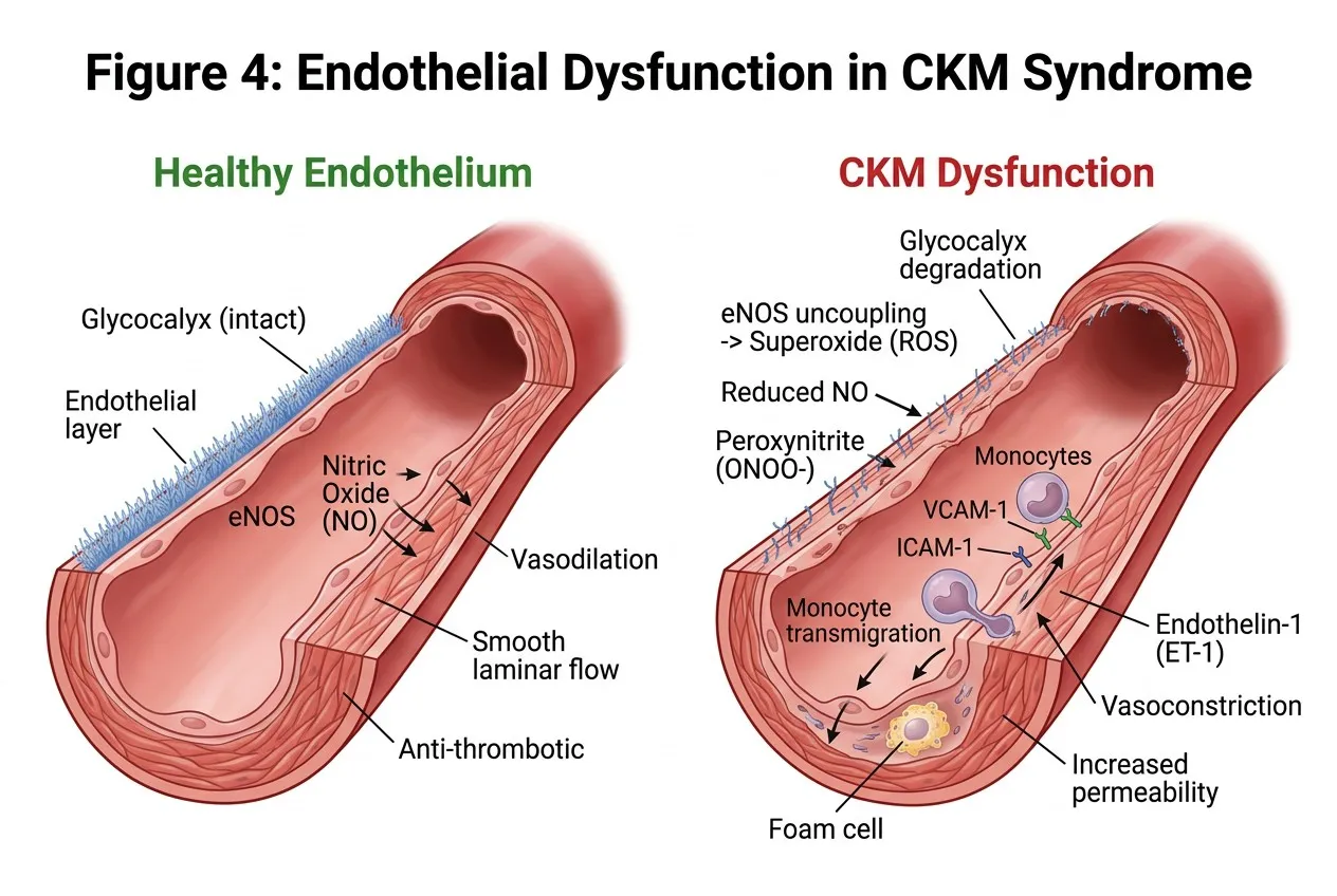
Figure 4: Endothelial Dysfunction in CKM Syndrome
Caption. The healthy endothelium maintains an intact glycocalyx and produces nitric oxide via eNOS, supporting vasodilation, laminar flow and an anti-thrombotic surface. In CKM, glycocalyx degradation, eNOS uncoupling and peroxynitrite formation reduce NO bioavailability, while VCAM-1/ICAM-1-mediated monocyte adhesion and transmigration, foam-cell formation, ET-1-driven vasoconstriction and increased permeability accelerate vascular and end-organ injury.
sorbitol and fructose, depleting NADPH and impairing BH4 synthesis; (ii) activation of PKCβ by diacylglycerol, promoting NADPH oxidase-mediated superoxide production; (iii) non-enzymatic glycation of proteins to form AGEs, which signal through RAGE to activate NF-κB, NAD (P)H oxidase, and TGF-β; and (iv) increased hexosamine pathway flux, O-GlcNAcylating eNOS at Ser1177 and thereby reducing its activity [86,87].
6.3 Oxidative Stress as Central Integrator
Reactive oxygen species (ROS) occupy the central position in the CKM crosstalk network, simultaneously promoting inflammation, impairing insulin signalling, and damaging the endothelium [69,70]. In CKM syndrome, excess ROS are generated by multiple sources: NADPH oxidase (NOX) isoforms (NOX2 in macrophages and endothelial cells; NOX4 in cardiomyocytes and renal tubular cells), mitochondrial electron transport chain leakage, xanthine oxidase (elevated in uricaemic CKD), uncoupled eNOS, and activated LOX/COX enzymes [69,70].
ROS suppress insulin signalling by oxidising the catalytic cysteine residue of PTEN phosphatase (which normally degrades PIP3 to limit Akt activation) — paradoxically, mild ROS can enhance insulin signalling, but the chronically elevated ROS in CKM overwhelms this effect and instead promotes net insulin resistance through JNK activation, IRS-1 serine phosphorylation, and GLUT4 carbonylation [69]. In the vasculature, ROS degrade NO (forming peroxynitrite), oxidise BH4 (uncoupling eNOS), oxidise LDL (generating pro-atherogenic oxLDL), and activate matrix metalloproteinases that degrade the glycocalyx and extracellular matrix, collectively perpetuating endothelial dysfunction [25,70].
The Nrf2 (nuclear factor erythroid 2-related factor 2) pathway represents the primary transcriptional antioxidant defence system and is significantly impaired in CKM syndrome. Under basal conditions, Nrf2 is sequestered in the cytoplasm by Keap1 (Kelch-like ECH-associated protein 1) and targeted for proteasomal degradation. Electrophilic ROS and dietary phytochemicals (e.g., sulforaphane, curcumin) modify cysteine residues on Keap1, releasing Nrf2 to translocate to the nucleus and activate the
antioxidant response element (ARE), driving transcription of phase II detoxification enzymes including HO-1, NQO1, GSH biosynthesis enzymes, and ferritin [69]. In CKM syndrome, impaired Nrf2 activation
— partly due to NF-κB-mediated suppression and AGE-induced Keap1 upregulation — reduces the cellular capacity to quench the inflammatory ROS burden, contributing to progressive organ damage [69,70].
7. KEY MOLECULAR PATHWAYS IN CKM SYNDROME
7.1 RAAS: Classical and Alternative Axes
The renin-angiotensin-aldosterone system (RAAS) is the single most important neurohormonal system in the pathogenesis of CKM syndrome, with pathological activation of the classical RAAS axis contributing to hypertension, cardiac fibrosis, glomerulosclerosis, and insulin resistance through multiple mechanisms [81,82]. Angiotensin II (Ang II), generated from angiotensin I by angiotensin-converting enzyme (ACE), activates AT1 receptors on vascular smooth muscle cells, cardiomyocytes, mesangial cells, and adrenal cortex, producing vasoconstriction, myocardial hypertrophy, aldosterone secretion, and renal sodium retention [50,81].
At the molecular level, Ang II–AT1R signalling activates multiple pro-inflammatory and pro-fibrotic pathways: NOX2/NOX4-mediated superoxide generation, NF-κB activation, TGF-β1 secretion, PDGF-mediated smooth muscle proliferation, and ERK1/2-dependent anti-apoptotic signalling in fibroblasts [50,81]. Ang II also directly impairs insulin signalling by promoting serine phosphorylation of IRS-1 through PKC and JNK activation, providing a mechanistic link between RAAS overactivation and insulin resistance in CKM syndrome [81].
The discovery of the 'alternative RAAS axis' — comprising ACE2, Angiotensin-(1–7) [Ang (1-7)], and the Mas receptor — has provided a conceptually important counterbalancing system with cardioprotective, reno-protective, and insulin-sensitising effects [82]. ACE2 cleaves Ang II to generate Ang (1-7), which acts on the Mas receptor to produce NO, activate AMPK, reduce oxidative stress via Nrf2, and inhibit the AT1R-mediated pro-fibrotic cascade [82]. Importantly, ACE2 is dramatically
downregulated by classical RAAS activation — creating a pathological shift towards Ang II dominance that perpetuates CKM progression [82]. SGLT2 inhibitors and GLP-1 receptor agonists have been shown to upregulate ACE2 expression and shift the RAAS balance towards the protective axis, partly explaining their pleiotropic organ-protective effects [93,94].
7.2 AMPK: The Master Energy Sensor
AMP-activated protein kinase (AMPK) functions as the cell's master energy sensor and metabolic regulator, activated by an increase in AMP:ATP ratio during energy deficit states such as exercise, caloric restriction, and ischaemia [35]. In CKM syndrome, AMPK activity is significantly reduced in the heart, skeletal muscle, liver, and vasculature — contributing to impaired mitochondrial biogenesis, excessive fatty acid synthesis, enhanced gluconeogenesis, and reduced autophagy [35].
AMPK activation in CKM-relevant tissues produces a comprehensive suite of protective effects: in skeletal muscle, AMPK phosphorylates AS160/TBC1D4 to promote GLUT4 translocation and glucose uptake independently of insulin; in the liver, AMPK phosphorylates and inactivates acetyl-CoA carboxylase (ACC), reducing malonyl-CoA and thereby relieving CPT-1 inhibition to enhance mitochondrial fatty acid oxidation and reduce hepatic lipid accumulation; in the vasculature, AMPK phosphorylates eNOS at Ser1177 to augment NO production and directly suppresses NF-κB activity by preventing IKK-β activation [35,36]. In cardiomyocytes, AMPK activates ULK1 to initiate mitophagy, clearing damaged mitochondria and reducing cardiomyocyte ROS production [35].
Metformin, the most widely used antidiabetic agent globally, exerts its primary anti-hyperglycaemic and cardiovascular-protective effects principally through AMPK activation (by inhibiting mitochondrial complex I and thereby increasing cellular AMP:ATP ratio), though AMPK-independent mechanisms involving the lysosomal AXIN/LKB1 complex are also important [35,56]. AMPK is also the principal pharmacological target of the pleiotropic protective effects attributed to caloric restriction mimetics and physical exercise in CKM syndrome.
7.3 mTOR Signalling
The mechanistic target of rapamycin (mTOR) exists in two functionally distinct complexes — mTORC1 and mTORC2 — that play opposing roles in CKM syndrome [34]. mTORC1 integrates signals from growth factors (via PI3K-Akt), amino acids (via Ragulator-RAG GTPase), and energy status (via AMPK-mediated TSC1/2 regulation) to control cell growth, protein synthesis, and autophagy [34]. Chronic overactivation of mTORC1 — driven by nutrient excess, insulin resistance, and Ang II in CKM syndrome
In contrast, mTORC2 is generally cardioprotective: it phosphorylates AKT at Ser473 to activate full AKT signalling, supports eNOS function in the vasculature, and maintains cardiomyocyte survival signalling [34]. Dysregulation of the mTORC1:mTORC2 balance in favour of mTORC1 dominance is a defining feature of metabolic overnutrition in CKM syndrome, and represents a therapeutic vulnerability that is partially addressed by caloric restriction, rapamycin analogues (in experimental settings), and metformin [34,35].
7.4 SIRT1 and the Longevity Axis
SIRT1, a NAD⁺-dependent class III histone deacetylase, occupies a pivotal position in the molecular pathology of CKM syndrome as both an energy status sensor and a master epigenetic regulator of inflammation, metabolism, and stress resistance [37,38]. SIRT1 activity is dependent on cellular NAD⁺ levels, which are significantly reduced in CKM syndrome due to chronic oxidative stress (which consumes NAD⁺ via PARP activation) and impaired NAD⁺ biosynthesis [87,88].
SIRT1 exerts its protective effects through deacetylation of multiple critical substrates: PGC-1α (activating mitochondrial biogenesis and fatty acid oxidation), FOXO3 (promoting antioxidant defence via SOD2 and catalase), NF-κB p65 (suppressing inflammatory transcription), eNOS (increasing NO bioavailability), LKB1 (activating AMPK), and PPARΑ (promoting lipid catabolism) [37,38]. SIRT1
activity is significantly reduced in adipose tissue, skeletal muscle, and the liver of obese and insulin-resistant individuals, and its deficiency has been directly linked to increased NF-κB-driven inflammation, impaired insulin signalling, and accelerated endothelial senescence [87].
SIRT3, the predominant mitochondrial sirtuin, deacetylates and activates multiple ETC subunits and antioxidant enzymes (including MnSOD/SOD2), and its downregulation in CKM syndrome contributes to mitochondrial superoxide accumulation and cardiomyocyte dysfunction [38]. The SIRT1-PGC-1α-SIRT3 axis thus represents an integrated mitochondrial-epigenetic regulatory network whose disruption in CKM syndrome perpetuates the bioenergetic and oxidative stress that drive multi-organ damage.
8. BIOMARKERS IN CKM SYNDROME
The identification of reliable, clinically accessible biomarkers for CKM syndrome is of paramount importance for early risk stratification, prognostication, treatment monitoring, and the identification of patients who will derive greatest benefit from multi-organ protective therapies. Biomarkers in CKM ideally should reflect the integrated pathophysiology of the syndrome — capturing elements of cardiac stress, renal injury, metabolic dysfunction, inflammation, and endothelial activation simultaneously [39,72].
8.1 Traditional Cardiac and Renal Biomarkers
B-type natriuretic peptide (BNP) and its N-terminal prohormone NT-proBNP are released from cardiomyocytes in response to increased wall stress and are the most widely validated cardiac biomarkers in heart failure and CKM syndrome [54,74]. Both markers independently predict all-cause mortality, heart failure hospitalisation, and CKD progression in patients with CKM, and elevated NT-proBNP (>125 pg/mL in non-acute settings) forms part of the diagnostic criteria for heart failure with preserved ejection fraction (HFpEF) — the cardiovascular phenotype most strongly associated with metabolic CKM syndrome [51,54].
High-sensitivity cardiac troponin T (hs-cTnT) and troponin I (hs-cTnI) detect subclinical myocardial injury and are chronically elevated in CKM syndrome even in the absence of acute coronary syndromes, reflecting ongoing cardiomyocyte necrosis and turnover driven by chronic inflammation and
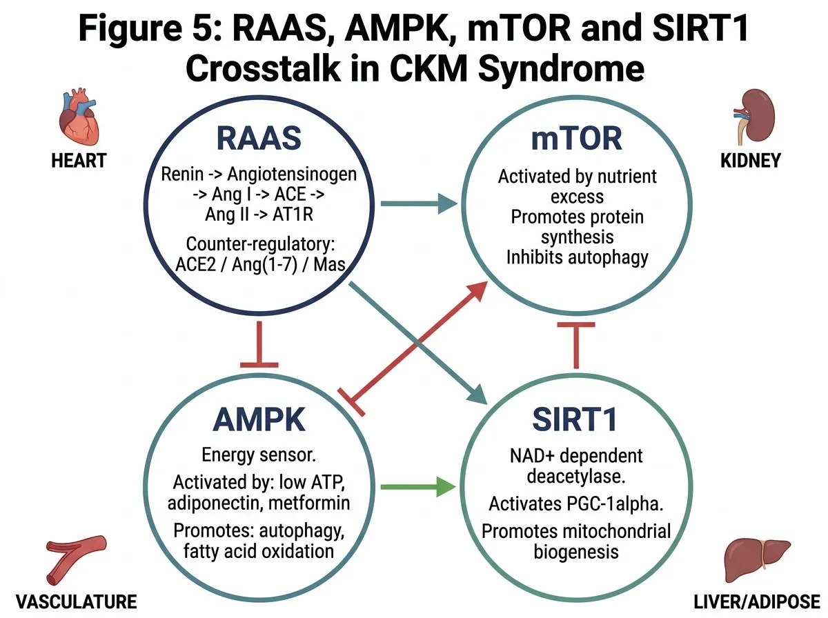
Figure 5: RAAS, AMPK, mTOR and SIRT1 Crosstalk in CKM
Caption. Four master regulators integrate metabolic and cardiorenal signals. Angiotensin II activates mTOR and inhibits AMPK, promoting fibrosis and proliferation. AMPK and SIRT1 (NAD■-dependent) reciprocally activate one another, suppress mTOR, and drive autophagy, fatty-acid oxidation and PGC-1-mediated mitochondrial biogenesis. The counter-regulatory ACE2/Ang (1–7)/Mas axis opposes the deleterious arm of RAAS.
ischaemia [74]. Estimated glomerular filtration rate (eGFR) and urinary albumin-to-creatinine ratio (UACR) remain the clinical cornerstones of CKD assessment, with UACR reflecting glomerular endothelial dysfunction and serving as an independent predictor of both cardiovascular and renal outcomes in CKM syndrome [40,41,100].
8.2 Novel Inflammatory and Fibrotic Biomarkers
Galectin-3, a β-galactoside-binding lectin secreted by activated macrophages, has emerged as a marker of myocardial and renal fibrosis in CKM syndrome, with elevated circulating galectin-3 independently predicting incident heart failure, CKD progression, and all-cause mortality [84]. Soluble ST2 (sST2), the soluble decoy receptor for IL-33, is elevated in heart failure and correlates with myocardial fibrosis and poor prognosis, particularly in HFpEF where it reflects the pericardial and myocardial inflammatory burden that characterises the metabolic heart failure phenotype [84].
Growth differentiation factor-15 (GDF-15), a stress-responsive cytokine of the TGF-β superfamily, is markedly elevated across all stages of CKM syndrome and reflects multiorgan stress — with its expression driven by mitochondrial dysfunction, inflammatory cytokines, and AMPK activation [72]. FGF-23 elevation, as discussed previously, reflects advanced CKD and serves as a mediator (not merely a marker) of LVH and mortality in the cardiorenal axis [72]. High-sensitivity C-reactive protein (hs-CRP) and IL-6 remain the most clinically validated systemic inflammatory biomarkers, with hs-CRP ≥ 2 mg/L associated with more than doubling of cardiovascular risk in metabolic syndrome patients [13,15].
8.3 Endothelial and Metabolic Biomarkers
Asymmetric dimethylarginine (ADMA), an endogenous eNOS inhibitor that is elevated in CKD, insulin resistance, and hypertension, provides a direct measure of impaired NO bioavailability and endothelial dysfunction with strong independent predictive value for cardiovascular events [72]. Circulating endothelial microparticles (EMPs) — membrane-bound vesicles released from activated or apoptotic endothelial cells — are elevated in CKM syndrome and carry pro-coagulant and pro-inflammatory cargo
that amplifies vascular injury in a paracrine fashion [71]. HbA1c and fasting insulin (or HOMA-IR) quantify chronic glucose exposure and insulin resistance severity, respectively, and remain essential components of CKM risk stratification panels.
Emerging multi-omics approaches — including plasma metabolomics (profiling branched-chain amino acids, short-chain acylcarnitines, ceramides, and trimethylamine N-oxide [TMAO] as markers of insulin resistance and gut–liver–cardiovascular axis dysfunction), proteomics (using proximity extension assay platforms to simultaneously profile hundreds of cardiovascular-relevant proteins), and circulating microRNA panels (miR-21, miR-133a, miR-155, miR-499 as cardiac and vascular injury biomarkers) — offer transformative potential for capturing the integrated molecular complexity of CKM syndrome at a systems level [54,72]. These platforms have already demonstrated the ability to predict incident heart failure, CKD progression, and major adverse cardiovascular events with superior performance to traditional single-biomarker approaches, and their integration into clinical decision-making pipelines represents a key frontier in CKM precision medicine [72,74].
9. THERAPEUTIC TARGETS AND PHARMACOLOGICAL STRATEGIES
The mechanistic understanding of CKM syndrome as an integrated inflammatory–metabolic–endothelial disorder has profound implications for therapeutic strategy. Rather than targeting individual risk factors in isolation, the most effective CKM pharmacotherapy achieves pleiotropic benefits by simultaneously modulating multiple nodes of the shared pathophysiological network [74,80]. The following section reviews the mechanistic basis of pharmacological agents with proven multi-organ benefit in CKM syndrome, organised by their primary molecular target.
9.1 SGLT2 Inhibitors
Sodium-glucose cotransporter 2 (SGLT2) inhibitors — including empagliflozin, dapagliflozin, and canagliflozin — have emerged as the most transformative class of agents in CKM pharmacotherapy, demonstrating consistent cardiovascular and renal protection across a spectrum of clinical phenotypes including T2DM, heart failure, and CKD [8,9,10]. While their primary pharmacological mechanism is
urinary glucose excretion through SGLT2 blockade in the renal proximal tubule, their organ-protective effects in CKM extend far beyond glycaemic control [92,94].
Mechanistically, SGLT2 inhibitors activate AMPK and inhibit mTORC1 — partly through glucose exclusion-mediated energetic sensing — promoting autophagy, mitophagy, and anti-inflammatory signalling in cardiomyocytes and renal tubular cells [93,94]. In the kidney, they restore tubuloglomerular feedback by increasing sodium delivery to the macula densa (reducing hyperfiltration), decrease intraglomerular pressure, and reduce proximal tubular hypertrophy and inflammation [40,100]. In the heart, they reduce preload and afterload through osmotic diuresis, shift myocardial substrate use towards ketone body oxidation (which is a more oxygen-efficient fuel than glucose or fatty acids), and suppress cardiac NLRP3 inflammasome activation and fibrotic TGF-β signalling [93,94]. SGLT2 inhibitors also upregulate the protective ACE2/Ang (1-7)/Mas axis and reduce EAT volume and inflammatory activity, adding cardioprotective benefits that extend beyond haemodynamic and metabolic effects [93].
Landmark clinical trials — EMPA-REG OUTCOME (empagliflozin), CANVAS (canagliflozin), DECLARE-TIMI 58 (dapagliflozin), DAPA-HF and EMPEROR-Reduced (heart failure with reduced ejection fraction), EMPEROR-Preserved and DELIVER (HFpEF), CREDENCE and DAPA-CKD (CKD with and without T2DM) — have collectively established SGLT2 inhibitors as a cornerstone of CKM pharmacotherapy across the full syndrome spectrum [8,9,10,41,51,52,53,100].
9.2 GLP-1 Receptor Agonists
Glucagon-like peptide-1 receptor agonists (GLP-1RAs) — including liraglutide, semaglutide (subcutaneous and oral), dulaglutide, and exenatide — exert direct cardioprotective effects through GLP-1R expressed on cardiomyocytes, endothelial cells, and macrophages, independent of their glucose-lowering and weight-reducing actions [11,58]. GLP-1R activation in cardiomyocytes activates the cAMP/PKA pathway, augmenting calcium handling and contractile function, reducing apoptosis via Bcl-2 upregulation, and attenuating ischaemia-reperfusion injury [58]. In endothelial cells, GLP-1R signalling
activates eNOS via PKA-dependent and PI3K-Akt-dependent pathways, increasing NO bioavailability and reducing endothelial oxidative stress [58].
GLP-1RAs reduce systemic inflammation by suppressing NF-κB and NLRP3 inflammasome activation in macrophages — an effect validated by the consistent reduction in hs-CRP, IL-6, and TNF-α observed across major cardiovascular outcome trials (LEADER with liraglutide, SUSTAIN-6 with semaglutide) [11,12,44]. In the kidney, GLP-1R activation reduces hyperfiltration, mesangial inflammation, and tubular injury partly through AMPK-mediated pathway activation and direct anti-inflammatory effects on glomerular podocytes [44]. The combination of SGLT2 inhibitors and GLP-1RAs is increasingly recognised as providing complementary and potentially synergistic multi-organ protection in CKM syndrome — an hypothesis currently being formally tested in combination trial designs [58].
9.3 Finerenone and Non-Steroidal MRA
Finerenone, a non-steroidal highly selective mineralocorticoid receptor antagonist (MRA), represents a major advance in CKM pharmacotherapy, addressing the specific pathophysiological role of aldosterone and mineralocorticoid receptor (MR) overactivation in driving cardiac and renal fibrosis, inflammation, and oxidative stress [42,43]. Unlike the steroidal MRAs spironolactone and eplerenone, finerenone demonstrates highly selective MR blockade without significant off-target sex hormone receptor effects, and its distinct binding mode to the MR ligand-binding domain confers superior anti-inflammatory and anti-fibrotic efficacy relative to pure receptor blockade [42,43].
Mechanistically, MR activation in cardiomyocytes and renal mesangial cells by aldosterone — as well as by cortisol and potentially other ligands — drives pro-fibrotic gene expression (TGF-β, CTGF, collagen I/III), NADPH oxidase activation (increasing ROS), and NF-κB-mediated inflammatory cytokine production [42,43]. Finerenone antagonises these effects and has demonstrated in both FIDELIO-DKD and FIGARO-DKD trials significant reductions in UACR, eGFR decline, cardiovascular death, and non-fatal
MI/stroke in patients with CKD and T2DM — establishing its position as a foundational agent in the pharmacological management of CKM syndrome [42,43].
9.4 Anti-inflammatory Strategies
The direct anti-inflammatory approach to CKM syndrome has been validated by the CANTOS trial with canakinumab (anti-IL-1β antibody), which demonstrated that reducing hs-CRP below 2 mg/L was associated with significant cardiovascular event reduction, supporting the causal role of inflammation in atherosclerotic events [13]. Colchicine — a tubulin polymerisation inhibitor that suppresses NLRP3 inflammasome activation and neutrophil chemotaxis — has demonstrated cardiovascular protection in both post-MI patients (COLCOT trial) and chronic coronary disease patients (LoDoCo2 trial) at low doses [63,64]. Its anti-CKM mechanism relates to NLRP3 suppression, reduction in IL-1β and IL-18, and inhibition of NF-κB activation through tubulin stabilisation in macrophages [63,64].
Statins, beyond their lipid-lowering effects, exert important pleiotropic anti-CKM actions: inhibition of small GTPase (Rho/Rac) prenylation reduces NOX2 assembly and superoxide production, improves eNOS coupling by maintaining BH4 levels, suppresses NF-κB activity, and attenuates RAAS-mediated vascular inflammation [80]. PCSK9 inhibitors — evolocumab and alirocumab — while primarily lipid-lowering agents, also reduce systemic inflammation as evidenced by hs-CRP reduction in FOURIER and ODYSSEY OUTCOMES trials, and have demonstrated benefit in patients with CKM features including insulin resistance and metabolic syndrome [61,62].
9.5 Lifestyle Interventions
Lifestyle modifications remain the foundation of CKM syndrome management, with robust evidence supporting the Mediterranean diet, DASH diet, moderate-intensity aerobic exercise, and intentional weight loss as interventions that modulate multiple pathways of the CKM network simultaneously [95,96,97]. Caloric restriction and the Mediterranean diet reduce systemic inflammation (lowering IL-6, TNF-α, CRP, and VCAM-1), improve insulin sensitivity (through AMPK activation and
reduction of visceral adiposity), and enhance endothelial function (via NO-mediated vasodilation and glycocalyx restoration) [95,96]. Exercise training, even without weight loss, consistently improves flow-mediated dilation, reduces ADMA, activates AMPK in cardiac and skeletal muscle, and promotes cardiac mitochondrial biogenesis — effects that are mechanistically complementary to pharmacological CKM intervention [97].
10. FUTURE PERSPECTIVES
The emerging understanding of CKM syndrome as a systems-level disorder with deeply interconnected molecular axes positions this field at the frontier of several transformative advances in biomedical science and clinical medicine.
10.1 Artificial Intelligence and Digital Health
Artificial intelligence (AI) and machine learning (ML) are poised to fundamentally transform CKM syndrome management through enhanced phenotyping, risk stratification, and treatment personalisation [38]. ML algorithms applied to electronic health record data, wearable biosensor outputs, imaging data, and multi-omics profiles are already demonstrating superior predictive performance for incident heart failure, CKD progression, and cardiovascular events compared to conventional risk scores [38,74]. Digital twin technology — computational models of individual patient cardiovascular and metabolic physiology that can be used to simulate treatment responses in silico before prescribing — holds particular promise for personalising combination pharmacotherapy in complex CKM patients [51,54].
10.2 Precision Medicine and Multi-Omics
The integration of genomics, transcriptomics, proteomics, metabolomics, and microbiomics — collectively referred to as 'multi-omics' — offers the prospect of molecularly subtyping CKM syndrome into mechanistically distinct endotypes that may respond differentially to specific therapeutic strategies [37,38]. For example, patients with CKM dominated by ceramide-driven inflammasome activation may derive particular benefit from sphingomyelin pathway-targeted therapies, while those with predominant
RAAS-aldosterone overactivation may be optimally managed with finerenone-based regimens [42,43,86]. Pharmacogenomics — particularly CYP2C19 polymorphisms affecting antiplatelet agent metabolism — is already influencing prescribing decisions in CKM patients with coronary artery disease and will increasingly guide multi-drug CKM regimens [80].
10.3 Emerging Therapeutic Horizons
Several emerging therapeutic strategies hold particular promise for CKM syndrome. The dual GIP/GLP-1 receptor agonist tirzepatide has demonstrated superior weight loss and metabolic improvement versus semaglutide in T2DM and is now being evaluated for cardiovascular and renal outcomes in dedicated CKM populations [59]. Senolytic agents targeting the clearance of senescent 'zombie cells' — which accumulate in the cardiovascular, renal, and metabolic tissues of ageing CKM patients and drive chronic SASP-mediated inflammation — represent a mechanistically novel approach to CKM reversal [4]. Gene-silencing technologies (siRNA, ASOs), exemplified by inclisiran for PCSK9 inhibition and vutrisiran for transthyretin amyloidosis, are increasingly being explored for CKM-relevant targets including angiotensinogen (for RAAS suppression) and apolipoprotein C-III (for hypertriglyceridaemia) [44,62]. Finally, the gut microbiome–cardiovascular axis — mediated principally through microbial metabolite production of TMAO, short-chain fatty acids, and uremic toxins — is emerging as a modifiable environmental determinant of CKM risk that may be targeted through dietary prebiotics, probiotics, and gut microbiome engineering strategies [12].
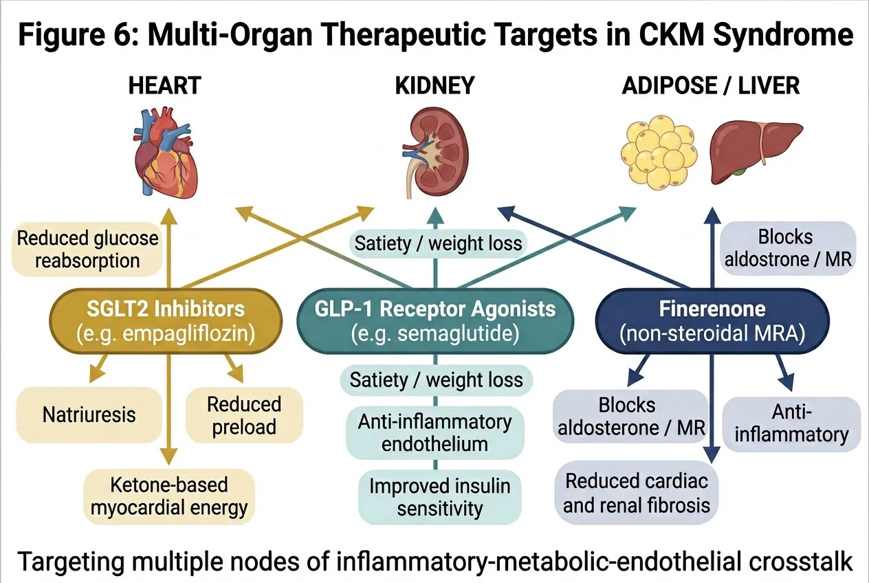
Figure 6: Multi-Organ Therapeutic Targets in CKM Syndrome
Caption. SGLT2 inhibitors (e.g. empagliflozin), GLP-1 receptor agonists (e.g. semaglutide) and the non-steroidal mineralocorticoid receptor antagonist finerenone exert pleiotropic benefits across heart, kidney and metabolic tissues. By simultaneously modulating natriuresis, preload, myocardial energetics, satiety, endothelial inflammation and fibrosis, these agents target several nodes of the inflammatory–metabolic–endothelial crosstalk that defines CKM syndrome.
CONCLUSION
CKM syndrome represents one of the most significant integrative challenges in contemporary medicine — a multisystem disorder arising from the convergence of metabolic dysfunction, cardiovascular disease, and chronic kidney disease that cannot be adequately understood or treated through the lens of any single organ system or molecular pathway.
This review has systematically demonstrated that inflammation, insulin resistance, and endothelial dysfunction are not merely co-morbid components of CKM syndrome, but are mechanistically interlinked axes that amplify each other through shared molecular mediators including NF-κB, ROS, ceramide, ADMA, and RAAS-derived Ang II. The molecular pathways of AMPK, mTOR, SIRT1, and the alternative ACE2/Ang (1-7)/Mas axis are consistently dysregulated across all three pathological axes and represent high-yield therapeutic targets that are already being pharmacologically exploited by SGLT2 inhibitors, GLP-1 receptor agonists, and finerenone — agents with demonstrated multi-organ protective effects in landmark cardiovascular and renal outcome trials.
The transition from organ-specific to integrated CKM management represents both a scientific imperative and a clinical opportunity. The co-prescription of SGLT2 inhibitors and GLP-1RAs — with or without finerenone, based on albuminuria and UACR — offers the most comprehensively validated pharmacological strategy for simultaneously addressing the cardiovascular, renal, and metabolic components of CKM syndrome available today. Complemented by structured lifestyle interventions targeting the Mediterranean diet pattern and regular aerobic exercise, this multi-modal approach provides mechanistically coherent, evidence-based management across all stages of CKM syndrome.
Looking forward, the integration of multi-omics biomarker profiling, AI-assisted phenotyping, digital twin modelling, and emerging gene-silencing technologies will enable the precision medicine paradigm necessary to individualise CKM management at a molecular level. The formal recognition of CKM syndrome as a distinct clinical entity by the American Heart Association in 2023 marks not merely a taxonomic advance, but a call to action for the biomedical community to align research, guideline
development, and clinical practice around the integrated systems biology that underpins this syndrome. It is only through this integrative lens — recognising the heart, kidneys, and metabolic organs as components of a single, interconnected physiological network — that we can hope to meaningfully reduce the immense cardiovascular and renal burden imposed by CKM syndrome on the global population.
REFERENCES





Mohammad Adnan, Bhavyadeep Verma, Kandiva Udhaya Devan, Ahmad Choudhry, Shivprasad Dhage, Inflammatory–Metabolic–Endothelial Crosstalk in Cardiovascular-Kidney-Metabolic (CKM) Syndrome: Molecular Mechanisms, Biomarkers, and Therapeutic Targets, Int. J. of Pharm. Sci., 2026, Vol 4, Issue 10, 2489-2511. https://doi.org/10.5281/zenodo.23414220
 10.5281/zenodo.23414220