

We use cookies to ensure our website works properly and to personalise your experience. Cookies policy
1,2,4Department of Microbiology, Vels Institute of Science, Technology & Advanced Studies, Chennai, Tamil Nadu, India.
3Department of Bio-Engineering, Vels Institute of Science, Technology & Advanced Studies, Chennai, Tamil Nadu, India.
Termite mounds offer a selective micro environment to Actinobacteria and are an under-explored source of bioactive microorganisms. Mound soils gathered at four sites in Ranipet District, Tamil Nadu in this study provided five morphologically different actinomycete isolates. Six-day submerged cultures were screened to obtain ethyl acetate extracts that were tested on antioxidant properties via the DPPH assay, where isolates TK4 and TK5 ultimately produced the strongest radical-scavenging activities (67.6% and 66.4%, respectively). TK4 was selected for further characterisation based on its bioactivity. Partially sequencing the 16S rRNA gene revealed the isolate as Streptomyces griseus TK4 (GenBank: PP774232). The extract exhibited concentration-related cytotoxicity towards MCF-7 human breast adenocarcinoma cells with an IC50 value of 38.1 µg / mL. Analysis using GC -MS showed that the metabolite profile was comprised of 2,4-di-tert-butylphenol along with L - (+)-ascorbic acid 2,6-dihexadecanoate and long-chain aliphatic hydrocarbons and a phthalate ester. These findings highlight termite mound-derived Streptomyces as a promising source of bioactive secondary metabolites for future bioassay-guided drug discovery.
The two therapeutic arithmetics have come to a common frustration. In 2021, 4.71 million deaths worldwide were attributed to bacterial antimicrobial resistance, 1.14 million of which were directly linked to resistance, and the modelled trend projects this trend to increase through 2050, with approximately two-thirds of the all-age mortality occurring in South Asia [1]. Oncology boasts an equivalent heft: it induced nearly 24 million cancer-related diagnoses worldwide in 2022, and female breast cancer was the most commonly diagnosed cancer in India [2]. Neither of the problems is ultimately of clinical delivery. Both are ultimately based on a dwindling supply of chemically novel small molecules that has been whittling away throughout the last forty years. A fantastic proportion of the molecules that solved the initial version of this problem were provided by filamentous Actinobacteria. Streptomycin, tetracycline, vancomycin, erythromycin, amphotericin B and the anthracycline antitumour agents are all derivatives of the same metabolically lavish order [3,4,5]. These organisms were highly productive; however, there is nothing mysterious about that. They live in nutritionally patchy, crowded habitats, where chemical antagonism is a standard competitive outcome and their genomes encode the biosynthetic machinery to implement it: multi-modular polyketide synthases, non-ribosomal peptide synthetases, terpene cyclases and the enzymes to tailor each scaffold. Eventually, productivity of that kind hit the law of diminishing returns. The same scaffolds are reported with greater regularity with repeated surveys of temperate agricultural topsoil sampled with much the same media and with much the same isolation strategy over decades. The percentage increase of truly novel compounds being prepared by screening on earth has decreased whilst the rate of rediscovery has increased. The full disillusionment has since been made clear by genome sequencing: in most streptomycete genomes, the biosynthetic gene clusters much larger than those expressed by the strains in standard laboratory settings are encoded within. Nevertheless, the practical reply has been to change the search and not just to strengthen it, and the focus has switched to habitats, which subject their resident Actinobacteria to aberrant selective regimes. That attention has been paid quite well by insect-associated Actinobacteria. In a defensive symbiosis in which a bacterium is involved, the intensity and selectivity of the chemistry engaged in a relationship is directly available to selection, and the products of this interaction are likely to be structurally unusual. Angucycline glycosides found in a termite-associated strain of Streptomyces lannensis, pentacyclic polyketides in the plant-ant symbiont S. formicae and macrolides in Actinobacteria of fungus-growing ants all exemplify this trend, which now extends over several insect lineages. It is no graver a matter than chemistry that these symbioses have become; it has become one of its more certain sources. The termite mounds are placed at a definite location in this landscape. A mound does not consist of an excavated heaped-in soil. It is an artificial and artificial habitat cemented with saliva and faecal matter, the water content, pH, organic carbon, clay fraction and exchangeable base status of which differ between the habitat and the surrounding matrix. Such deviations have microbiological implications. Cultured dependent and culture-independent surveys focus on the observation that mounds host specialised communities different than both termite gut communities and surrounding soils, that the Actinobacteria are overrepresented in them, and that the assemblages are generally less rich but more even than the soils which have been recruited, in agreement with strong environmental [6]. On the one hand, a habitat selective to Actinobacteria and forbidding its competitors is an attractive location to seek the metabolites which mediate such selective exclusion. In comparison, the Indian dry tropical mound systems are thinly sampled in comparison to the African savanna and Southeast Asian Macrotermes mounds. It is the mosaic of landscapes in Northern Tamil Nadu that is worth studying based on that reason alone. Ranipet district is in the rain-shadow of the Eastern Ghats, and has a mosaic of irrigated agro-ecosystems and dry deciduous reserve forest within a few kilometers of each other, with rich termitic mound-building activity in both. The juxtaposition of that into differences that could be recovered in the secondary metabolism of Actinomycetes is an open question, and one that was not previously considered in this district. The survey documented here was modeled after a strategically music lean screening rationality. The first strategy was the evaluation of radical-scavenging capacity, against a stable free radical, 2,2-diphenyl-1-picrylhydrazyl, as a cheap surrogate measure of the occurrence of redox-active and phenolic secondary metabolites in a crude extract. Those isolates successfully screened on that screen were then taken into a mammalian cytotoxicity screen, as well as chemical profiling, via gas chromatography-mass spectrometry, on the basis that an extract rich in phenolics is a more rational starting point in antiproliferative screening, as opposed to a random one. It is not a mechanistic study, but a screening cascade, and is so. There is one methodological commitment that should be stated at the beginning. The most commonly used first tool to identify Actinobacterial is the 16S rRNA gene, which is sufficient to assign an isolate to a genus. It is in itself insufficient to place one in a species of Streptomyces. The argument has been made many times: S. griseus 16S clade members are not reliably separable based on ribosomal sequence, and multilocus analysis of atpD, gyrB, recA, rpoB and trpB is necessary to separate them, with strain pairs sharing greater than 99.9% pairwise 16S identity since revealing a different genomic species. Taxonomic assignments in cases of partial 16S data only are thus not meaningful. The isolate that is described below is thus reported as Streptomyces griseus. TK4 throughout. It was on that basis that the aims of this work were fourfold: to reculture cultivable Actinomycetes as soil samples of termite mounds in four sites across agricultural and forest ecosystems in Ranipet district; to rank crude ethyl acetate extracts of the isolates in terms of their DPPH radical-scavenging capacity; to establish, of the best-performing isolate, the dependence of MCF-7 breast adenocarcinoma viability loss on concentration and the half-maximal inhibitory concentration; and to establish a preliminary constituent profile of the active extract by gas chromatography-mass spectrometry, in order to identify candidate compounds to be purified by further bioassay [7,8,9].
MATERIALS AND METHODS
Research site and sampling of the soil.
The material of the termite mounds was sampled at 4 sampling sites in Ranipet district located in the north of Tamil Nadu, India. 1 of the sites was in an irrigated agro-ecosystem (Sengainatham) and 3 in a dry deciduous region (Thagarakuppam, Sekkadikuppam and Ammur). Sampling was conducted in the dry season, where individual sites were recorded by geotagged photography at the site of collection. Intact mound structures at each site were scraped with a sterile stainless-steel spatula, and material stripped off an estimated 10 cm below the upper surface of the mound, without taking into profile the outer weathered crust. These were then placed in pre-labelled sterile polyethylene bags with one spatula per sample to avoid cross-contamination and transported to the laboratory at temperatures of 4°C [10,11].
Testing and culturing of Actinomycetes.
In a 250 mL Erlenmeyer flask, 10 g of soil was suspended in 100 mL sterile distilled water and was agitated at 150 rpm for 1 h to dislodge soil aggregates and soil spores and cells. Serial dilutions were made to 10-6 and aliquots of 100 µL were placed on actinomycete isolation agar, with and without: soluble starch 10 g, sodium caseinate 0.3 g, KNO3 2 g, K2HPO4 2 g, MgSO4.7H2O 0.05 g, NaCl 2 g, CaCO3 0.02 g, FeSO4.7H2O 0.01 g and agar 15 g and adjusted to pH 7.0 ± 0.2. Once the medium had cooled down below 50 °C, nystatin (5 mg/mL) and nalidixic acid (20 µg/mL) was added to suppress the growth of fungi and non-filamentous bacteria, respectively [12,13]. Plates were kept in the dark at 37 °C for 10 days [14]. Colonies that had the appearance of actinomycetes dry, chalky, adherent, with aerial pigmentation were selected, subculture to purity on fresh actinomycete isolation agar, and maintained on slants at 4°C. The isolates are identifiable here by the dilution in which they were recovered and the site code, with the appearance being TK 2, TK 3, TK 4, SK 3 and SK 5 [15].
Morphological and biochemical characterisation.
The smears were put on clean glass slides and allowed to dry and fix utilizing heat and afterwards Gram-stained: crystal violet 1 min, decolourisation using 95% ethanol and counter-staining using safranin. Slides were studied using oil immersion [16]. The bio specific analysis was done using standard diagnostic procedures. Emulsification was evaluated with the change of effervescence when adding 3% hydrogen peroxide [17]. Oxidase was determined by the use of the p-phenylenediamine discs, whereby a deep purple colour was seen in less than 1 min was considered positive. Mucoid thread formation was detected by solubility in 3% potassium hydroxide. The use of citrate was tested on Simmons citrate agar; a positive result was achieved on the shift of blue to green [18]. The use of urease agar resulted in a yellow to pink transition being read. Methyl red test was carried out in glucose phosphate peptone broth. Voges-Proskauer test was determined using alpha-naphthol and potassium hydroxide that gave a pink-red colour signifying the generation of acetoin. The presence of indole in peptone water was tested on Kovacs reagent. Triple sugar iron agar was used to record carbohydrate fermentation, hydrogen sulphide production and gas formation [19].
Genomic DNA extraction, amplification and sequencing
To cultivate isolate TK4, 72 h of culture of isolate TK4 in actinomycete broth was carried out at 28°C and biomass was collected by centrifugation. A modified cetyltrimethylammonium bromide technique was used to prepare genomic DNA [20]. The cells were in liquid nitrogen, having an approximate mass of 0.2 g, ground to a fine paste. 500 µL of extraction buffer containing 2% cetyltrimethylammonium bromide, 1.5 M NaCl, 100 mM Tris HCl pH 8.0, 50 mM EDTA as well as 1% polyvinylpyrrolidone was added, and 0.2% 2-mercaptoethanol was added just before use. Proteinase K (10 µL at 100 mg/mL) and RNase A (2 µL of 10 mg/mL) were put in the mixture and incubated at 65°C for 30 min, then allowed to cool at room temperature and then rested on ice for 10 min. The mixture was centrifuged at 12,000 rpm for 10 min to remove the debris. The aqueous solution was then mixed with an equivalent of chloroform-isoamyl alcohol (24:1) and centrifuged at 12,000 rpm at room temperature over 15 min and nucleic acid precipitated by the addition of an equivalent volume of ice-cold isopropanol during 15 min at room temperature, then centrifuged at 12,000 rpm at room temperature over 15 min followed by a 70% ethanol wash, dried under air and resolvated in 50 µL TE buffer. B27F (5′-AGAGTTGATCMTGGCTCAG-3) and 1525R (5-AAGGAGGTGWTCCARCC-3′) were the primers used to amplify the 16S rRNA gene. Reaction: A 20 µL PCR mixture contained 50 ng template DNA, 0.5 µM of each primer, 2 µL dNTP mix (2 mM each), 2 µL 10× Taq buffer, 0.5 µL Taq DNA polymerase, and nuclease-free water to volume. The estimated procedure included cycling, initial denaturation of the starch, and 35 repeats of initial denaturation at 95°C, 30 s at 95°C, 30 s at 55°C, and 90 s at 72°C; followed by loading the starch onto a 1% agarose gel, on a Sengene gel documentary [21]. The purification of the amplicons was performed in QIAquick PCR purification kit (Qiagen, Netherlands) and sequenced in an ABI 3130xl Genetic Analyzer (Applied Biosystems, USA) [22]. The sequence was submitted to GenBank in accession PP774232.
Submerged culture and extraction
The cultures we isolated were purified and then inoculated in 200 mL of actinomycotic broth in a 1000 mL Erlenmeyer flask and incubated at 30°C for 6 days on an orbital shaker at 200 rpm. The filtration process was used to remove biomass. Cell-free supernatant was twice partitioned against the same quantities of ethyl acetate and the combined organic phases were concentrated to dryness under reduced pressure on a rotary evaporator [23]. Dried crude extracts were stored at 4°C until use.
DPPH radical-scavenging screen
Radical-scavenging capacity was determined in accordance with the Yen and Chen (1995) method. An aliquot (2.0 mL) of extract was mixed with 2.0 mL of 0.16 mM DPPH in methanol and shaken with a vortex mixer for 1 min and incubated in the dark at room temperature for 30 min [24]. Scavenging was calculated as,
Scavenging (%) = [(A control -A sample) / A control] × 100
The concentration in this screen was used as a ranking exercise with just one extract concentration amongst the five isolates. It is not a determination of concentration-response, and there is no IC50 of scavenging.
MTT cytotoxicity assay
The human breast adenocarcinoma cells MCF-7 were acquired at the National Centre of Cell Science, Pune, India, and grown in Dulbecco modified Eagle medium, supplemented with 10% fetal bovine serum, penicillin (100 U/mL) and streptomycin (100 µg/mL) at 37°C in a Humidified atmosphere containing 5% CO₂.
Cells were seeded at 1 x 10⁵ in 24-well plates and allowed to proliferate. Extract was placed over a 2-fold dilution series of 7.8–1000 µg/mL and allowed to incubate for 24 h. Phosphate-buffered saline (pH 7.4) was then added and 100 µL of 3 (4,5-dimethylthiazol-2-yl)-2, 5-diphenyltetrazolium bromide at 5 mg/mL. After 4 h, solubilisation of formazan in 1 mL dimethyl sulphoxide was done and the absorbance was measured at 570 nm relative to a dimethyl sulphoxide blank [25]. Viability was as follows:
Viability (%) = (A570 treated / A570 untreated control) × 100
The least-squares regression of viability versus the log10-transformed series of concentrations was done over the entire 8-point series, and the half-maximal inhibitory concentration is given with the coefficient of determination of that regression. Isolate TK4 is the only to have been brought into cytotoxicity testing, due to its performance on the radical-scavenging screen.
Gas chromatography-mass spectrometry
This TK4 ethyl acetate extract was examined in a PerkinElmer Clarus 680 gas chromatograph, coupled with a Clarus 600 mass selective detector in electron impact mode operating on TurboMass version 5.4.2 in Chennai at the Sathyabama Institute of Science and Technology. Separation by using an Elite-5MS fused silica column, which contained helium carrier gas under a constant flow of 1 mL/min 30 m length, 0.25 mm internal diameter, and 0.25 µm film thickness). Split-mode injection was employed (10:1), injector temperature was maintained at 260°C, and injector volume at 1 µL. Oven temperature was 60°C for 2 minutes, ramped to 300°C at 10°C/min, and held at 300°C for 6 min. Transfer line temperature 230°C, ion source temperature 230°C, ionisation energy 70 eV, and holding time 6 min. Spectra were recorded over m/z 50 to 600. Comparison to the NIST 2008 mass spectral library was done to assign the constituents. Assignment was done through library matching [26]. There was no calculation of retention indices versus an n-alkane series, or co-injection of authentic standards. Reported constituent identifications are thus temporary, strictly speaking, and are so all the way through.
RESULTS
Sampling sites and isolate recovery
Termite mound samples were collected from four locations in Ranipet District, Tamil Nadu, representing two contrasting ecosystems: one agricultural site (Sengainatham; 13.045504° N, 79.341965° E) and three reserve forest sites, namely Thagarakuppam (13.043756° N, 79.313941° E), Sekkadikuppam (13.057233° N, 79.349548° E), and Ammur (13.045078° N, 79.343653° E). Geotagged photography was used to record the geographic coordinates of geo tagging of each sampling site because of the importance of having proper documentation and to literally map where the sampling took place (Fig.1). By sampling these opposing habitat settings representative termite mound material was available to further isolation and characterization of fungi. Five cultivable actinomycete isolates were successfully recovered from four termite mound sampling sites for subsequent characterization
[Figure - see the formatted PDF]
Figure 1: Photographs of Termite Mound Sampling Sites in Ranipet District, Tamil Nadu, India. Representative Photographs of the Termite Mound Sampling Sites Used for the Isolation of Actinomycetes. a, Sekkadikuppam (Reserve Forest); B, Thagarakuppam (Reserve Forest); C, Sengainatham (Agricultural Ecosystem); D, Ammur Road Site.
Biochemical characterisation
All five isolates were Gram-positive and were negative in the 3% potassium hydroxide (KOH) string test, as per that assignment. Catalase activity was negative, whereas oxidase activity was positive. All isolates were negative in indole production and the Voges-Proskauer test. The isolates varied only in three tests. Four out of the isolates tested positive in methyl red with SK 5 testing negative. The use of citrate was only positive in TK 3 and SK 5. All isolates but SK 5 were positive for urease. The set was further separated on triple sugar iron agar with reactions as: TK 2, TK 3 and SK 3 giving an alkaline slant over an alkaline butt containing hydrogen sulphide and TK 4 produced an acid slant and acid butt without hydrogen sulphide and SK 5 acidifying but not further reacting (Table 1). Overall, the biochemical characteristics supported the assignment of all isolates to Gram-positive actinomycetes while demonstrating minor metabolic variability.
Table 1: Biochemical Characteristics of Actinomycete Isolates Recovered from Termite Mound Soil.
| Test | TK2 | TK3 | TK4 | SK3 | SK5 |
|---|---|---|---|---|---|
| Gram staining | + | + | + | + | + |
| KOH solubility | − | − | − | − | − |
| Catalase | + | + | + | + | + |
| Oxidase | − | − | − | − | − |
| Indole | − | − | − | − | − |
| Methyl red | + | + | + | + | − |
| Voges–Proskauer | − | − | − | − | − |
| Citrate utilisation | − | + | − | − | + |
| Urease | + | + | + | + | − |
| TSI (slant/butt) | Alk/Alk | Alk/Alk | A/A | Alk/Alk | A |
| H2S production | + | + | − | − | − |
Radical-scavenging assay
Table 2. Shows that at one concentration that was tested, the five isolates decolourised DPPH to some extent using ethyl acetate extracts. Scavenging ranged from 29.2% for TK3 to 67.6% for TK4. SK5 did a similar job to that of TK4, with a 0.664% variation at 66.4. Because the screen was operated as a one determination (not replicated by any clue) per isolate, the separation of TK4 and SK5 is well within the range that would be generated by random variation of assays, and no statement is here that TK4 performed better than SK5. TK4 was chosen to do more work on that footing, along with its rediscovery in the forest-site series and its abundance of extract, not on the footing that it was different. TK4 exhibited the highest radical-scavenging activity and was therefore selected for further analyses.
Table 2: DPPH Radical-Scavenging Activity of Crude Ethyl Acetate Extracts of Actinomycete Isolates.
| Isolate | Control absorbance at 517 nm | Sample absorbance at 517 nm | Scavenging (%) |
|---|---|---|---|
| TK2 | 0.902 | 0.436 | 51.66 |
| TK3 | 0.902 | 0.639 | 29.15 |
| TK4 | 0.902 | 0.292 | 67.62 |
| SK3 | 0.902 | 0.481 | 46.67 |
| SK5 | 0.902 | 0.303 | 66.40 |
Molecular identification of isolate TK4
The 16S rRNA gene of TK4 genomic DNA was amplified using the B27F and 1525R primer pair, which resulted in a single amplicon of the predicted size (Fig. 2). Sequencing yielded a partial sequence of around 993 nucleotides. Comparison of databases left the sequence within the genus Streptomyces without a doubt. This sequence was found in GenBank with accession PP774232. No assignment is made, and not available at less than genus rank. A fragment of the 1500 nucleotide gene has been found to cover approximately one-quarter of the genome size; a fragment as small as 993 nucleotides was used to obtain a fragment of a single 16S sequence, and even a complete higher-quality 16S sequence would not differentiate reliably between species of Streptomyces, especially in the instance of the S. griseus clade. However, because partial 16S rRNA gene sequences do not reliably resolve species within the S. griseus clade, this taxonomic assignment should be regarded as provisional and may require confirmation by multilocus sequence analysis or whole-genome sequencing.
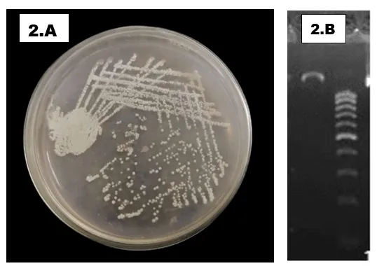
Figure 2: A. Representative Purified Culture of Streptomyces griseus TK4 on Actinomycete Isolation Agar. the Isolate Was Obtained Following Successive Streak Purification and Incubated to Obtain a Pure Culture. B. Agarose Gel Electrophoresis of the 16S rRNA Gene PCR Product Amplified from Streptomyces griseus TK4.
TK4 extract on MCF-7 viability
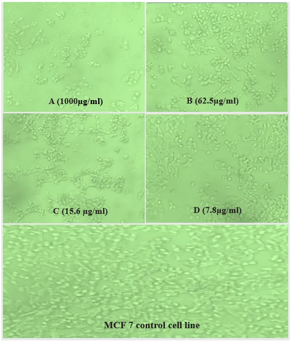
Figure 3: Morphological Changes in MCF-7 Cells Following Treatment with the Ethyl Acetate Extract of Streptomyces griseus TK4.
In the monolayers of MCF-7, the TK4 ethyl acetate extract was applied after 24 h, and a graded effect on formazan conversion was observed over the entire concentration range used. The loss of metabolic signal was accompanied by morphological alteration: treated monolayers rounded, detached, and had decreased confluence compared to the untreated controls (Fig. 3). The decrease of viability was monotonous between 62.4 % (7.8 µg/mL) and 24.0 % (1000 µg/mL) (Table. 3). A regression of the viability on the log10 concentration gave viability = -18.23 log10C + 78.83 with a coefficient of determination value of 0.9997. The half-maximal inhibitory concentration of 38.1 µg/mL is obtained by solving that relation with a 50% viability. Linear interpolation between the two bracketing points, 52.06% viability at 31.2 µg/mL, and 45.80% viability at 62.5 µg/mL, has the same value of 39.2 µg/mL on a logarithmic concentration scale (Fig. 4). There are two characteristics of the response that are interesting. The curve has not leveled off at either extreme of the range of testing: it was not, at the lowest concentrations of the range tried, nor is it yet, at the highest, at control levels. A four-parameter logistic model is then fitted to these data, which necessarily does not converge to biologically meaningful upper and lower asymptotes, and instead the log-linear description is the one that is more defensible of what was measured. Second, at 125 µg/mL, viability was 40.54, which is far below the half-maximal point, indicating that the IC50 is contained solidly below the half-maximal point. The extract exhibited concentration-dependent cytotoxicity against MCF-7 cells with an IC₅₀ of 38.1 µg/mL.
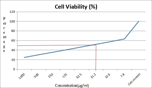
Figure 4: Concentration-dependent Effect of the Ethyl Acetate Extract of Streptomyces griseus TK4 on the Viability of MCF-7 Cells. Cell Viability Was Determined by the MTT Assay After 24 H Exposure to Increasing Concentrations of the Ethyl Acetate Extract and Expressed as a Percentage Relative to the Untreated Control. the Estimated IC₅₀ Value Derived from Regression Analysis Was 38.1 Μg/ml.
Table 3: Concentration-dependent Effect of the Ethyl Acetate Extract of Streptomyces griseus TK4 on the Viability of MCF-7 Cells After 24 H of Exposure.
| Concentration (µg/mL) | Dilution | A570 | Viability (%) |
|---|---|---|---|
| 1000 | Neat | 0.169 | 24.03 |
| 500 | 1:1 | 0.210 | 29.87 |
| 250 | 1:2 | 0.246 | 34.99 |
| 125 | 1:4 | 0.285 | 40.54 |
| 62.5 | 1:8 | 0.322 | 45.80 |
| 31.2 | 1:16 | 0.366 | 52.06 |
| 15.6 | 1:32 | 0.401 | 57.04 |
| 7.8 | 1:64 | 0.439 | 62.44 |
| Untreated control | — | 0.703 | 100.00 |
GC-MS profile of the TK4 extract
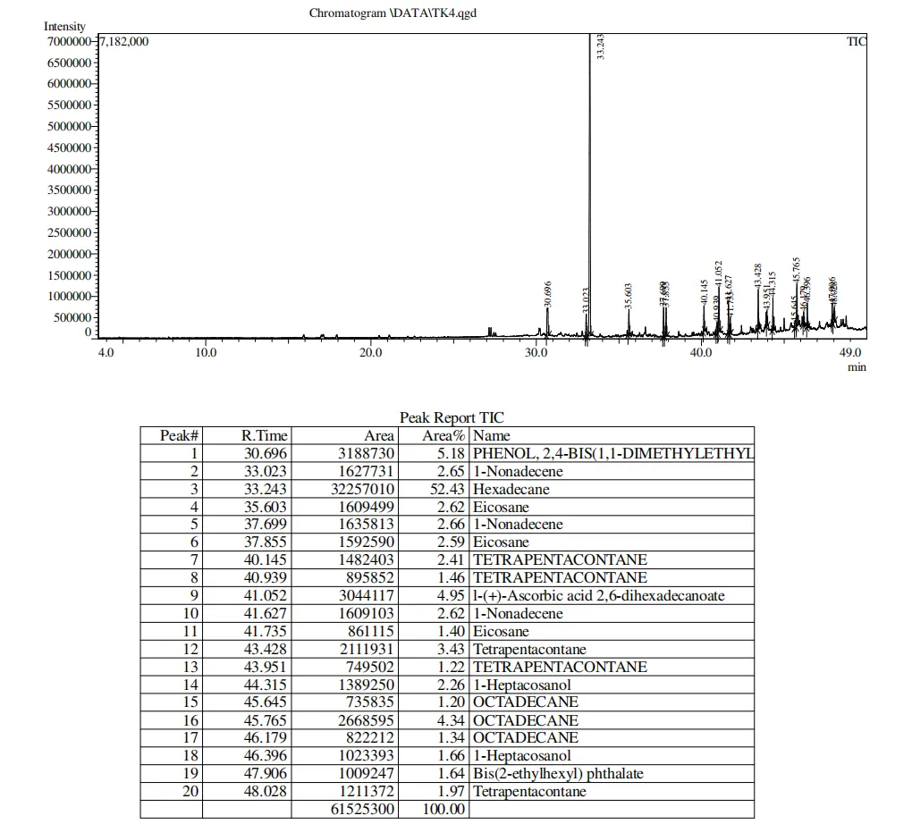
Figure 5: Total Ion Chromatogram (TIC) of the Ethyl Acetate Extract Obtained from Streptomyces griseus TK
The TK4 ethyl acetate extract analyzed by gas chromatography-mass spectrometry yielded a pattern of constituents-rich during a run of up to 49 min, where most of the assignments appeared beyond 30 min (Fig. 5). The instrument peak report was made using 20 peaks which collectively represent a total ion current area of 61,525,300. The various metabolites found included hexadecane (the dominant component, with a retention time of 33.243 min) and made up 52.43% of the total chromatographic peak area. The second most abundant was 2, 4-bis (1, 1- dimethyl ethyl) phenol at 30.696 min, which was a 5.18 % of the total peak area with L-(+)-ascorbic acid 2,6-dihexadecanoate (41.052 min; 4.95 %) and octadecane (45.765 min; 4.34 %) almost following it. Tetrapentacontane was observed in various retention times (40.145, 40.939, 43.428, 43.951 and 48.028 min), with individual peak areas of 1.22-3.43 and these hydrocarbons could be gained in the fraction obtained in several retention times. Similarly, 1-nonadecene and eicosane were detected at various retention times with a relative abundance ranging between 1.40 and 2.66 which show the presence of structurally similar aliphatic hydrocarbons in the extract. Minor constituents were 1-heptacosanol (2.26% and 1.66%), bis (2-ethylhexyl) phthalate (1.64%), and other octadecane peaks (1.201.34%). All in all, the chromatographic profile was dominated by the long-chain alkanes, alkenes, alcohols, and phenolic derivatives, indicating the high level of chemical diversity of the fungal metabolites found in the extract. GC-MS analysis indicated that the extract was dominated by long-chain hydrocarbons together with minor phenolic constituents (Table. 4).
Table 4: Tentatively Identified by NIST Library Matching Compounds of Ethyl Acetate Extract of Streptomyces griseus TK4 by GC-MS Analysis
| S. No | RT (min) | Peak Area | Peak Area (%) | Compound Identified |
|---|---|---|---|---|
| 1 | 30.696 | 3,188,730 | 5.18 | Phenol, 2,4-bis(1,1-dimethylethyl)- |
| 2 | 33.023 | 1,627,731 | 2.65 | 1-Nonadecene |
| 3 | 33.243 | 32,257,010 | 52.43 | Hexadecane |
| 4 | 35.603 | 1,609,499 | 2.62 | Eicosane |
| 5 | 37.699 | 1,635,813 | 2.66 | 1-Nonadecene |
| 6 | 37.855 | 1,592,590 | 2.59 | Eicosane |
| 7 | 40.145 | 1,482,403 | 2.41 | Tetrapentacontane |
| 8 | 40.939 | 895,852 | 1.46 | Tetrapentacontane |
| 9 | 41.052 | 3,044,117 | 4.95 | l-(+)-Ascorbic acid 2,6-dihexadecanoate |
| 10 | 41.627 | 1,609,103 | 2.62 | 1-Nonadecene |
| 11 | 41.735 | 861,115 | 1.40 | Eicosane |
| 12 | 43.428 | 2,111,931 | 3.43 | Tetrapentacontane |
| 13 | 43.951 | 749,502 | 1.22 | Tetrapentacontane |
| 14 | 44.315 | 1,389,250 | 2.26 | 1-Heptacosanol |
| 15 | 45.645 | 735,835 | 1.20 | Octadecane |
| 16 | 45.765 | 2,668,595 | 4.34 | Octadecane |
| 17 | 46.179 | 822,212 | 1.34 | Octadecane |
| 18 | 46.396 | 1,023,393 | 1.66 | 1-Heptacosanol |
| 19 | 47.906 | 1,009,247 | 1.64 | Bis(2-ethylhexyl) phthalate |
| 20 | 48.028 | 1,211,372 | 1.97 | Tetrapentacontane |
DISCUSSION
The strongest aspect of this study is the cytotoxicity data, and this data is the right pivot of interpretation. MCF-7 monolayer viability dropped monotonically at seven doubling dilutions, without any reversal, and regression of viability versus an interval transformation of concentration characterized the series with a regression coefficient of 0.9997. Closeness to a log-linear relation implies that a dilution series has been done well, and implies the data on which it is based is internally consistent. The obtained half-maximal inhibitory concentration of 38.1 µg/mL positions the unfractionated extract in a credible range of values for crude microbial material, and comfortably below the value of approximate one-hundred µg/mL, a value with HeLa cells against a crude Streptomyces griseus extract [27]. The chemistry underlying that activity is to be interpreted requiring a more sceptical interpretation of the chromatographic data on which such a profile is traditionally based. The entire peak report dissolves 20 constituents, and their distribution is impressive. 88.26% of the total ion current is saturated and monounsaturated long-chain hydrocarbons combined with a long-chain primary alcohol; 1.64% is a phthalate ester; only two oxygenated compounds, 2,4-di-tert-butylphenol at 5.18 % and an ascorblylhexadecanoate diester at 4.95 %, belong to the categories of compounds that microbial secondary metabolism could be expected to supply. Nine-tenths of the chromatogram is of material therefore whose provenance cannot be supposed to be biological. There are three internal inconsistencies that put the assignments of the aliphatic into grave doubt. On a 5% diphenyl, 95% dimethylpolysiloxane stationary phase, n-alkanes come out in a strict ascending order of carbon number, such a regularity being so reliable that it is the foundation of the Kováts retention index system [28]. It is contravened by the current assignments. The octadecane (C18) is assigned to three peaks between 45.645 and 46.179 min and the eicosane (C20) to a peak at 35.603, 37.855 and 41.735 min; a C18 alkane will not elute after a C20 alkane on a non-polar column. Tetrapentacontane is a C54 hydrocarbon with a nominal retention index of 5400 and a boiling point far beyond the physical operating limit of the mentioned procedure of 300°C, but assigned to five separate peaks spread across the same window, and physically impossible to elute under the conditions specified. To reinforce the two observations, there are five different names that are repeated at two to five retentions that give a peak in a pure compound. The search of the library is matching on the generic saturated-hydrocarbon fragment series of m/z 57, 71, 85 and 99 and is almost exactly the same across the corresponding homologous series and contains minimal structural information. These are not identifications, but rather the library recovering its nearest neighbour to an uninformative range. Hexadecane (52.43% of total area of one peak at 33.243 min) deserves special remark. A single n-alkane that contributes to over half the ion current of an ethyl acetate extract of culture supernatant is much better attributed to stationary phase degradation, solvent impurity, solvent residue in sample vials or sample carryover than to secondary metabolism, and n-hexadecane is commonly used in the laboratory as a keeper solvent and as a chromatographic standard. An additional inconsistency exacerbates the alarm. The reported oven programme is stopped at 32 min, but nineteen of the twenty peaks or 94.85% of the integrated area is measured beyond that. It can either be misreported, or the programme was preceded by an unreported isothermal hold, which, until fixed, the retention data cannot be matched with the assignments in any given event. What comes out of this examination is a smaller and far more interesting extract. The second most prolific constituent is the 2,4-di-tert-butylphenol, with the only significant independent literature. Isolated from Streptomyces griseus KCA1 it exhibited anti-bacterial and anti-proliferative effects; it was discovered in a Saharan Streptomyces mutabilis isolate it inhibited Candida albicans and other pathogenic fungi; and repurposed by bioassay-directed fractionation of Nocardiosis sp. strain LC-8, raising an MCF-7 IC50 of 17.5 ± 1.5 µg/mL in addition to evidence of apoptosis induction [29]. The quantitative association is educative. An extract of crude material containing this compound in approximately 5% of the detected substance had an IC50 of 38.1 µg/mL, nearly 2.2-fold greater than the purified product [30]. It is a ratio of the kind that a small component produces a substantial portion of action, but does not account for all of it and much more compatible with a phenolic action than with one that could be ascribed to long-chain alkanes, which are virtually inert at these levels. It is a consistency argument and not a demonstration that needs to be translated into evidence by bioassay-guided fractionation [31]. Mechanistic rationale creates out of structure. A bulkier tert-butyl substituent on the ortho and para to the hydroxyl group is a good hydrogen-atom donor, which explains the antioxidant activity of synthetic hindered phenols like butylated hydroxytoluene [32], and it can explain directly the DPPH decolourisation observed here. It is not as linearly associated with cytotoxicity. Phenolic antioxidants may reverse to prooxidant behavior within the cell, especially at high concentration and when present in the presence of transition metals, to produce phenoxy radicals and downstream reactive oxygen species; tumour cells with a high basal oxidative load and low reserve antioxidant capacity are particularly susceptible to such a change. The antibacterial situation has independently implicated dihydrofolate reductase and DNA gyrase via docking. Neither of the two propositions are proven in the current work because no quantification of reactive oxygen species, the determination of glutathione, annexin V staining or caspase assay was done. A more guarded reading corresponds to the ascorbic hexadecanoate diester of 4.95%. Ascorbyl palmitate and the related but markedly distinct 6-hexadecanoate are well-known lipophilic antioxidants, the latter a characterised hyaluronidase inhibitor, and either might have contributed to the radical-scavenging signal, so far detected in this extract. The question whether such esters are due to fermentation or to the extraction environment is rarely dealt with in reports of the kind, nor can it be determined by the data given. On its ecological presupposition, the study is anticipation-consistent, but not, as yet, tested. The termite mounds form different kinds of habitat whose water regime, pH, organic carbon and clay fraction are not related to the surrounding matrix, and the communities they choose are either enriching Actinobacteria in general and usually less abundant yet more even than the soils where they are recruited, the hallmark of environmental filtering instead of random sampling [33]. Such material is consistent with the recovery of an actinomycete that has antiproliferative chemistry that can be measured. To determine that the mound isolates are chemically unique, however, it is necessary to sample with a paired mound and adjacent non-mound sampling, which was not done here. The remedial work is cheap compared to the investments that have been made. An uninoculated medium extract treated identically through the workflow would downgrade some 90% of the constitutive table in a single day of instrument time, would confirm directly that hexadecane is metabolite and not artefact. The natural next study is bioassay-guided fractionation following the activity of MCF-7, independent of the maximum abundance, the active fraction being defined by nuclear magnetic resonance, and library matching, with a library matching experiment being halted. The GC–MS compound identifications reported in this study are tentative and based on NIST library matching. Future work will involve purification of the major bioactive metabolites and structural confirmation using NMR spectroscopy and complementary spectroscopic techniques to verify their chemical identities and biological significance with the replicates. The remaining structural limitations would be overcome by triplicate determination of the 2 assays in the presence of a Trolox standard and a non-tumorigenic comparator line, and complete-length 16S sequencing, which can be further complemented by multilocus analysis of the standard housekeeping loci. It is as much to be feared as it is to be hoped, the merit of the entire full report is that it reduces the genuine chemistry of this extract, sharpens it and makes it much easier to hunt down.
CONCLUSIONS
This study demonstrated that termite mound soils from Ranipet District, Tamil Nadu, are a valuable source of cultivable actinomycetes with promising bioactive potential. Five isolates were recovered and screened for antioxidant activity, among which Streptomyces griseus strain TK4 exhibited the highest DPPH radical-scavenging activity and was therefore selected for further investigation. The ethyl acetate extract of TK4 showed concentration-dependent cytotoxic activity against MCF-7 human breast adenocarcinoma cells, with an IC₅₀ value of 38.1 µg/mL, indicating appreciable antiproliferative potential for a crude microbial extract. GC-MS analysis revealed a chemically diverse extract containing 2,4-di-tert-butylphenol and L-(+)-ascorbic acid 2,6-dihexadecanoate among the detected constituents, although the chromatographic assignments require confirmation by retention index determination and spectroscopic characterization. The findings support termite mound-derived actinomycetes as a promising source of biologically active metabolites while emphasizing the need for bioassay-guided fractionation, structural elucidation, and mechanistic studies to identify the compounds responsible for the observed biological activities and evaluate their therapeutic potential.
Funding:
The authors received no specific funding for this work.
Acknowledgments:
The authors sincerely thank the Department of Microbiology and Department of Bio-Engineering, Vels Institute of Science, Technology & Advanced Studies (VISTAS), Chennai, Tamil Nadu, India, for providing the laboratory facilities and technical support required to carry out this research.
Author contributions: Manikandan: Conceptualization, methodology, validation, investigation, resources, writing-review and editing, supervision, and project administration. Sundar S: Methodology and investigation. Pravin Kumar: Conceptualization, methodology, validation, formal analysis, investigation, data curation, writing-original draft preparation, writing-review and editing, visualization. All authors have read and agreed to the published version of the manuscript
Conflict of interest:
The authors declare that they have no competing interests.
Data availability: All other data supporting the findings of this study are included within the article. Additional data are available from the corresponding author upon reasonable request.
REFERENCES


Manikandan, Sundar S., Eswari S., Pravin Kumar S., Isolation and Characterization of a Cytotoxic and Antioxidant Streptomyces Griseus from Termite Mound Soil of Ranipet District, Tamil Nadu, India., Int. J. of Pharm. Sci., 2026, Vol 4, Issue 10, 2460-2473. https://doi.org/10.5281/zenodo.23410620
 10.5281/zenodo.23410620