

We use cookies to ensure our website works properly and to personalise your experience. Cookies policy
Gokhale Education Society's Sir Dr. M.S. Gosavi College of Pharmaceutical Education and Research, Nashik, Maharashtra, India.
Liposomes are spherical, concentric vesicles originating from greek words lipos meaning fat and soma meaning body. The science of liposomolgy was founded by British scientist alec bangham and associate at babraham Cambridge in mid-1960, who in 1961 accidently scattered phosphatidylcholine molecules in water and discovered closed bilayer structure in 1964 liposomes are self –assembling (phosphor) lipid –based drug vesicles that enclose a core aqueous compartment in a bilayer (uni-lamellar) or a concentric series of several bilayer multilamellar. They are colloidal carries with diameter ranging from 0.01 to 5.0 um with phospholipid bilayer thickness 4-5 nm ranging from 30 nm to micrometre scale, though for drug delivery application typically unilamellar with diameter 50-150 nm as bigger liposomes are quickly eliminated from blood stream .they are tiny, spherical artificial vesicles made from natural nontoxic phospholipid and cholesterol. The main structural elements are phospholipid – phophatidylcholine (lecithin) pc being most widely utilized amphipathic molecule consisting of hydrophilic polar head group phosphocholine Glycerol Bridge and two acyl hydrophobic polar head group.
The Greek terms lipos, which means fat, and soma, which means body, were the source of the spherical, concentric vesicles known as liposomes [1]. In 1961, Bangham et al. developed the first liposome by accidentally scattering phosphatidyl choline molecules in water. He discovered that the molecules were creating a closed bilayer structure with an aqueous phase that were trapped by a lipid bilayer [2]. Liposomes are particularly helpful since they can carry a wide range of medications with possible therapeutic effects or other qualities. Liposomes are colloidal carriers with diameters ranging from 0.01 to 5.0 μm. Because the drug must be released from the liposome prior to metabolism and excretion, drugs encapsulated in liposomes achieve therapeutic levels for extended periods of time [3]. These are tiny, spherical artificial vesicles that can be made from natural, non-toxic phospholipids and cholesterol. Liposomes are attractive drug delivery devices because of their size, hydrophobic and hydrophilic nature, and biocompatibility [4]. Liposomes are appealing drug delivery methods for both hydrophilic and hydrophobic medicines because of their special capacity to entrap pharmaceuticals in both the aqueous and lipid phases.
Another definition of liposomes is artificial tiny vesicles with an aqueous compartment encircled by one or more concentric layers of phospholipid. More substances, including peptides, proteins, hormones, enzymes, antibiotics, antifungals, and anticancer chemicals, are contained within the sphere-like liquid interior. Liposomes are tiny synthetic spherical vesicles that can produce phospholipid, which is naturally non-toxic, and cholesterol. Size, hydrophobicity, and hydrophilicity are the determining factors. A liposome is a spherical vesicle with a minimum of one lipid bilayer. Both pharmacological medications and minerals are administered through it. It exhibits both traits:
1) Hydrophilic head 2) Lipophilic
Liposomes are self-assembling (phospho) lipid-based drug vesicles that enclose a core aqueous compartment in a bilayer (uni-lamellar) or a concentric series of several bilayers (multilamellar) [5]. The phospholipid bilayer is 4–5 nm thick, while liposomes range in size from 30 nm to the micrometer scale [6]. The British scientist Alec Bangham and associates at Babraham Cambridge founded the science of liposomology in the middle of the 1960s [7], and in 1964 they reported the structure of liposomes [8]. Since then, liposomes have been extensively studied as delivery systems for proteins, nucleic acids, small molecules, and imaging agents [9].
One or more concentric lipid bilayers that encapsulate an interior aqueous volume or volumes make up liposomes. Liposomes are typically unilamellar and have a diameter of between 50 and 150 nm for drug delivery applications. Bigger liposomes are quickly eliminated from the bloodstream.
DIAGRAM:
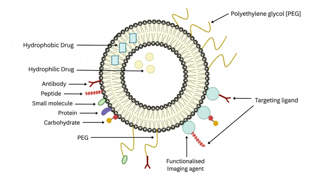
Figure 1. Schematic representation of Liposomes.
COMPONENTS:
The structural components are:
The main structural elements of liposomes are phospholipids. Phosphatidylcholine (PC8) is the most widely utilized phospholipid in liposomal preparation. The amphiphatic molecule phosphatidylcholine is made up of • Phosphocholine, a hydrophilic polar head group A bridge made of glycerol • Two acyl hydrocarbon chains that are hydrophobic The composition of spontaneously occurring The glycerol component of phosphatidylcholine is joined to two acyl chains, which can be either unsaturated or saturated. The packing of the lipid molecules' hydrocarbon chains determines the stability of the liposome membrane. Bilayer characteristics including elasticity and phase behavior are caused by the characteristics of the fatty acid in the lipid molecule, such as the quantity of double bonds in the chain [10]. Liposomes are made from phospholipids, which are common in nature and include choline. Phospholipids include, for example: • Phosphatidyl serine (PS) • Phosphatidyl ethanolamine (Cephalin)-PE • Phosphatidyl choline (Lecithin) PC • PG, or phosphatidyl glycerol
It is a sterol that is frequently utilized. Stability and stiffness functions are modulated by the presence of sterols. It doesn't create a bilayer structure on its own [11]. Up to a 1:1 or 2:1 molar ratio of cholesterol to phosphatidyl choline, it is integrated into phospholipids at extremely high concentrations. The lipid bilayer's stability is improved and a highly organized, stiff membrane structure is formed when cholesterol is present [12]. Cholesterol increases the fluidity and durability of cellular membranes while decreasing the permeability of water-soluble substances. Cholesterol inhibited the interaction and instability of liposomes [13].
CLASSIFICATION CHART:
Lyposomes based on vesicle type24:
|
Vesicle Type |
Diameter size |
No. of lipid Layer |
|
Multi lamellar large vesicles [MLV] |
More than 0.5 µm |
5-25 |
|
Oligo lamellar vesicles [OLV] |
0.1-1.0 µm |
Approx 0.5 |
|
Uni lamellar vesicles [UV] |
All size ranges |
1 |
|
Small Uni lamellar vesicles [SUV] |
20-100 nm |
1 |
|
Medium sized uni lamellar vesicles [MUV] |
More than 100nm |
1 |
|
Large uni lamellar vesicle [LUV] |
More than 100nm |
1 |
|
Giant uni lamellar vesicle [GUV] |
More than 1.0 µm |
1 |
|
Multi vesicular vesicle [MVV] |
More than 1.0 µm |
Multi compartmental structure |
Lyposomes based on vesicle type:
|
Vesicle Type |
Method of preparation |
|
REV |
Prepared by Reverse phase evaporation method |
|
MLV |
By Reverse phase evaporation method |
|
VET |
Prepared by extrusion method |
|
FUV |
Vesicles prepared by fusion |
|
FPV |
Vesicles prepared by French press |
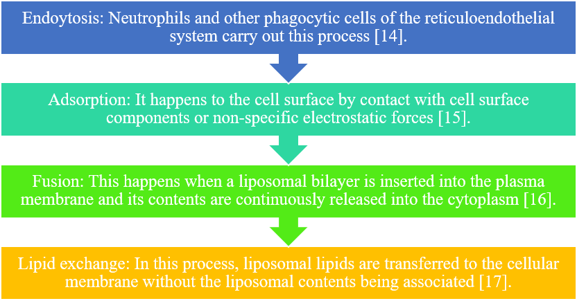
MECHANISM OF ACTION:
Liposomes work through four distinct mechanisms. They are as follows:
METHOD OF PREPARATION:
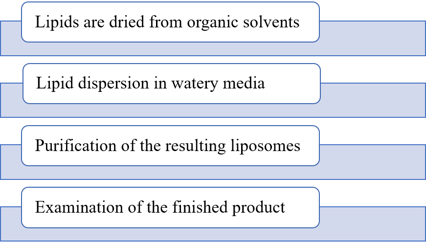
The manufacture of liposomes involves various techniques. General preparation method: There are four processes involved in making liposmes [18].
Micro emulsification: SLV is prepared using this technique. It can be accomplished by employing high shearing stress produced by a high pressure homogenizer to microemulsify lipid mixtures. Reconstituted dried vesicles: This technique involves adding liposomes to a drug-containing aqueous solution or mixing them with lyophilized protein, then dehydrating the mixture. Freeze-thaw method: SUVs were quickly frozen using a gradual thawing technique. This is the cause of unilamellar formation.
In this method lipid and drug were coated onto a soluble carrier to form free flowing granular material in pro-liposome which forms an isotonic liposomal suspension on hydration [20]. Lyophillization: The removal of water from products in a frozen state at a reduced pressure is called Lyophillization. This method is generally used to dry the products that are thermolabile.
MARKETING PRODUCT:
Market product of liposomes:
|
Sr. No |
PRODUCT NAME |
DRUG |
COMPANY |
|
1. |
Ambisome |
Amphotericin B |
NeXstar pharmaceuticals Inc.CO |
|
2. |
Abelcet |
Amphotericin B |
The Liposome company N.J. |
|
3. |
Amphocil |
Amphotericin B |
Sequus pharmaceuticals Inc.CA |
|
4. |
Doxil |
Doxorubicin |
Sequus pharmaceuticals Inc.CA |
|
5. |
DaunoXome |
Daunorubicin |
NeXstar pharmaceuticals Inc.CO |
|
6. |
Mikasome |
Amikacin |
NeXstar pharmaceuticals Inc.CO |
|
7. |
DC99 |
Doxorubicin |
Liposome CO.,NJ,USA |
|
8. |
Epaxel |
Hepatitis A vaccine |
Swiss Serum Institue,Switzerland |
|
9. |
ELA max |
Lidocaine |
Biozone Labs,CA,USA |
STABILITY:
Therapeutic efficacy of drug molecule is governed by stability of liposomes. There are two types of stability-
Physical Stability- There are various physical processes which effects the shelf life of liposomes like fusion , aggregation and shape and size .The general problem that occurs is leakage of drug material. The morphology and size distribution are important parameters for accessing stability. Physical stability can be maintained by avoiding excess unsaturation in the phospholipids. They must be stored at 4◦C with no freezing and light exposure [21].
Chemical Stability- Phospholipids are unsaturated fatty acids prone to hydrolysis alter the stability of drug product. Liposomes can be prevented from oxidative degradation by adding antioxidants such as butylated hydoxy anisole [22].
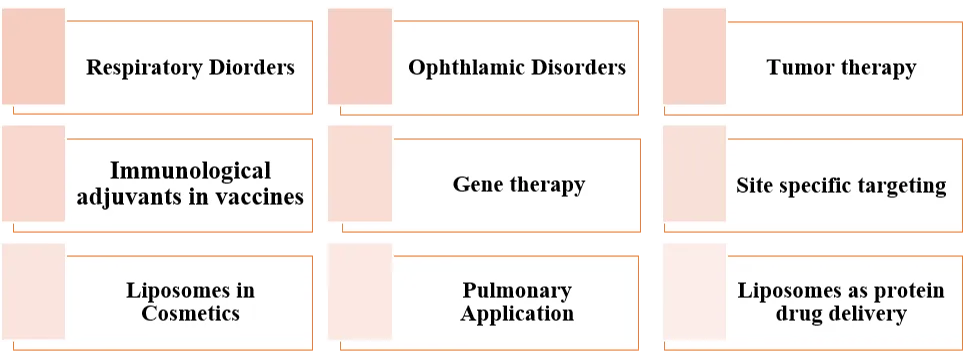
APPLICATION:
REFERENCES




Pradnya Patil, Bhagyesh Varule, Gargi Kulkarni, Dr. Vipulata Yeole, Liposomes as a Novel Drug Delivery System: A Comprehensive Review on Component, Classification, Preparation Methods, Mechanism of Action, Stability, Marketed Formulations and Therapeutic Application, Int. J. of Pharm. Sci., 2026, Vol 4, Issue 10, 2278-2285. https://doi.org/10.5281/zenodo.23424924
 10.5281/zenodo.23424924