

We use cookies to ensure our website works properly and to personalise your experience. Cookies policy
Department of Pharmacy, Pioneer Pharmacy College, Vadodara, Gujarat, India.
Lysosomal storage disorder (LSDs), a collection of heterogeneous disorders caused by abnormalities in lysosomal enzymes which are vital to cellular function preventing any damage on a substantial scale. Due to variability, they are individually rare however together, their incidence rate appears relatively higher in comparison. Malfunctions found in the lysosome result in the buildup of degraded substrates which contain lipids, carbohydrates, and other macromolecules often intensifying cellular impairment and tissue damage. This causes secondary mechanisms such as inflammation, apoptosis, oxidative stress and other mechanisms to lead up to disease progression. Significant organs are damaged due to the clinical manifestations of these disorders including the brain, liver, spleen, skeletal system, kidneys and others. The diagnostic processes involved are biochemical enzyme assays, biomarker analysis and molecular genetic testing. Recent therapies focus on alterations to enzymes and substrate nevertheless, other approaches involve advancements towards improved pharmaceuticals and stem-cell transplantations. Future research aims to study genome editing, identification of sensitive biomarkers, individualized medicine and ability to cross the blood-brain barrier. This review explores the current research conducted on LSDs including their diagnosis, present treatment available and future direction of LSD research
Lysosomal storage disorders (LSDs) cover 70 different diseases. These diseases happen because of dysfunction. Lysosomal Storage Diseases (LSDs) are inherited from both parents. This group of lysosomal storage diseases is passed down in an autosomal recessive pattern. (1)
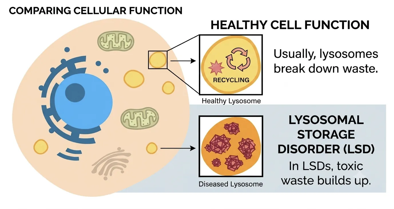
Figure 1. Healthy lysosome within a cell compared with a diseased one.
Lysosomes are cellular organelles whose primary functions include intracellular digestion, macromolecule degradation, and maintenance of cellular homeostasis. The discovery was reported by Christian de Nuve; his findings contributed to the physiological understanding of metabolic diseases. Lysosomal storage disorders are caused by genetic mutations in single genes that encode a particular enzyme; however, any mismatches occurring lead to inactive enzymes. In parallel, defective activators are a result of mutations in activator genes. These mutations culminate in a deficiency of the particular enzymes responsible for the breakdown of certain lipids (fats) and/or carbohydrates (sugars) within the lysosomes. The absence or insufficiency of these enzymes means targeted fats or sugars are not appropriately degraded and recycled. These are the cumulative effects of toxins that interfere with normal cell function, which then cause cell, tissue, and organ damage. (2,3) The presentation of LDS is extremely variable; therefore, a new classification has been introduced which groups the disorder by severity. The demonstration by Hershey in 1963 of the correlation between enzyme deficiency and a storage disease provided the basis to establish an intracellular biology of these enzymes and their substrates, resulting in success with the treatment of GD. (4) Although no cure exists, the nature of disease development is variable among individuals, but this is all being challenged with the development of novel therapies that target the disease process directly. (5) Figure 1 shows the healthy lysosome within a cell compared with a diseased one.
EPIDEMIOLOGY:
While, the instances of individual LSDs remain generally to be uncommon each affects the infantile by and large, as near to 1 LSD is found in as many as 5000 live birth.(6) The most common forms of LSDs around the world are those of FD, GD, MLD and PD. It can also be observed how, also ethnical and geographical differences play a very important role in the epidemiology and demonstrate large discrepancies. (7) Hence frequency of the LSD mutation that caused it, arises further on and that likely many changes would occur within epidemiological data on LSD due to the increasing immigration in Western countries from heterogeneous people.(8) Table 1 consist of global prevalence of different type of LSDs.
Table 1. Global incidence of specific types of Lysosomal Storage Disorders.
|
Disorder |
Approximate global presence |
|
Gaucher disease |
~1.5 per 1,00,000 |
|
Fabry disease |
~1 in 40,000-1,20,000 |
|
Metachromatic leukodystrophy |
~1 in 40,000-1,60,000 |
|
Krabbe disease |
~1 in 1,00,000 |
|
MPS I |
~1 in 1,00,000 |
|
MPS II |
~1-2 per 1,00,000 (male births) |
|
Pompe disease |
Highly variable by population and screening method |
Current data is likely to be an underestimation, due to cases of unrecorded diagnosis of the disease. However, epidemiology is rapidly evolving due to newborn screening, which provides advantages towards diagnosis of the diseases, specifically for patients which have late onset symptoms or no family history. Data suggests a considerable increase in the true prevalence of disease, as in comparison due to previous data there have been 5-80 times more cases identified after newborn screening. This is essential for early diagnosis of the disease which can lead appropriate management strategies and treatment options for patients.(9)
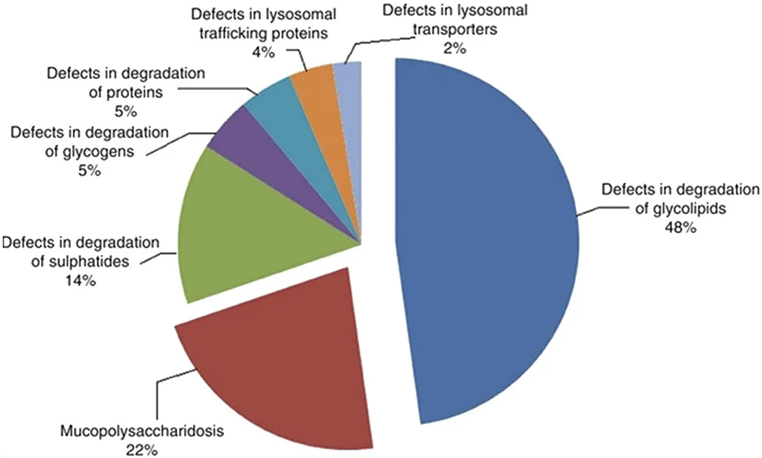
Figure 2. Categories of origin of different storage disorders across India.(10)
ETIOLOGY:
There are 40 different acid hydrolases found in the lysosomes, which are encoded by genes at certain chromosomal loci. A cysteine protease gene, cathepsin K, was highly expressed in osteoclasts and mapped to the pycnodysostosis region. The patients had nonsense, missense, and stop codon mutations in the gene that encodes cathepsin K. Transient expression of the complementary DNA with the stop codon mutation produced messenger RNA but no protein detectable immunologically. Thus, pycnodysostosis results from gene defects in a lysosomal protease that is expressed most highly in osteoclasts.(11) These disorders are divided into 3 main types of LSDs, which depend on the enzyme lacking. These types comprise lipidoses, mucopolysaccharidoses, and sphingolipidoses. In lipidosis, the enzyme that breaks down fats is deficient, so the diseases that commonly occur due to this deficiency are cholesterol ester storage disease and Wolman disease. The deficiency of enzymes that break down mucopolysaccharides (complex molecules like sugar) causes diseases such as Hurler disease and Hunter syndrome. Sphingolipidoses, the most common type, are due to insufficiency of an enzyme that breaks down sphingolipids; disorders include FD, GD, Krabbe, MLD, etc.(12) Additionally, epigenetics also plays a role in the course of progression of the disorder. Changes in DNA methylation and histone modifications. Recent studies demonstrate a strong correlation between epigenetics and certain mechanisms involved in LSDs.(13) In contrast, with the progress of technology, high-throughput multiplex methods are now available for mass screening of several LSDs. NBS has been performed in Latin America since the 1970s, but so far has only been incorporated as a public health program in a few countries of the region.(14) The deficiencies that cause LSD are of soluble lysosomal proteins which are located in the lumen of the lysosome; in addition, a minority of the defects are present in the lysosomal membrane proteins. Table 2 below presents LSDs and their corresponding defective proteins and storage materials.
Table 2. LSDs with their corresponding defective proteins and storage materials.(15)
|
Disease |
Defective protein |
Storage materials |
|
Mucopolysaccharidoses (MPS) |
||
|
MPS I (Hurler, Scheie, Hurler/Scheie) |
α-Iduronidase |
Dermatan sulphate and heparan sulphate, GM2, GM3, SCMAS |
|
MPS II (Hunter) |
Iduronate-2-sulphatase |
Dermatan sulphate and heparan sulphate, GM2, GM3, SCMAS |
|
Spingolipidoses |
||
|
Fabry |
α-Galactosidase A |
Globotriaosylceramide, galabiosylceramide, globotriaosylsphingosine, blood-group-B glycolipids |
|
Gaucher |
β-Glucosidase |
Glucosylceramide, GM1, GM2, GM3, GD3, glucosylsphingosine |
|
Globoid cell leukodystrophy (Krabbe) |
Galactocerebroside β-galactosidase |
Galactosylceramide, psychosine lactosylceramide, globotriaosylceramide, globotetraosylceramide, fucosylneolactotetraosylceramide |
|
Metachromatic leukodystrophy |
Arylsulphatase A |
Sulphatide, 3-O-sulpholactosylceramide, lysosulphatide, seminolipid, gangliotetraosylceramide-bis-sulphate, GM2 |
|
Niemann–Pick A and B |
Sphingomyelinase |
Sphingomyelin, cholesterol, bismonoacylglycerophosphate, GM2, GM3, glucosylceramide, lactosylceramide, globotriaosylceramide, globotetraosylceramide |
|
GM1 gangliosidosis |
β-Galactosidase |
GM1, GA1, GM2, GM3, GD1A, lyso-GM1, glucosylceramide, lactosylceramide, oligosaccharides, keratan sulphate |
|
GM2 gangliosidosis (Tay–Sachs) |
β-Hexosaminidase A |
GM2, GD1aGalNac, GA2, lyso-GM2 |
|
GM2 gangliosidosis (Sandhoff) |
β-Hexosaminidase A and B |
GM2, GD1aGalNac, globoside, oligosaccharides, lyso-GM2 |
|
Oligosaccharidoses and glycoproteinoses |
||
|
Schindler disease |
α-N-Acetylgalactosaminidae |
Glycopeptides with N- or O-linked oligosaccharides, oligosaccharides |
|
Glycogenosis |
||
|
Pompe (glycogen-storage-disease type II) |
α-Glucosidase |
Glycogen |
PATHOPHYSIOLOGY:
The basic mutations that cause most of the diseases in this category are understood better than the cellular processing that causes the storage of the substrate, with resultant cellular and organ defects. Current studies demonstrate the significance of inflammation, apoptosis, modifications to signal transduction, and transport in disease development. Previous studies proposed a relationship between oxidative stress and the pathogenesis of several inborn errors of metabolism, including LSD (16)
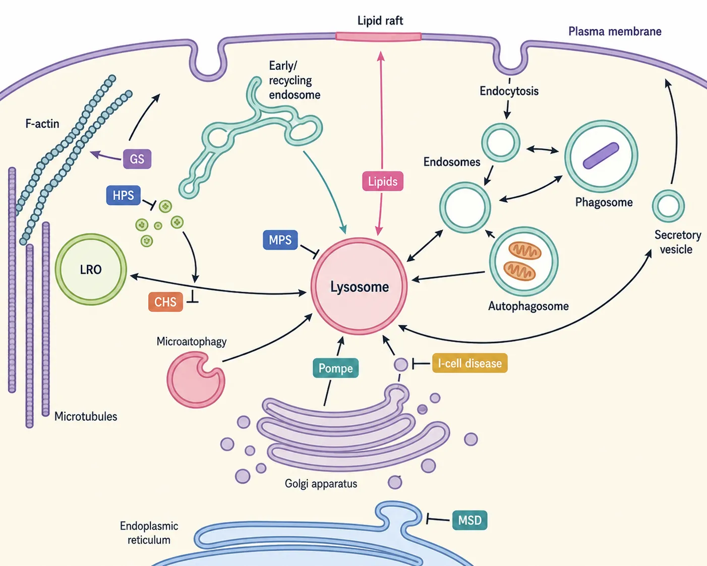
Figure 2. Stylised cell presenting function which are disrupted in LSDs.
Still obscure for LSD patients: the pathogenic mechanism of the disease progresses to a clinical problem. (17) Accumulated substrate in the lysosome leads to inflammatory signalling and, consequently, cell oxidation, ultimately stimulating ROS, chemokines, and growth factors, as well as neuronal degeneration. (18) The conclusion of the oncologic study of LSD patients is mixed, but the clinicians found that patients who have a pathogenic mutation in the LSD gene may have an increased risk of some cancers. Any inflammatory response activated over time progresses in parallel with the storage accumulation. Recent studies indicate that, despite inflammation being a downstream mechanism, it is found to be a target for auxiliary therapy in several LSDs, as shown in a mouse model used for Sandhoff disease with NSAID therapy. (19,20) Alterations in calcium homeostasis are a key mechanism in follow-up to a lysosomal storage disorder in accordance with certain calcium channels; specifically, higher calcium release often relates to Gaucher’s disease. In contrast, reduction in calcium uptake denotes Sandhoff's disease and Niemann-Pick A disease. Another pathway disrupted by LSD is autophagy, which is responsible for the removal of damaged proteins and cellular organelles; it is a vital part, as autophagosomes fuse with lysosomes for maintenance of cellular homeostasis. Therefore, its impairment contributes to the pathogenesis of LSD. (21) Furthermore, other consequences of lysosomal buildup include cellular dysfunction directing towards tissue damage. Particularly in the liver, hypertrophy and spleen with the enlarging mass impairing functional activity, and bone due to various anomalies involving skeletal architecture plus multifarious other organs in which various abnormalities would arise- functionally as well as structurally. LSD can also cause multi-organ failures.
DIAGNOSIS:
Biochemistry and Genetic testing: Referral for biochemical and genetic investigation to specialized labs is necessary to confirm the clinical diagnosis of an LSD. This is usually done via screening of blood, urine, amniotic fluid, skin fibroblasts, and, in some cases, tissue biopsy. (22) The first process of diagnosis of a lysosomal storage disease is assumed and suspected by the clinician. Followed by enzyme activity assays; in particular, the use of fluorometry and MS/MS methods in newborn screening programs enables diagnosis prior to any symptoms. (23) The secondary diagnostic step includes molecular analysis, which mainly involves the measurement of enzymatic activity. It may be the morphology of a cell that is disrupted by lysosomes being absent, and from this, other proteins and organelles are affected, which may be interpreted as being a symptom. Disorders can be attributed to tests carried out at the molecular level. Currently, tests that are commonly used for diagnosis are categorised as urinary oligosaccharides, urinary glycosaminoglycans, assessment of specific substrates, assessment of lysosomal enzyme activity, and indirect biomarkers. The foundation of diagnosing most LSDs post clinical assessment is based on detection of certain enzymatic deficiencies; increased accuracy is available with molecular genetic testing. After identification of the genotype of a patient, genetic counselling is recommended. This enables an understanding of possible phenotype and recognition of carriers within the family who are at risk. (24)
CLINICAL FEATURES:
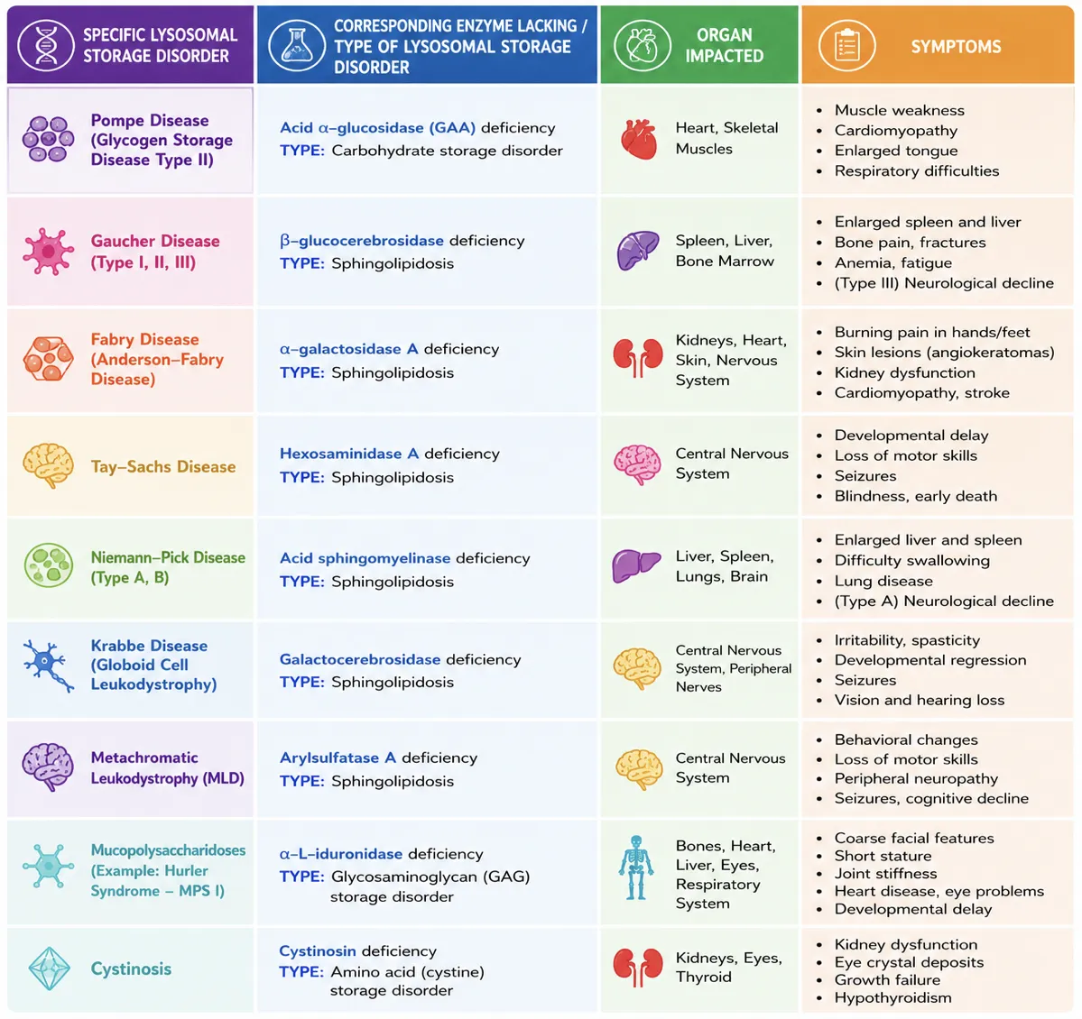
Lysosomal storage disorders are a group of inherited diseases causing malfunction of lysosomes, which affect multiple body organs including the brain, heart, spleen, liver, bone, and joints. Though symptoms of such diseases vary according to the form of the disease, there are some universal symptoms like seizures, an enlarged heart that goes on to affect the spleen for deposition and spleen enlargement, and lung consolidation and abnormalities of the bone. (25) The clinical signs listed generally appear in late adulthood, although present normally at infancy and can change in degree of enzyme deficiency. (26)
Table 3. How different LSDs affect different organs across the body.
TREATMENT
Following the latest clinical research, there have now been developed two novel licensed treatments in LSD. (27) Among them are ERT (enzyme replacement therapy) particularly effective for the treatment of the disorders (GD, FD, MPS types I, II, IV, Pompe). (28) The efficiency and safety of ERT are widely acknowledged in the majority of cases. But there are disadvantages in the application of ERTs: immune response, incorrect targets, and difficulty in the tissues. ERT can be administered directly into the brain and across the blood-brain barrier. (29) The second major type of therapy involved the use of substrate reduction therapy (SRT), which is mediated by the use of small molecules such as miglustat and eliglustat. They act as competitive inhibitors of ceramide glycosyltransferase, therefore decreasing the level of glucocerebroside, the ganglioside substrate, and allowing it to be degraded by the enzyme β-glucosidase. (30) Also, there are other promising CNS-targeted treatments for neurological LSDs that are in development. Stem cell gene therapies are also undergoing clinical trials. (31) There have been promising results found with the usage of viral gene therapies in the management of LSDs.(32)
DISCUSSION
Furthermore, investigations into LSDs will continue to approach identifying a particular mechanism by which individual LSDs result in the development of chronic illness. Investigation into sensitive biomarkers can improve earlier diagnosis in patients, decreasing the toxic accumulation of substrate and disease progression. (33) Advancements in genetic engineering to code functional genes specifically via a CRISPR-based solution could lead to long-term positive outcomes. (34) An obstacle that requires further study is the inability to cross the blood-brain barrier, as nervous system impairment is a commonality present in all lysosomal storage disorders. Personalised medicines for patients can potentially increase cure rates and protect against damage to more susceptible organs. (35)
CONCLUSION
Lysosomal storage diseases, is a group of rare monogenic diseases caused by defects in a single gene that encodes a specific enzyme. In this manner, the enzyme fails, ultimately producing abnormalities in various systems within the body. Symptoms usually vary among the different types of diseases, but commonly affect the nervous system, producing a variety of symptoms such as seizures and slow development. Although difficulties arise in the diagnosis of the specific disorder, a range of clinical assessments are used. Ongoing enzyme-replacement and substrate reduction technologies can be expanded and optimized in the future through personalized and gene therapies.
REFERENCES

Ved Patel, Sapna Desai, Mayank Panchal, Yagnesh Modi, Satyajit Sahoo, Tejas Patel, D.B Meshram, Lysosomal Storage Disorders: From Molecular Defects to Clinical Complexity, Int. J. of Pharm. Sci., 2026, Vol 4, Issue 8, 5152-5161, https://doi.org/10.5281/zenodo.22202847
 10.5281/zenodo.22202847