
We use cookies to ensure our website works properly and to personalise your experience. Cookies policy
Department of Pharmaceutics, Laureate Institute of Pharmacy, Kathog, Jawalamukhi, Kangra, India
Cutaneous leishmaniasis (CL) is one of the most prevalent forms of leishmaniasis and remains a significant public health challenge in many tropical and subtropical regions. The disease is caused by intracellular protozoan parasites of the genus Leishmania and is transmitted through the bite of infected female sandflies. Although several therapeutic agents, including amphotericin B, miltefosine, paromomycin, and pentavalent antimonial, are available for the treatment of CL, their clinical effectiveness is often limited by severe toxicity, poor patient compliance, prolonged treatment duration, high treatment costs, emerging drug resistance, and inadequate penetration into infected skin tissues. Furthermore, the intracellular localization of parasites within macrophages presents a major obstacle to achieving effective drug concentrations at the site of infection. Recent advances in nanotechnology have opened new opportunities for the development of targeted and more efficient therapeutic approaches. Various nanocarrier systems, including liposomes, lipid nanoparticles, polymeric nanoparticles, metallic nanoparticles, Nano emulsions, microneedles, exosomes, and nanohydrogels, have demonstrated promising potential in enhancing drug delivery and improving therapeutic outcomes. Among these systems, macrophage-targeted nanohydrogels have emerged as particularly attractive platforms due to their ability to combine controlled drug release, enhanced skin penetration, prolonged retention at the site of infection, and selective intracellular delivery to infected macrophages. This review comprehensively discusses the global burden and pathophysiology of cutaneous leishmaniasis, current therapeutic limitations, and recent advances in nanotechnology-based drug delivery systems. Special emphasis is placed on macrophage-targeted nanohydrogel systems, receptor-mediated targeting strategies, and smart stimuli-responsive nanocarriers as next-generation therapeutic platforms. The review also highlights translational challenges, clinical perspectives, and future research directions required for the successful development of precision nanomedicine-based therapies for CL. The integration of targeted drug delivery, bioactive nanohydrogels, and green nanotechnology may provide innovative solutions for improving treatment efficacy and reducing the global burden of cutaneous leishmaniasis.
Cutaneous leishmaniasis (CL) is a vector-borne parasitic disease caused by various species of the genus Leishmania and transmitted through the bite of infected female phlebotomine sandflies. It is one of the most common clinical manifestations of leishmaniasis and represents a major public health challenge in tropical, subtropical, and Mediterranean regions (WHO, 2025; Corman et al., 2023). The disease is characterized by localized skin lesions that may progress into chronic ulcers, resulting in permanent scarring, cosmetic disfigurement, and significant psychosocial consequences. Although CL is rarely fatal, it contributes substantially to morbidity and reduced quality of life, particularly in resource-limited populations (Van Bocxlaer & Croft, 2021; Corman et al., 2023). Leishmania parasites exist in two developmental forms during their life cycle: promastigotes within the sandfly vector and amastigotes within mammalian host macrophages. Following transmission, promastigotes invade host macrophages and differentiate into intracellular amastigotes, where they replicate and establish infection. The intracellular localization of parasites within macrophages poses a significant challenge for effective treatment because therapeutic agents must overcome multiple biological barriers before reaching the target site (Van Bocxlaer & Croft, 2021). The management of cutaneous leishmaniasis remains difficult despite the availability of several antileishmanial drugs. Conventional therapeutic options such as amphotericin B, miltefosine, paromomycin, and pentavalent antimonials have shown clinical effectiveness; however, their use is often associated with severe adverse effects, poor patient compliance, high treatment costs, prolonged treatment duration, and the emergence of drug-resistant strains (Corman et al., 2023; Van Bocxlaer & Croft, 2021). In recent years, nanotechnology-based drug delivery systems have emerged as promising alternatives for improving the treatment of CL. Nanocarriers can enhance drug solubility, improve bioavailability, increase skin penetration, reduce systemic toxicity, and enable targeted delivery to infected cells. Various nanosystems, including liposomes, lipid nanoparticles, polymeric nanoparticles, metallic nanoparticles, nanoemulsions, and hydrogels, have demonstrated encouraging results in preclinical studies (Registre et al., 2023; OpenNano Review, 2023). Among these approaches, nanohydrogels have attracted considerable attention due to their unique ability to combine the advantages of nanoparticles and hydrogels within a single platform. Nanohydrogels are three-dimensional cross-linked polymeric networks capable of absorbing large amounts of water while maintaining structural integrity. These systems provide sustained drug release, improved skin retention, enhanced biocompatibility, and localized delivery of therapeutic agents (Bhattacharya et al., 2023). More importantly, nanohydrogels can be engineered to actively target infected macrophages through receptor-mediated mechanisms, thereby increasing intracellular drug accumulation and improving therapeutic efficacy. The incorporation of targeting ligands such as mannose, folic acid, peptides, and antibodies has further expanded the potential of nanohydrogels for precision treatment of CL (Ahmed et al., 2023; Bashir et al., 2023). The growing interest in macrophage-targeted nanocarriers reflects a shift from conventional passive drug delivery toward more sophisticated and selective therapeutic strategies. By exploiting specific receptors expressed on macrophages, targeted nanocarriers can improve cellular uptake, minimize off-target effects, and enhance parasite clearance (Ahmed et al., 2023). Despite significant progress in nanomedicine, the clinical translation of these technologies remains limited. Challenges related to large-scale manufacturing, formulation stability, regulatory approval, long-term safety, and cost-effectiveness continue to hinder commercialization (Registre et al., 2023).
2. Global Burden of Cutaneous Leishmaniasis (with citations)
Cutaneous leishmaniasis is one of the most widespread neglected tropical diseases worldwide and continues to impose a significant burden on public health systems, particularly in developing countries. The disease is endemic in more than 90 countries across Asia, Africa, the Middle East, South America, and parts of Southern Europe (WHO, 2025). According to global surveillance reports, hundreds of thousands of new cases are reported annually, although the true incidence is believed to be substantially higher due to underreporting, inadequate surveillance systems, and limited access to healthcare facilities in endemic regions (WHO, 2025). Countries such as Afghanistan, Syria, Iran, Iraq, Saudi Arabia, Pakistan, India, Brazil, Colombia, Peru, and Algeria account for a substantial proportion of global cases. Political instability, armed conflicts, forced migration, and displacement of populations have further contributed to disease transmission in several endemic regions (WHO, 2025). The burden of CL extends beyond its direct clinical manifestations. Although the disease is generally not life-threatening, chronic skin ulcers and permanent scars can have profound social and psychological consequences. Patients often experience stigma, social exclusion, reduced self-esteem, depression, and diminished quality of life (Van Bocxlaer & Croft, 2021). Environmental and climatic factors play an important role in the transmission dynamics of CL. Rising global temperatures, changes in rainfall patterns, deforestation, urbanization, and land-use modifications have altered the geographic distribution of sandfly vectors and reservoir hosts, increasing the risk of disease emergence in previously non-endemic regions (WHO, 2025). Poverty remains one of the strongest determinants of CL prevalence. Poor housing conditions, inadequate sanitation, malnutrition, and limited access to healthcare services significantly increase susceptibility to infection (WHO, 2025). The emergence of drug-resistant Leishmania strains has become an increasing concern in several endemic countries. Resistance to conventional therapies complicates disease management, contributes to treatment failure, and increases healthcare costs (Corman et al., 2023). Given the persistent global burden of CL and the limitations of current treatment approaches, there is growing interest in advanced drug delivery systems capable of enhancing therapeutic efficacy while minimizing toxicity. Nanotechnology-based formulations, particularly macrophage-targeted nanohydrogels, represent promising strategies for improving intracellular drug delivery and clinical outcomes (Registre et al., 2023; Bashir et al., 2023).
3. Pathophysiology of Cutaneous Leishmaniasis and Therapeutic Targets
Cutaneous leishmaniasis begins when an infected female phlebotomine sandfly introduces Leishmania promastigotes into the host skin during a blood meal (Burza et al., 2018; WHO, 2025). These extracellular parasites are rapidly recognized and phagocytosed by macrophages, dendritic cells, and neutrophils, which constitute the first line of host immune defense against infection (Kaye & Scott, 2020; Martínez-López et al., 2018). Within macrophages, promastigotes transform into amastigotes, the intracellular stage responsible for disease establishment and progression. The amastigotes multiply within phagolysosomes and evade host immune responses through several mechanisms, including inhibition of antigen presentation, modulation of cytokine production, suppression of oxidative burst, and interference with macrophage signaling pathways (Kaye & Scott, 2020; Corman et al., 2023). Persistent replication of parasites results in chronic inflammation, tissue destruction, ulcer formation, and eventual scarring, which are characteristic clinical manifestations of cutaneous leishmaniasis (Van Bocxlaer & Croft, 2021; WHO, 2025). The severity of disease is influenced by parasite species, host immune status, genetic susceptibility, and local inflammatory responses (Mou et al., 2022). Because macrophages serve as the primary host cells for parasite survival and replication, they represent an attractive therapeutic target for advanced drug delivery systems (Ahmed et al., 2023; Bashir et al., 2023). Nanocarriers capable of selectively delivering therapeutic agents to infected macrophages can significantly improve intracellular drug accumulation, enhance parasite clearance, prolong drug retention at the site of infection, and minimize systemic toxicity. Furthermore, macrophage-targeted nanocarriers can exploit receptor-mediated endocytosis pathways, such as mannose receptor and folate receptor targeting, to improve therapeutic efficacy and reduce off-target effects (Ahmed et al., 2023; Registre et al., 2023).
Table 1. Conventional Drugs Used for the Treatment of Cutaneous Leishmaniasis and Their Mechanisms of Action
|
Drug |
Drug Class |
Mechanism of Action |
Route of Administration |
Advantages |
Limitations |
|
Amphotericin B |
Polyene antifungal |
Binds to ergosterol-like sterols in parasite membrane causing membrane disruption and cell death |
Intravenous |
High efficacy, broad-spectrum activity, effective against resistant strains |
Nephrotoxicity, infusion-related reactions, high cost, hospitalization required |
|
Liposomal Amphotericin B |
Lipid-based amphotericin formulation |
Enhanced macrophage uptake and targeted delivery to infected tissues |
Intravenous |
Reduced toxicity, improved efficacy, targeted delivery |
Very expensive, limited accessibility |
|
Miltefosine |
Alkyl phosphocholine |
Disrupts membrane phospholipid metabolism and induces apoptosis-like cell death
|
Oral |
Convenient administration, improved patient compliance |
Gastrointestinal toxicity, teratogenicity, emerging resistance |
|
Paromomycin |
Aminoglycoside antibiotic |
Inhibits protein synthesis by binding to ribosomal RNA |
Topical/Injectable |
Reduced systemic toxicity, affordable, suitable for localized lesions |
Skin irritation, limited penetration, variable efficacy |
|
Sodium Stibogluconate |
Pentavalent antimonial |
Interferes with parasite energy metabolism |
Intramuscular/Intravenous |
Long history of clinical use, effective against several species |
Cardiotoxicity, hepatotoxicity, resistance development |
|
Meglumine Antimoniate |
Pentavalent antimonial |
Inhibits essential metabolic pathways in parasites |
Intramuscular/Intravenous |
Widely available in endemic countries |
Painful injections, prolonged therapy, severe adverse effects |
Table 2. Major Limitations of Conventional Antileishmanial Therapy
|
Limitation |
Description |
Impact on Treatment |
|
Toxicity |
Severe adverse effects including nephrotoxicity, hepatotoxicity, cardiotoxicity, and gastrointestinal disturbances |
Reduced patient compliance and increased treatment discontinuation |
|
Drug Resistance |
Emergence of resistant Leishmania strains due to prolonged drug exposure |
Therapeutic failure and increased disease burden |
|
Poor Skin Penetration |
Inadequate delivery through the stratum corneum into infected tissues |
Reduced therapeutic efficacy |
|
Intracellular Localization |
Parasites reside inside macrophages where drugs cannot easily reach therapeutic concentrations |
Incomplete parasite clearance |
|
Long Treatment Duration |
Therapy often extends from weeks to months |
Poor adherence and increased healthcare costs |
|
High Treatment Cost |
Expensive formulations and hospitalization requirements |
Limited accessibility in low-income regions |
|
Poor Patient Compliance |
Frequent dosing, painful injections, and adverse effects |
Increased relapse and treatment failure |
4. Challenges in Drug Delivery for Cutaneous Leishmaniasis
Effective treatment of cutaneous leishmaniasis remains challenging due to multiple biological and physicochemical barriers. The stratum corneum, which is the outermost layer of the skin, acts as a highly efficient barrier that restricts drug penetration into deeper tissues. In addition, Leishmania parasites reside within macrophages located in the dermis, making intracellular drug delivery particularly difficult. Many antileishmanial drugs exhibit poor aqueous solubility, limited bioavailability, and rapid clearance from infected tissues, resulting in subtherapeutic drug concentrations. Furthermore, systemic administration often leads to toxicity and adverse effects, reducing patient compliance. Nanotechnology-based drug delivery systems have emerged as promising approaches to overcome these challenges by enhancing skin penetration, improving intracellular targeting, prolonging drug retention, and enabling controlled release at the site of infection.
Table 3. Clinical Challenges Associated with Current Cutaneous Leishmaniasis Therapy
|
Challenge |
Cause |
Consequence |
Potential Nanotechnology-Based Solution |
|
Low Drug Bioavailability |
Poor aqueous solubility and rapid clearance |
Reduced therapeutic efficacy |
Liposomes, nano emulsions, SLNs |
|
Inadequate Skin Penetration |
Stratum corneum barrier |
Insufficient drug reaching infected tissue |
Transferosomes, Ethosomes, microneedles |
|
Limited Macrophage Uptake |
Lack of cell-specific targeting |
Poor intracellular drug concentration |
Mannose-targeted nanoparticles |
|
Systemic Toxicity |
Non-specific drug distribution |
Organ damage and side effects |
Targeted nanocarriers |
|
Drug Resistance |
Genetic adaptation of parasites |
Treatment failure |
Combination nano therapy |
|
Frequent Dosing |
Short drug retention time |
Poor patient compliance |
Sustained-release nanohydrogels |
Table 4. Comparison of Conventional and Nanotechnology-Based Drug Delivery Approaches for CL
|
Parameter |
Conventional Therapy |
Nanotechnology-Based Therapy |
|
Drug Penetration |
Limited |
Enhanced |
|
Macrophage Targeting |
Absent |
Possible |
|
Drug Release |
Immediate |
Controlled/Sustained |
|
Systemic Toxicity |
High |
Reduced |
|
Patient Compliance |
Moderate to Poor |
Improved |
|
Treatment Duration |
Long |
Potentially Shorter |
|
Therapeutic Efficacy |
Variable |
Enhanced |
|
Precision Medicine Potential |
Low |
High |
5. 5. Liposomal Drug Delivery Systems for Cutaneous Leishmaniasis
Liposomes are among the most extensively investigated nanocarriers for the treatment of infectious diseases, including cutaneous leishmaniasis (CL) (Registre et al., 2023; Doroud et al., 2021). These vesicular systems consist of phospholipid bilayers capable of encapsulating both hydrophilic and lipophilic therapeutic agents. Conventional liposomes improve drug solubility and reduce toxicity by protecting encapsulated drugs from degradation while facilitating targeted drug delivery to infected tissues (Registre et al., 2023; Van Bocxlaer & Croft, 2021). However, their penetration through the skin barrier remains limited. To address this issue, advanced liposomal systems such as transferosomes and ethosomes have been developed. Transferosomes contain edge activators that increase membrane flexibility and allow penetration through narrow skin pores, whereas ethosomes incorporate high concentrations of ethanol that disrupt skin lipid organization and facilitate transdermal drug transport (Touitou et al., 2020; Ahmed et al., 2023). Several studies have demonstrated that liposomal formulations of amphotericin B and paromomycin enhance drug accumulation within macrophages and improve therapeutic outcomes against intracellular Leishmania parasites (Doroud et al., 2021; Registre et al., 2023). Despite these advantages, issues related to physical instability, storage conditions, limited shelf-life, and production costs continue to restrict their widespread clinical application (Van Bocxlaer & Croft, 2021).
6. Lipid Nanoparticles for Cutaneous Leishmaniasis Therapy
Lipid nanoparticles have emerged as attractive alternatives to conventional liposomes due to their superior stability and controlled drug release properties (Mendes et al., 2022; Registre et al., 2023). Solid lipid nanoparticles (SLNs) are composed entirely of solid lipids and provide prolonged drug release, enhanced physicochemical stability, and improved bioavailability of encapsulated drugs (Naseri et al., 2021). However, the highly ordered lipid matrix of SLNs may restrict drug loading capacity and contribute to drug expulsion during storage. Nanostructured lipid carriers (NLCs), which contain a mixture of solid and liquid lipids, were developed to overcome these limitations. The imperfect crystalline structure of NLCs allows greater drug incorporation and improved storage stability (Mendes et al., 2022; Müller et al., 2020). Several investigations have reported improved antileishmanial activity using amphotericin B-loaded SLNs and NLCs due to enhanced skin penetration, sustained drug release, and efficient macrophage uptake (Registre et al., 2023; Pereira et al., 2022). Furthermore, lipid nanoparticles can reduce dosing frequency and improve patient compliance through prolonged drug retention at the infection site. Their excellent biocompatibility, scalability, and ease of industrial production make them promising candidates for future clinical translation (Naseri et al., 2021; Müller et al., 2020).
7. Critical Comparison of Liposomal and Lipid Nanoparticle Systems
A comparison of liposomal and lipid nanoparticle systems reveals that each platform possesses unique advantages and limitations (Registre et al., 2023; Mendes et al., 2022). Liposomes exhibit excellent biocompatibility and macrophage-targeting capabilities; however, they often suffer from stability issues, leakage of encapsulated drugs, and relatively low drug-loading capacity (Van Bocxlaer & Croft, 2021). Transferosomes and ethosomes provide superior skin penetration and enhanced transdermal delivery but may be associated with increased formulation complexity and storage challenges (Touitou et al., 2020; Ahmed et al., 2023). In contrast, SLNs and NLCs offer enhanced physical stability, controlled drug release, and higher drug-loading efficiency (Müller et al., 2020). Among lipid nanoparticles, NLCs have demonstrated the greatest potential because of their ability to accommodate larger amounts of therapeutic agents while maintaining formulation stability and sustained release characteristics (Mendes et al., 2022). Nevertheless, both liposomal and lipid nanoparticle systems have shown considerable promise for improving CL treatment and may serve as valuable components of future macrophage-targeted nanohydrogel formulations (Registre et al., 2023).
8. Polymeric Nanoparticles for Cutaneous Leishmaniasis
Polymeric nanoparticles have emerged as one of the most promising nanocarrier systems for the treatment of cutaneous leishmaniasis due to their excellent biocompatibility, biodegradability, controlled drug release properties, and ability to target infected macrophages (Makoni et al., 2021; Registre et al., 2023). These nanoparticles are generally prepared using natural polymers such as chitosan, alginate, gelatin, and dextran, or synthetic polymers such as poly(lactic-co-glycolic acid) (PLGA), polycaprolactone (PCL), and polyethylene glycol (PEG) (Makoni et al., 2021; Anwar et al., 2023). Their nanoscale size enables efficient penetration through biological barriers and facilitates intracellular drug delivery to infected macrophages. Furthermore, polymeric nanoparticles can be functionalized with targeting ligands such as mannose, folic acid, peptides, and antibodies to enhance receptor-mediated uptake by macrophages and improve therapeutic efficacy (Ahmed et al., 2023; Anwar et al., 2023). Several studies have demonstrated enhanced antileishmanial activity using drug-loaded PLGA and chitosan nanoparticles due to improved intracellular retention, sustained release behavior, and reduced systemic toxicity. These advantages make polymeric nanoparticles highly attractive platforms for developing next-generation macrophage-targeted therapies against cutaneous leishmaniasis (Makoni et al., 2021; Registre et al., 2023).
8.1. Preparation Methods
Several techniques have been employed for the preparation of polymeric nanoparticles, including emulsion-solvent evaporation, nanoprecipitation, ionic gelation, spray drying, solvent diffusion, and double emulsion methods (Makoni et al., 2021; Crucho & Barros, 2017). Among these methods, nanoprecipitation and ionic gelation are the most commonly utilized because of their simplicity, reproducibility, low energy requirements, and ability to produce nanoparticles with narrow particle-size distributions and high drug entrapment efficiency (Fessi et al., 2018; Anwar et al., 2023). The choice of preparation method significantly influences nanoparticle size, surface charge, encapsulation efficiency, drug release behavior, and biological performance (Makoni et al., 2021).
8.1.2. Mechanism of Action
Polymeric nanoparticles act as drug reservoirs that protect encapsulated therapeutic agents from premature degradation and facilitate controlled and sustained drug release (Makoni et al., 2021; Registre et al., 2023). Following topical administration, nanoparticles penetrate the skin and accumulate within infected tissues. Due to their nanoscale dimensions and surface characteristics, macrophages readily internalize these particles through phagocytosis and endocytosis pathways, resulting in enhanced intracellular drug concentrations (Ahmed et al., 2023; Anwar et al., 2023). Sustained release of antileishmanial drugs within macrophages improves parasite eradication while reducing systemic toxicity and dosing frequency (Registre et al., 2023; Van Bocxlaer & Croft, 2021).
8.2. PLGA Nanoparticles
Poly(lactic-co-glycolic acid) (PLGA) is one of the most extensively investigated biodegradable polymers approved by regulatory agencies, including the U.S. Food and Drug Administration (FDA) and the European Medicines Agency (EMA), for biomedical applications (Danhier et al., 2019; Makoni et al., 2021). PLGA nanoparticles have demonstrated excellent encapsulation efficiency for antileishmanial drugs such as amphotericin B, miltefosine, and paromomycin, enabling controlled drug release and enhanced intracellular delivery to infected macrophages (Registre et al., 2023; Doroud et al., 2021).
8.3. Chitosan Nanoparticles
Chitosan is a naturally occurring polysaccharide derived from the deacetylation of chitin and has gained considerable attention as a nanocarrier material for topical drug delivery (Kumar et al., 2020; Anwar et al., 2023). Due to its cationic nature, chitosan exhibits strong mucoadhesive properties and enhanced interaction with negatively charged biological membranes, facilitating improved skin retention and cellular uptake (Kumar et al., 2020). Chitosan nanoparticles have demonstrated promising antileishmanial activity owing to their ability to improve drug penetration and macrophage targeting (Registre et al., 2023).
9. Antileishmanial Efficacy
Several studies have reported that amphotericin B-loaded PLGA nanoparticles and chitosan nanoparticles significantly reduce parasite burden in infected macrophages and experimental models of cutaneous leishmaniasis (Doroud et al., 2021; Registre et al., 2023). Enhanced intracellular delivery, prolonged drug retention, macrophage targeting, and sustained drug release contribute to superior therapeutic efficacy compared with conventional formulations (Ahmed et al., 2023; Van Bocxlaer & Croft, 2021). Furthermore, polymeric nanoparticles have demonstrated the ability to improve drug bioavailability, reduce systemic toxicity, and enhance treatment outcomes, making them promising candidates for future macrophage-targeted nanohydrogel formulations (Makoni et al., 2021; Registre et al., 2023).
Table 5. Polymeric Nanoparticles for Cutaneous Leishmaniasis
|
Nanoparticle Type |
Polymer |
Preparation Method |
Advantages |
Limitations |
|
PLGA Nanoparticles |
PLGA |
Nanoprecipitation |
Sustained release, biodegradability |
Costly production |
|
Chitosan Nanoparticles |
Chitosan |
Ionic gelation |
Mucoadhesion, skin penetration |
Stability concerns |
|
Alginate Nanoparticles |
Alginate |
Emulsion crosslinking |
Biocompatibility |
Lower drug loading |
|
PEGylated Nanoparticles |
PEG derivatives |
Solvent evaporation |
Longer circulation time |
Complex synthesis |
10. Metallic Nanoparticles for Cutaneous Leishmaniasis
Metallic nanoparticles have gained significant attention for the treatment of cutaneous leishmaniasis (CL) because of their intrinsic antiparasitic, antimicrobial, anti-inflammatory, and immunomodulatory properties (Diniz et al., 2024; Registre et al., 2023). Unlike conventional drug carriers, metallic nanoparticles may exhibit direct leishmanicidal activity in addition to serving as efficient drug delivery systems. Their small particle size, large surface area, and tunable physicochemical properties enable enhanced interaction with infected macrophages and intracellular parasites (Khan et al., 2022; Diniz et al., 2024).
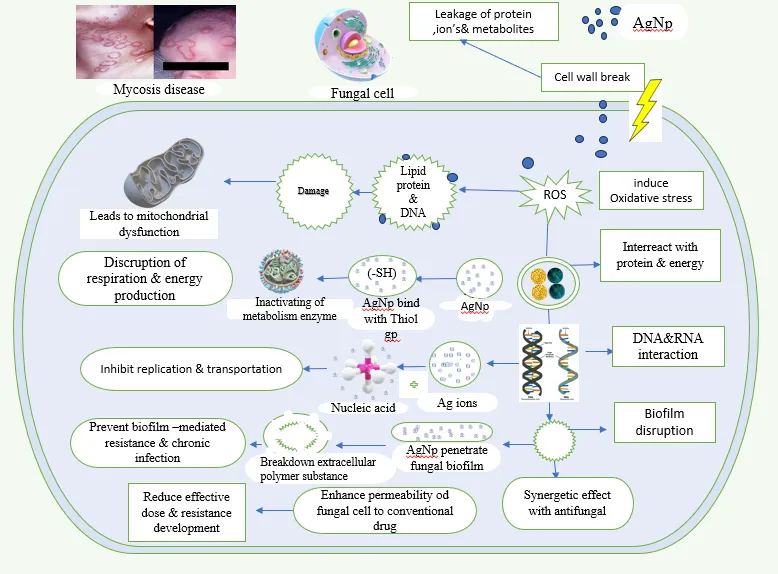
10.1. Silver Nanoparticles (AgNPs)
Silver nanoparticles (AgNPs) are among the most extensively studied metallic nanoparticles for leishmaniasis treatment due to their potent antiparasitic activity and relatively simple synthesis procedures (Diniz et al., 2024; Salehi et al., 2022). They can be synthesized through chemical, physical, or green synthesis approaches using plant extracts such as garlic (Allium sativum), neem (Azadirachta indica), aloe vera (Aloe barbadensis), and other medicinal plants. Green synthesis methods are particularly attractive because they are environmentally friendly, cost-effective, and avoid the use of toxic reducing agents (Iravani et al., 2021; Salehi et al., 2022).
10.1.2. Mechanism of Action
10.2. Gold Nanoparticles (AuNPs)
Gold nanoparticles possess unique optical, electronic, and surface properties that make them suitable for targeted drug delivery, biosensing, and photothermal therapy applications (Anand et al., 2021; Khan et al., 2022). AuNPs can be functionalized with drugs, antibodies, peptides, and targeting ligands to improve selective delivery to infected macrophages. Their excellent biocompatibility and low toxicity make them attractive nanocarriers for antileishmanial therapy; however, their high production cost remains a significant limitation (Anand et al., 2021).
10.3. Iron Oxide Nanoparticles
Iron oxide nanoparticles have emerged as promising theranostic agents due to their superparamagnetic properties, which allow simultaneous diagnosis and therapy (Khan et al., 2022; Diniz et al., 2024). These nanoparticles can be directed toward infected tissues using external magnetic fields, thereby enhancing drug accumulation and reducing systemic exposure. Furthermore, iron oxide nanoparticles can serve as contrast agents for magnetic resonance imaging (MRI), enabling real-time monitoring of treatment responses (Khan et al., 2022).
Table 6. Metallic Nanoparticles Investigated for CL
|
Nanoparticle |
Mechanism |
Advantages |
Limitations |
|
Silver Nanoparticles |
ROS generation, membrane damage |
Strong antiparasitic activity |
Cytotoxicity concerns |
|
Gold Nanoparticles |
Drug delivery and photothermal effects |
Biocompatibility |
High cost |
|
Iron Oxide Nanoparticles |
Magnetic targeting |
Theranostic potential |
Complex synthesis |
10.4. Critical Analysis
Among metallic nanoparticles, silver nanoparticles have demonstrated the most significant antileishmanial activity owing to their ability to generate oxidative stress within parasites. Furthermore, green-synthesized silver nanoparticles align well with current trends toward sustainable nanomedicine and have particular relevance to bioactive hydrogel-based formulations.
11. Nano emulsions and Microemulsions
nano emulsions and microemulsions are colloidal dispersions consisting of oil, water, surfactants, and co-surfactants. These systems have attracted considerable attention because they improve drug solubility, enhance skin penetration, increase drug retention, and facilitate topical delivery of poorly water-soluble antileishmanial drugs (Shakeel et al., 2021; Registre et al., 2023).
11.1. Nano emulsions
Nano emulsions typically possess droplet sizes ranging from 20 to 200 nm and exhibit kinetic stability due to their small droplet size and large interfacial surface area (Gupta et al., 2021). Their nanoscale dimensions facilitate improved drug permeation through the stratum corneum and enhance localization of therapeutic agents within infected tissues (Shakeel et al., 2021).
11.1.2. Preparation Methods
Several methods are employed for nano emulsion preparation, including:
These techniques enable the formation of stable nano-sized droplets with uniform size distribution and high drug-loading capacity.
11.2. Microemulsions
Microemulsions are thermodynamically stable, isotropic systems that form spontaneously when appropriate proportions of oil, water, surfactants, and co-surfactants are combined (Shah et al., 2022). Compared with nano emulsions, microemulsions possess superior thermodynamic stability and can significantly enhance the solubility of hydrophobic antileishmanial drugs (Shakeel et al., 2021).
11.3. Antileishmanial Efficacy
Several studies have reported enhanced efficacy of amphotericin B, miltefosine, paromomycin, and essential oil-loaded nano emulsions against Leishmania parasites due to improved skin penetration, prolonged retention at the site of infection, and enhanced intracellular uptake by macrophages (Registre et al., 2023; Shah et al., 2022). Nano emulsion-based formulations have demonstrated improved bioavailability and reduced toxicity compared with conventional formulations, making them promising candidates for topical CL therapy (Gupta et al., 2021; Shakeel et al., 2021).
Table 7. Nano emulsion and Microemulsion Systems
|
System |
Droplet Size |
Advantages |
Limitations |
|
Nano emulsion |
20–200 nm |
Enhanced penetration |
Kinetic instability |
|
Microemulsion |
10–100 nm |
Thermodynamic stability |
High surfactant content |
|
Drug-loaded Nano emulsion |
Variable |
Increased efficacy |
Formulation complexity |
12. Hydrogels and Nanohydrogels
Hydrogels are three-dimensional polymeric networks capable of absorbing and retaining large amounts of water while maintaining their structural integrity. Due to their excellent biocompatibility, flexibility, moisture-retaining capacity, and ability to provide localized drug delivery, hydrogels have become attractive platforms for the topical treatment of cutaneous leishmaniasis (CL) (Ahmed et al., 2022; Hoare & Kohane, 2020). Their hydrated structure mimics biological tissues and provides a favorable microenvironment for wound healing and controlled drug release. Nanohydrogels represent an advanced generation of hydrogel systems in which nanoparticles are incorporated within the hydrogel matrix. This combination integrates the advantages of nanoparticles and hydrogels into a single multifunctional delivery platform, enabling enhanced drug loading, controlled release, improved skin retention, and targeted intracellular delivery (Hamidi et al., 2021; Sharma et al., 2023). Nanohydrogels have demonstrated significant potential for improving the therapeutic efficacy of topical formulations against intracellular pathogens such as Leishmania spp. (Registre et al., 2023).
12.1. Preparation Methods
Nanohydrogels can be prepared through various approaches, including:
The selection of the preparation method significantly influences the mechanical properties, swelling behavior, drug loading efficiency, and release characteristics of the resulting nanohydrogel formulation (Sharma et al., 2023).
12.1.1. Mechanism of Action
Following topical administration, nanohydrogels form a hydrated matrix over the lesion surface, maintaining a moist environment that promotes wound healing and enhances drug permeation (Ahmed et al., 2022). Encapsulated nanoparticles gradually release therapeutic agents, maintaining effective drug concentrations for prolonged periods. The hydrogel matrix improves skin retention and residence time, while embedded nanoparticles facilitate skin penetration and intracellular drug delivery into infected macrophages (Hamidi et al., 2021; Registre et al., 2023). This dual mechanism enables improved parasite eradication while minimizing systemic toxicity.
12.2. Macrophage-Targeted Nanohydrogels: The Core Novelty
Macrophage-targeted nanohydrogels represent one of the most promising future approaches for CL treatment because macrophages serve as the primary host cells for Leishmania parasites (Kaye & Scott, 2020; Ahmed et al., 2023). These systems can be functionalized with targeting ligands such as:
to facilitate receptor-mediated uptake by infected macrophages (Ahmed et al., 2023; Anwar et al., 2023). Targeted delivery enhances intracellular drug accumulation, improves parasite clearance, reduces off-target toxicity, and may significantly improve therapeutic outcomes compared with conventional formulations.
12.3. Future Perspective
The future of CL therapy is likely to involve smart macrophage-targeted nanohydrogels capable of:
Stimuli-responsive nanohydrogels capable of responding to pH changes, oxidative stress, temperature variations, and enzymatic activity within infected tissues are expected to play a major role in next-generation precision nanomedicine (Sharma et al., 2023; Hamidi et al., 2021).
13. Microneedle-Based Drug Delivery Systems for Cutaneous Leishmaniasis
Microneedle-based drug delivery systems have emerged as innovative transdermal platforms capable of overcoming the limitations associated with conventional topical formulations (Donnelly et al., 2021; Ita, 2022). Microneedles are micron-sized projections, typically ranging from 25 to 2000 μm in length, designed to painlessly penetrate the stratum corneum and create transient microchannels for enhanced drug delivery. Unlike hypodermic needles, microneedles do not reach deep nerve endings, thereby minimizing pain and improving patient acceptance (Donnelly et al., 2021). In cutaneous leishmaniasis, parasites reside within macrophages located in the dermal layer. Therefore, direct delivery of therapeutic agents into the skin using microneedles offers a significant advantage over conventional creams and ointments that often fail to penetrate sufficiently (Ita, 2022; Registre et al., 2023). Microneedle systems can be fabricated from silicon, metals, ceramics, or biodegradable polymers and can be loaded with antileishmanial drugs, nanoparticles, vaccines, or immunomodulators (Donnelly et al., 2021).
13.1. Types of Microneedles
13.1.1. Solid Microneedles
Solid microneedles are primarily used to create micropores in the skin followed by the application of a drug formulation, enhancing transdermal drug transport (Donnelly et al., 2021).
13.1.2. Coated Microneedles
Coated microneedles carry a thin layer of therapeutic agent on their surface and release the drug rapidly upon insertion into the skin (Ita, 2022).
13.1.3. Dissolving Microneedles
Dissolving microneedles are fabricated from biodegradable polymers that dissolve within the skin and release encapsulated drugs in a controlled manner (Donnelly et al., 2021; Ita, 2022).
13.1.4. Hydrogel-Forming Microneedles
Hydrogel-forming microneedles absorb interstitial fluid after insertion and swell to create pathways for sustained and controlled drug release (Donnelly et al., 2021).
13.2. Antileishmanial Potential
Microneedle-assisted delivery of amphotericin B, miltefosine, and nanoparticle-loaded formulations has shown promising results in preclinical studies owing to improved skin penetration, enhanced drug localization, and increased intracellular delivery to infected macrophages (Registre et al., 2023; Ita, 2022). Their ability to bypass the stratum corneum barrier and deliver drugs directly into infected dermal tissues makes microneedles particularly attractive for localized treatment of CL lesions. Furthermore, combining microneedles with nanocarrier-loaded hydrogels may offer a highly effective strategy for future macrophage-targeted therapies.
Table 8. Microneedle-Based Delivery Systems
|
Microneedle Type |
Mechanism |
Advantages |
Limitations |
|
Solid Microneedles |
Skin pretreatment |
Simple design |
Two-step process |
|
Coated Microneedles |
Drug coating release |
Rapid delivery |
Limited drug loading |
|
Dissolving Microneedles |
Polymer dissolution |
No sharp waste |
Mechanical fragility |
|
Hydrogel Microneedles |
Swelling-mediated release |
Controlled delivery |
Complex fabrication |
14. Exosome-Based Drug Delivery Systems
Exosomes are naturally occurring extracellular vesicles with diameters ranging from 30 to 150 nm. They are secreted by various cell types and play a crucial role in intercellular communication through the transfer of proteins, lipids, RNA, and signaling molecules (Kalluri & LeBleu, 2020; Pegtel & Gould, 2019). Due to their biological origin, excellent biocompatibility, low immunogenicity, and inherent cell-targeting capabilities, exosomes have attracted considerable interest as next-generation drug delivery vehicles (Bunggulawa et al., 2018; O'Brien et al., 2020). Exosome-based delivery systems offer several advantages over synthetic nanoparticles, including high biological stability, prolonged circulation time, efficient cellular uptake, and intrinsic targeting ability (Kalluri & LeBleu, 2020). These properties make exosomes highly suitable for delivering antileishmanial agents directly to infected macrophages.
14.1. Mechanism of Action
Exosomes interact with recipient cells through several cellular uptake mechanisms, including endocytosis, membrane fusion, receptor-mediated uptake, and phagocytosis (O'Brien et al., 2020; Bunggulawa et al., 2018). These pathways enable efficient internalization of exosomes by macrophages and other immune cells involved in the pathogenesis of cutaneous leishmaniasis. Once internalized, exosomes release their encapsulated therapeutic cargo, including drugs, proteins, nucleic acids, and bioactive molecules, directly into the cytoplasm of recipient cells. This targeted intracellular delivery enhances therapeutic efficacy while minimizing off-target effects (Kalluri & LeBleu, 2020).
Table 9. Exosome-Based Delivery Systems
|
Parameter |
Exosomes |
|
Size Range |
30–150 nm |
|
Origin |
Natural extracellular vesicles |
|
Cellular Uptake |
Excellent |
|
Immunogenicity |
Low |
|
Drug Loading |
Moderate |
|
Clinical Translation |
Emerging |
|
Major Challenge |
Large-scale production |
15. Stimuli-Responsive Nanocarriers
Stimuli-responsive nanocarriers, also known as smart nanocarriers, are advanced drug delivery systems capable of responding to specific biological or environmental triggers (Torchilin, 2018; Sharma et al., 2023). These systems release therapeutic agents selectively at disease sites, thereby improving therapeutic precision and minimizing systemic toxicity. Because cutaneous leishmaniasis lesions exhibit altered pH, elevated oxidative stress, and inflammatory microenvironments, stimuli-responsive nanocarriers have emerged as attractive platforms for targeted therapy (Hamidi et al., 2021; Sharma et al., 2023).
15.1. pH-Responsive Nanocarriers
The phagolysosome compartments within infected macrophages exhibit acidic conditions (approximately pH 4.5–5.5), providing an ideal trigger for pH-responsive drug delivery systems (Kaye & Scott, 2020; Sharma et al., 2023).
15.1.1. Mechanism of pH-Responsive Nanocarriers
pH-responsive nanocarriers are designed to remain stable under physiological conditions but rapidly release drugs under acidic environments. Drug release may occur through:
These mechanisms facilitate selective intracellular drug release within infected macrophages, improving therapeutic efficacy while minimizing systemic side effects (Hamidi et al., 2021; Sharma et al., 2023).
15.2. Temperature-Responsive Nanocarriers
Temperature-responsive systems utilize polymers that undergo reversible phase transitions at predetermined temperatures (Torchilin, 2018).
15.2.1. Mechanism
Changes in temperature alter polymer conformation and trigger drug release through modifications in polymer hydration and permeability (Torchilin, 2018).
15.3. ROS-Responsive Nanocarriers
Reactive oxygen species (ROS) levels are significantly elevated within infected macrophages and inflammatory lesions, making ROS-responsive systems particularly attractive for CL therapy (Diniz et al., 2024).
15.3.1. Mechanism
ROS-sensitive linkers undergo oxidative degradation in high-ROS environments, resulting in controlled release of encapsulated drugs specifically within infected tissues (Sharma et al., 2023).
Table 10. Stimuli-Responsive Nanocarriers for CL
|
System |
Stimulus |
Mechanism |
Advantages |
|
pH-Responsive |
Acidic pH |
Polymer degradation |
Targeted intracellular release |
|
Temperature-Responsive |
Temperature changes |
Phase transition |
Controlled release |
|
ROS-Responsive |
Oxidative stress |
Linker cleavage |
High specificity |
|
Multi-Responsive Systems |
Multiple stimuli |
Combined mechanisms |
Enhanced precision |
16. Macrophage-Targeted Smart Nanocarriers: The Major Novelty of This Review
Macrophages are the primary host cells for Leishmania parasites and therefore represent the most important therapeutic target in cutaneous leishmaniasis (Kaye & Scott, 2020; Van Bocxlaer & Croft, 2021). Conventional therapies often rely on passive diffusion and fail to achieve sufficient intracellular drug concentrations. Consequently, macrophage-targeted smart nanocarriers have emerged as next-generation platforms capable of actively delivering therapeutic agents directly to infected cells (Ahmed et al., 2023; Anwar et al., 2023).
16.1. Receptor-Mediated Targeting
Macrophages express several receptors that can be exploited for targeted drug delivery.
16.1.1. Mannose Receptors
Mannose receptors are highly expressed on macrophage surfaces and facilitate receptor-mediated endocytosis of mannose-functionalized nanoparticles, significantly enhancing intracellular drug accumulation (Ahmed et al., 2023; Anwar et al., 2023).
16.2. Antibody-Mediated Targeting
Monoclonal antibodies can be conjugated to nanoparticles to achieve highly specific macrophage recognition and targeted drug delivery. Antibody-functionalized nanocarriers have demonstrated enhanced cellular uptake and improved therapeutic outcomes in preclinical studies (Anwar et al., 2023).
Table 11. Macrophage-Targeting Ligands and Their Applications
|
Ligand |
Target Receptor |
Mechanism |
Advantages |
|
Mannose |
Mannose receptor |
Receptor-mediated endocytosis |
Excellent macrophage targeting |
|
Folic Acid |
Folate receptor |
Cellular uptake enhancement |
High specificity |
|
Antibodies |
Surface antigens |
Specific binding |
Precision targeting |
|
Peptides |
Various receptors |
Active targeting |
Versatile functionalization |
17. Green-Synthesized Metallic Nanoparticles for Cutaneous Leishmaniasis Therapy
Green nanotechnology has emerged as an environmentally friendly and sustainable approach for nanoparticle synthesis. Unlike conventional chemical and physical methods, green synthesis utilizes biological resources such as plant extracts, microorganisms, fungi, algae, and biomolecules as reducing and stabilizing agents (Iravani et al., 2021; Salehi et al., 2022). This approach minimizes the use of toxic chemicals, reduces environmental hazards, and improves nanoparticle biocompatibility. The increasing prevalence of drug resistance and toxicity associated with conventional antileishmanial therapies has stimulated growing interest in green-synthesized metallic nanoparticles as alternative therapeutic agents (Diniz et al., 2024). Among various metallic nanoparticles, silver nanoparticles have received particular attention because of their potent antimicrobial, antiparasitic, anti-inflammatory, and wound-healing properties (Salehi et al., 2022).
17.1. Garlic-Mediated Silver Nanoparticles
Garlic (Allium sativum) contains bioactive compounds such as allicin, diallyl sulfide, diallyl disulfide, ajoene, and other organosulfur compounds that serve as reducing and stabilizing agents during nanoparticle synthesis (Iravani et al., 2021; Borlinghaus et al., 2019).
17.2. Future Role of Green Nanoparticles
Green-synthesized nanoparticles represent a promising strategy for developing multifunctional formulations capable of simultaneously providing:
Table 11. Green-Synthesized Nanoparticles Investigated for Leishmaniasis
|
Plant Source |
Nanoparticle Type |
Major Bioactive Compounds |
Reported Activity |
|
Garlic (Allium sativum) |
Silver nanoparticles |
Allicin, sulfur compounds |
Strong antileishmanial activity |
|
Aloe vera |
Silver nanoparticles |
Aloin, polysaccharides |
Wound healing and antiparasitic activity |
|
Neem (Azadirachta indica) |
Silver nanoparticles |
Azadirachtin, flavonoids |
Antimicrobial and antiparasitic effects |
|
Green Tea |
Gold nanoparticles |
Catechins, polyphenols |
Antioxidant and therapeutic activity |
18. Translational Challenges and Clinical Perspectives
Despite remarkable advances in nanotechnology-based therapies for cutaneous leishmaniasis (CL), relatively few formulations have progressed beyond laboratory investigations and reached clinical application (Registre et al., 2023; Ventola, 2021). Although numerous nanocarrier systems have demonstrated promising antileishmanial activity in vitro and in animal models, several scientific, regulatory, manufacturing, and economic barriers continue to hinder successful clinical translation (Sharma et al., 2023; Ventola, 2021).
18.1. Scale-Up and Manufacturing Challenges
Many nanoparticle formulations exhibit excellent performance under laboratory conditions but encounter significant challenges during large-scale manufacturing (Mitchell et al., 2021; Ventola, 2021). Reproducibility, batch-to-batch consistency, particle size control, surface characteristics, encapsulation efficiency, and formulation stability become increasingly difficult to maintain as production volumes increase. Furthermore, scale-up processes often require specialized equipment and optimization strategies to ensure compliance with Good Manufacturing Practice (GMP) standards (Mitchell et al., 2021).
18.2. Stability Issues
Nanoparticle-based formulations are susceptible to several physicochemical stability challenges, including:
These factors may significantly affect therapeutic efficacy, biodistribution, drug-release behavior, and shelf life (Sharma et al., 2023; Makoni et al., 2021). Environmental factors such as temperature fluctuations, humidity, pH changes, and prolonged storage conditions may further compromise nanoparticle stability and clinical performance (Ventola, 2021).
18.3. Regulatory Challenges
Regulatory approval of nanomedicines remains particularly complex because of the unique physicochemical characteristics of nanoscale materials (Ventola, 2021; Fadeel et al., 2018). Major regulatory concerns include:
Comprehensive evaluation of safety, efficacy, biodistribution, pharmacokinetics, immunogenicity, and environmental impact is generally required before commercialization and clinical adoption (Fadeel et al., 2018; Ventola, 2021).
18.4. Economic Constraints
Many advanced nanomedicines require costly raw materials, sophisticated manufacturing facilities, specialized instrumentation, and extensive quality-control procedures (Mitchell et al., 2021). These factors substantially increase production costs and may limit accessibility in low- and middle-income countries where CL is most prevalent (WHO, 2025; Registre et al., 2023). Cost-effectiveness therefore remains an important consideration for the successful implementation of nanotechnology-based therapies in endemic regions.
18.5. Clinical Trial Limitations
The number of clinical trials evaluating nanoparticle-based therapies for cutaneous leishmaniasis remains limited (Registre et al., 2023; Van Bocxlaer & Croft, 2021). Most available studies are currently restricted to:
Although preclinical findings have demonstrated promising therapeutic outcomes, additional well-designed clinical studies are required to establish long-term safety, therapeutic efficacy, optimal dosing regimens, and patient acceptability in humans (Van Bocxlaer & Croft, 2021; Registre et al., 2023). Furthermore, comparative clinical studies evaluating different nanocarrier platforms are needed to identify the most effective and economically viable strategies for CL management. Overall, overcoming manufacturing, regulatory, economic, and clinical barriers will be essential for translating promising nanotechnology-based therapies from the laboratory to clinical practice. Future efforts should focus on developing scalable, cost-effective, and regulatory-compliant nanomedicines capable of addressing the unmet therapeutic needs associated with cutaneous leishmaniasis.
Table 12. Translational Challenges and Potential Solutions
|
Challenge |
Impact |
Potential Solution |
|
Scale-up difficulties |
Manufacturing inconsistency |
Process optimization |
|
Stability issues |
Reduced efficacy |
Advanced formulation design |
|
Regulatory barriers |
Delayed approval |
Standardized guidelines |
|
High production cost |
Limited accessibility |
Cost-effective materials |
|
Limited clinical data |
Slow translation |
Multicenter clinical trials |
19. Future Perspectives
The future of cutaneous leishmaniasis treatment is likely to be driven by the convergence of nanotechnology, biomaterials science, molecular medicine, and artificial intelligence. Advanced drug delivery systems are expected to become increasingly sophisticated, enabling personalized and precision-based therapeutic approaches.
19.1 Smart Stimuli-Responsive Nanohydrogels
Future formulations may incorporate:
These technologies could ensure drug release only at infected sites, minimizing systemic toxicity.
19.2 Artificial Intelligence-Assisted Formulation Design
Machine learning and artificial intelligence may facilitate:
19.3 Theranostic Nanomedicine
Theranostic systems combine therapy and diagnosis within a single platform.
Potential applications include:
19.4 Precision Macrophage Targeting
Future nanocarriers may utilize:
to achieve highly selective intracellular delivery.
19.5 Multifunctional Nanohydrogel Systems
A next-generation therapeutic platform may simultaneously provide:
CONCLUSION
Cutaneous leishmaniasis remains a significant global health challenge due to its increasing prevalence, complex pathophysiology, and limitations associated with conventional therapies. Current treatment options, including amphotericin B, miltefosine, paromomycin, and pentavalent antimonial, are often constrained by toxicity, poor skin penetration, prolonged treatment duration, high costs, and emerging drug resistance. Nanotechnology-based drug delivery systems have demonstrated substantial potential for overcoming these limitations. Liposomes, lipid nanoparticles, polymeric nanoparticles, metallic nanoparticles, nano emulsions, microneedles, exosomes, and stimuli-responsive nanocarriers have all shown encouraging results in improving drug delivery and therapeutic outcomes. Among these approaches, macrophage-targeted nanohydrogels represent one of the most promising next-generation platforms for CL treatment. By combining controlled drug release, enhanced skin retention, receptor-mediated cellular targeting, and improved intracellular delivery, these systems address several key barriers associated with conventional therapy. Furthermore, green-synthesized metallic nanoparticles, particularly garlic- and aloe vera-mediated silver nanoparticles, offer additional advantages due to their intrinsic antiparasitic activity, sustainability, and potential synergistic therapeutic effects. The integration of these nanoparticles within smart nanohydrogel matrices may provide a highly effective strategy for localized and targeted treatment. Although significant challenges remain regarding large-scale production, regulatory approval, and clinical translation, continued advances in nanomedicine, biomaterials science, and precision drug delivery are expected to accelerate the development of clinically relevant therapies. Future research should focus on multifunctional macrophage-targeted nanohydrogel systems capable of combining targeted delivery, controlled release, wound healing, and immunomodulation within a single platform. The development of such advanced therapeutic systems may ultimately transform the management of cutaneous leishmaniasis and contribute significantly to reducing the global burden of this neglected tropical disease.
REFERENCES


Abhishek Bhardwaj*, Vinay Pandit, Abhinav Kaushal, Pravin Kumar, Mahendra Singh Ashawat, Macrophage-Targeted Nanohydrogel Systems for Cutaneous Leishmaniasis: Current Progress and Future Perspectives, Int. J. of Pharm. Sci., 2026, Vol 4, Issue 5, 4870-4894. https://doi.org/10.5281/zenodo.20757457
 10.5281/zenodo.20757457