

We use cookies to ensure our website works properly and to personalise your experience. Cookies policy
All India Shri Shivaji Memorial Society's College of Pharmacy, Pune-411001
Diabetes mellitus is a chronic metabolic disorder characterized by persistent hyperglycemia caused by impaired insulin secretion, insulin resistance, or both. The global prevalence of diabetes continues to rise rapidly and currently affects more than 463 million adults worldwide, with projections suggesting that this number will reach 700 million by 2045.[1] Conventional treatment strategies mainly involve subcutaneous insulin injections and oral hypoglycemic agents. Although these therapies are effective, they present several limitations including injection-associated pain, needle phobia, poor patient compliance, gastrointestinal degradation of peptide drugs, and extensive hepatic first-pass metabolism.[2–4] Microneedle (MN) technology has emerged as a minimally invasive transdermal drug delivery approach capable of overcoming many of these challenges. Microneedles are microscopic needle-like projections that penetrate the stratum corneum and create temporary microchannels for drug transport across the skin barrier.[5] Because these structures typically measure 25–2000 ?m in length, they penetrate only superficial skin layers without stimulating deep dermal pain receptors, enabling painless drug administration.[6] Advances in biomaterials, polymer science, and microfabrication technologies have accelerated the development of various microneedle systems, including solid, coated, dissolving, and hollow microneedles.[7–9] Additionally, emerging additive manufacturing approaches such as 3D printing are enabling rapid production of customizable microneedle arrays with improved structural precision and reproducibility.[10] This review summarizes the skin barrier physiology, microneedle classifications, materials used in microneedle fabrication, and fabrication technologies including 3D printing. The potential of microneedle-based systems for diabetes management is also discussed.
Diabetes mellitus is one of the most common chronic metabolic disorders worldwide and represents a major global health challenge. The disease is characterized by persistent hyperglycemia resulting from defects in insulin secretion, insulin action, or both.[1] Over the past few decades, the incidence of diabetes has increased dramatically due to factors such as urbanization, sedentary lifestyles, aging populations, and increasing obesity rates.[2]
According to epidemiological reports from the International Diabetes Federation, approximately 463 million adults aged 20–79 years were living with diabetes in 2019, and this number is expected to rise to 700 million by 2045.[1] Diabetes is associated with several serious complications including diabetic neuropathy, nephropathy, retinopathy, and cardiovascular diseases, which significantly contribute to morbidity and mortality.[3]
Insulin therapy remains the primary treatment option for individuals with type 1 diabetes and for many patients with advanced type 2 diabetes. Traditionally, insulin is administered through subcutaneous injections, which may require multiple daily doses.[4] Although this method effectively regulates blood glucose levels, repeated injections may cause pain, local tissue damage, and lipodystrophy at injection sites.[5] Furthermore, needle phobia affects a considerable proportion of diabetic patients and may lead to poor treatment adherence.[6]
Oral antidiabetic medications are widely used in the management of type 2 diabetes; however, peptide drugs such as insulin cannot be effectively delivered orally because they are rapidly degraded by digestive enzymes including pepsin, trypsin, and chymotrypsin.[7] In addition, many orally administered drugs undergo extensive hepatic first-pass metabolism, which significantly reduces systemic drug availability.[8]
Transdermal drug delivery systems have been explored as an alternative strategy to overcome these limitations. Conventional transdermal patches offer advantages such as improved patient compliance, controlled drug release, and avoidance of gastrointestinal degradation.[9] However, the outermost layer of the skin, known as the stratum corneum, acts as a strong barrier that restricts the penetration of most drug molecules.[10]
Microneedle technology has emerged as a promising solution to overcome this barrier. Microneedles are microscopic needle-like projections capable of creating temporary microchannels in the stratum corneum, thereby enabling drug molecules to reach deeper skin layers.[5] Because microneedles penetrate only superficial skin layers, they avoid stimulation of deeper pain receptors and therefore provide painless drug administration.[6]
Recent advances in microfabrication techniques and biomaterials have enabled the development of various microneedle systems with improved mechanical strength, drug loading capacity, and delivery efficiency.[11] Furthermore, emerging technologies such as 3D printing are increasingly being explored for microneedle fabrication, enabling rapid prototyping and customizable design.[12]
2. LITERATURE SURVEY
Several researchers have explored microneedle-based systems as promising alternatives to conventional drug delivery methods.
Prausnitz et al. demonstrated that microneedles can create microscopic pathways through the stratum corneum, allowing efficient transdermal delivery of therapeutic molecules that normally cannot penetrate the skin barrier.[4]
Kim et al. further highlighted the advantages of microneedles for drug and vaccine delivery, reporting that these systems provide painless administration and improved delivery efficiency compared with conventional injections.[5]
Davis et al. investigated the mechanical insertion of microneedles into skin tissue and showed that appropriately designed microneedles can penetrate the stratum corneum without causing significant tissue damage.[6]
Gill and Prausnitz developed drug-coated microneedles capable of delivering therapeutic molecules directly into the skin. Their findings demonstrated that drug coatings rapidly dissolve after insertion, resulting in efficient drug release.[17]
Lee et al. introduced dissolving polymeric microneedles composed of biodegradable polymers. These microneedles dissolve completely in the skin after insertion, eliminating the risk of sharp medical waste.[18]
Sullivan et al. further investigated dissolving microneedle patches for vaccine delivery and demonstrated their ability to deliver therapeutic molecules efficiently while improving patient compliance.[19]
Park et al. reported the development of biodegradable polymeric microneedles capable of controlled drug release through transdermal delivery.[23]
Larrañeta et al. reviewed recent advances in microneedle technology and emphasized the importance of material selection and fabrication methods in determining microneedle performance.[21]
Recent research has also focused on 3D-printed microneedles. Zhai et al. demonstrated that additive manufacturing technologies enable precise control over microneedle geometry and fabrication.[24]
Makvandi et al. reported that microneedle patches can significantly enhance drug delivery efficiency and patient compliance, particularly for chronic diseases such as diabetes.[33]
Furthermore, Gu et al. developed glucose-responsive microneedle patches capable of releasing insulin in response to elevated blood glucose levels.[42]
These studies collectively highlight the significant potential of microneedle technology as a minimally invasive and efficient platform for transdermal drug delivery.
3. SKIN BARRIER AND NEED FOR MICRONEEDLES
The skin is the largest organ of the human body and serves as the primary protective barrier against environmental hazards including pathogens, chemicals, and physical damage.[13]
Structurally, the skin consists of three main layers:
Among these layers, the stratum corneum plays the most critical role in regulating skin permeability.[14]
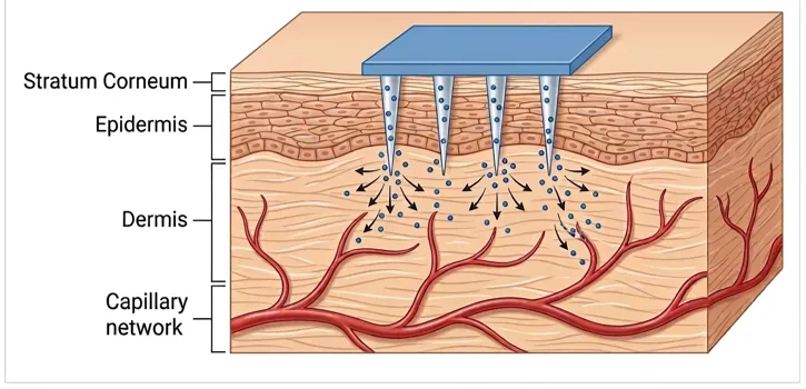
Figure 1. Mechanism of microneedle-mediated transdermal drug delivery across skin layers.
The stratum corneum consists of dead keratinized cells known as corneocytes, embedded within a lipid matrix composed mainly of ceramides, cholesterol, and fatty acids.[15] This structure is commonly described as the “brick-and-mortar” model.[16]
Due to its highly organized structure, the stratum corneum restricts the penetration of most drug molecules. Passive transdermal drug delivery is typically limited to molecules with molecular weights below 500 Da.[17]
However, therapeutic molecules such as insulin and GLP-1 analogues are large macromolecules that cannot penetrate the skin barrier through passive diffusion.[18] Therefore, several techniques have been developed to enhance transdermal drug delivery, including iontophoresis, ultrasound, chemical enhancers, and microneedles.[19]
Among these approaches, microneedles provide an effective method to bypass the stratum corneum while maintaining minimal invasiveness.[5]
4. TYPES OF MICRONEEDLES
Microneedles are classified into four main types based on their drug delivery mechanism:
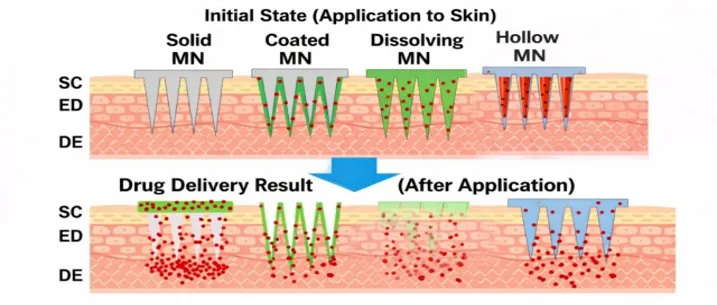
Figure 2. Schematic illustration of different microneedle types and their drug delivery mechanisms. SC stratum corneum, ED epidermis, DE dermis, MN micro needles.
Solid Microneedles
Solid microneedles are commonly fabricated from silicon, stainless steel, or titanium.[21] These microneedles create microchannels in the skin through a poke-and-patch technique, after which drug formulations are applied over the treated skin surface.[22]
Coated Microneedles
Coated microneedles consist of solid needles coated with a thin drug layer that dissolves rapidly upon insertion into the skin.[23]
Dissolving Microneedles
Dissolving microneedles are fabricated from biodegradable polymers containing drugs within the needle matrix.[24] After insertion, the polymer dissolves gradually and releases the encapsulated drug.[25]
Hollow Microneedles
Hollow microneedles resemble miniature hypodermic needles and allow liquid drugs to be injected directly through internal channels.[26]
5. MATERIALS USED IN MICRONEEDLES
The selection of materials plays an important role in determining the mechanical strength, drug loading capacity, and biocompatibility of microneedle systems. Microneedles can be fabricated from metals, silicon, polymers, and biodegradable sugars depending on the type of microneedle design and its intended application.[27]
Metallic microneedles are commonly fabricated from stainless steel or titanium, which provide excellent mechanical strength and durability.[28] These materials are often used in solid and hollow microneedles because they can penetrate the skin effectively without bending or breaking.
Silicon was one of the earliest materials used for microneedle fabrication due to its compatibility with semiconductor microfabrication techniques.[29] Silicon microneedles provide high structural precision; however, their brittle nature may lead to fracture during insertion.
Polymeric materials are widely used in dissolving microneedle systems because they are biocompatible and biodegradable.[30] Commonly used polymers include:
Among these materials, hyaluronic acid and PVA-based polymers are frequently used for dissolving microneedles because they dissolve rapidly in interstitial fluid and allow efficient drug release.[31]
Carbohydrate-based materials such as maltose and trehalose have also been explored for microneedle fabrication due to their excellent biocompatibility and ability to stabilize proteins.[32]
Table 2. Materials Used in Microneedle Fabrication
|
Material Type |
Examples |
Advantages |
|
Metals |
Stainless steel, titanium |
High mechanical strength |
|
Silicon |
Silicon wafers |
High precision fabrication |
|
Polymers |
PVA, PVP, HA, PLA |
Biocompatible and biodegradable |
|
Sugars |
Maltose, trehalose |
Safe and dissolvable |
6. FABRICATION TECHNIQUES FOR MICRONEEDLES
Various microfabrication techniques have been developed for the production of microneedles. These fabrication methods influence the geometry, mechanical strength, and reproducibility of microneedle arrays.[33]
One of the most commonly used techniques is micro-molding, which involves casting polymer solutions into microneedle molds followed by drying or curing.[34] This technique is widely used for dissolving microneedles because it allows efficient encapsulation of drugs.
Another widely used fabrication method is photolithography, which originates from semiconductor manufacturing processes. In this technique, microneedle structures are fabricated on silicon wafers using photoresist materials and etching processes.[35]
Laser cutting and etching methods have also been used to fabricate metallic microneedles with high precision.[36]
Although these conventional techniques are effective, they often require complex manufacturing processes and expensive equipment. Therefore, recent research has focused on the development of 3D printing technologies for microneedle fabrication.
Table 3. Fabrication Techniques Used for Microneedles
|
Fabrication Method |
Principle |
Advantages |
|
Micro-molding |
Polymer casting into molds |
Simple and scalable |
|
Photolithography |
Semiconductor fabrication |
High precision |
|
Laser cutting |
Laser material removal |
Suitable for metal MN |
|
Etching |
Chemical/plasma removal |
Accurate microstructures |
|
3D Printing |
Layer-by-layer fabrication |
Rapid prototyping |
7. 3D PRINTING IN MICRONEEDLE FABRICATION
Additive manufacturing technologies have recently emerged as promising approaches for the fabrication of microneedle (MN) systems due to their ability to produce highly precise, customizable, and reproducible microstructures. Unlike conventional fabrication techniques such as photolithography, laser cutting, and micro-molding, 3D printing enables layer-by-layer construction of complex microneedle geometries without requiring expensive clean-room facilities or complicated multi-step manufacturing processes.[37]
One of the major advantages of 3D printing is the ability to precisely control microneedle dimensions, including needle height, base diameter, tip sharpness, spacing, and array density. These parameters significantly influence skin penetration efficiency, mechanical strength, drug loading capacity, and transdermal drug delivery performance.[38] The digital nature of additive manufacturing also enables rapid design modification and easy optimization of microneedle structures for specific therapeutic applications.
Several 3D printing technologies have been investigated for microneedle fabrication, including:
Among these methods, stereolithography and digital light processing are the most widely used because they provide the high resolution required for fabrication of sharp and mechanically strong microneedles.[39]
7.1 Stereolithography (SLA)
Stereolithography is a photopolymerization-based technique in which ultraviolet laser light selectively cures liquid photopolymer resin layer-by-layer to form three-dimensional microneedle structures. SLA technology provides excellent dimensional accuracy and smooth surface morphology, making it highly suitable for fabricating microneedles with sharp tips and complex geometries.[40]
SLA-fabricated microneedles have been investigated for insulin delivery, vaccine delivery, and controlled drug release applications. Studies have demonstrated that these microneedles can efficiently penetrate the stratum corneum while causing minimal tissue damage and pain.[41]
The major advantages of SLA include:
However, SLA also presents certain limitations, including relatively high equipment cost, limited availability of biocompatible resins, and the requirement for post-curing treatment to remove residual monomers that may cause cytotoxicity.[42]
7.2 Digital Light Processing (DLP)
Digital Light Processing is another photopolymerization-based additive manufacturing technique that uses a digital projector to cure an entire resin layer simultaneously. Compared with SLA, DLP offers faster printing speed and improved manufacturing efficiency.[43]
DLP enables fabrication of highly reproducible microneedle arrays with excellent structural uniformity and mechanical strength. This technology has been widely explored for the fabrication of dissolving and hollow microneedles using biocompatible polymers such as polyethylene glycol diacrylate (PEGDA), gelatin methacryloyl (GelMA), and methacrylated hyaluronic acid.[44]
The major advantages of DLP include:
Despite these advantages, DLP systems may still face limitations in achieving extremely sharp nanoscale microneedle tips required for highly efficient painless insertion.[45]
7.3 Two-Photon Polymerization (TPP)
Two-Photon Polymerization is an advanced ultra-high-resolution 3D printing technique capable of fabricating micro- and nanoscale structures with exceptional precision. In this method, femtosecond laser pulses induce localized polymerization only at the focal point of the laser beam, allowing fabrication of highly detailed microneedle architectures.[46]
TPP technology enables the fabrication of:
Because of its extremely high resolution, TPP is considered one of the most advanced techniques for next-generation microneedle fabrication. However, its practical application remains limited due to slow printing speed, high operational cost, and limited scalability for mass production.[47]
7.4 Fused Deposition Modeling (FDM)
Fused Deposition Modeling is one of the most accessible and cost-effective 3D printing technologies. In FDM, thermoplastic polymers are melted and deposited layer-by-layer through a heated nozzle to form microneedle structures.[48]
Commonly used thermoplastic materials include:
FDM offers several advantages such as low manufacturing cost, simple operation, and wide availability of printable materials. However, due to its relatively low printing resolution, FDM-fabricated microneedles often possess blunt tips and rough surfaces, which may reduce skin penetration efficiency.[49] Therefore, additional post-processing methods such as chemical etching or thermal treatment are frequently required to improve microneedle sharpness and performance.
7.4 Materials Used in 3D-Printed Microneedles
Material selection is an important factor in determining the mechanical properties, biocompatibility, biodegradability, and drug release behavior of 3D-printed microneedles. Various materials have been explored for additive manufacturing of microneedles, including photopolymer resins, biodegradable polymers, and natural biomaterials.[50]
Commonly investigated materials include:
Biocompatible and biodegradable materials are preferred because they minimize toxicity and reduce the risk associated with sharp biomedical waste after drug administration.
7.5 Post-Processing of 3D-Printed Microneedles
Post-processing plays an important role in improving the quality and functionality of 3D-printed microneedles. Common post-processing methods include:
These techniques improve mechanical strength, surface smoothness, and tip sharpness of the microneedles. In coated microneedle systems, post-fabrication drug coating can also be performed to achieve rapid drug release following skin insertion.
7.6 Future Perspectives
Future research in 3D-printed microneedles is focused on the development of smart and multifunctional systems capable of simultaneous sensing and drug delivery. Emerging technologies such as glucose-responsive microneedles, 4D printing, biofabrication, and AI-assisted design optimization are expected to significantly improve the performance and clinical applicability of microneedle systems.
Advances in printable biomaterials, nanotechnology, and personalized medicine are also expected to accelerate commercialization of 3D-printed microneedle patches for diabetes management and other chronic diseases.
8. APPLICATIONS OF MICRONEEDLES IN DIABETES MANAGEMENT
Microneedle-based systems have been extensively investigated for transdermal delivery of insulin and other antidiabetic drugs.[43]
Studies have demonstrated that microneedle-mediated insulin delivery can provide faster drug absorption and improved pharmacokinetic profiles compared with traditional subcutaneous injections.[44]
In addition to drug delivery, microneedles can also be used for continuous glucose monitoring by extracting interstitial fluid from the skin.[45]
Recent advances have led to the development of glucose-responsive microneedle patches, which release insulin in response to elevated glucose levels.[46]
These smart drug delivery systems have the potential to improve glycemic control while reducing the risk of hypoglycemia.
9. ADVANTAGES AND LIMITATIONS
Microneedle-based drug delivery systems offer several advantages over conventional administration methods.
Advantages
Limitations
Despite these advantages, several challenges remain. Microneedle systems may face issues such as limited drug loading capacity, manufacturing complexity, and concerns regarding long-term safety.[48]
Further research is required to address these challenges and improve microneedle technology for large-scale clinical applications.
10. FUTURE DIRECTIONS
Future research in microneedle technology is focused on the development of smart and responsive drug delivery systems.[49]
Integration of microneedles with biosensors may enable real-time glucose monitoring and automated insulin delivery systems. Advances in materials science and fabrication technologies are expected to improve the scalability and commercialization of microneedle-based devices.
Furthermore, 3D printing technologies are expected to play a significant role in the next generation of microneedle-based drug delivery systems.
11. CONCLUSION
Microneedle-based transdermal drug delivery systems represent a promising alternative to conventional drug administration techniques. These systems provide painless drug delivery, improved patient compliance, and enhanced drug bioavailability.
Advances in biomaterials, microfabrication technologies, and additive manufacturing have significantly improved the design and performance of microneedle systems. With continued research and technological development, microneedle-based systems have the potential to revolutionize drug delivery for chronic diseases such as diabetes.
ACKNOWLEDGEMENT
The authors would like to thank the management and Principal of AISSMS college of Pharmacy, for providing the necessary facilities and support to carry out the research..
REFERENCES




Pavan Gaikwad, Ashwini Madgulkar, Reshma Mirajkar, Ishan Puranik, Microneedle-Based Transdermal Drug Delivery Systems for Diabetes Management: Materials, Fabrication Techniques and Emerging 3D-Printing Approaches, Int. J. of Pharm. Sci., 2026, Vol 4, Issue 8, 2853-2865. https://doi.org/10.5281/zenodo.22022521
 10.5281/zenodo.22022521