

We use cookies to ensure our website works properly and to personalise your experience. Cookies policy
Lokmanya Tilak Institute of Pharmacy, Navi Mumbai, Maharashtra, India.
The stratum corneum acts as a strict rate-limiting barrier that limits the cutaneous penetration of large hydrophilic macromolecules and bio-actives exceeding 500 Da. While conventional topical vehicles often fail to deliver therapeutic concentrations into deeper skin layers for localized action, invasive microneedling modalities frequently cause post-treatment discomfort and structural trauma. To address these limitations, microspicules derived from marine and freshwater sponges (such as Haliclona sp. and Spongilla) have emerged as a promising physical penetration enhancement technology for targeted cutaneous treatments. When applied topically with a gentle massage, these microscopic, needle-like structures partially pierce the epidermis to form transient microchannels (30–60 µm deep), facilitating the controlled and sustained release of complex payloads like peptides, siRNA, and stem cell secretomes directly within the skin tissue. This review systematically evaluates the biological origins, mineral classifications, and distinct physical mechanisms of microspicule-assisted topical delivery. Furthermore, we analyze diverse formulation strategies ranging from conventional hydrogels to advanced niosome-liposome hybrid systems, while addressing critical variables like manufacturing reproducibility, microbial purification, and regulatory considerations for localized skin therapies. Ultimately, microspicule technology provides a highly biocompatible, efficient, and patient-acceptable platform for advanced topical drug delivery and cosmetology.
1.1 Skin Barrier Limitations
The largest organ in the human body, the skin is made up of layers of soft, flexible tissues that are primarily used for sensing, regulation, and protection. Most significantly, it functions as the main defense against the external environment. The epidermis occupies the outer layers of the skin, which forms a protective barrier over the body's surface, responsible for helping the body keep enough water and preventing pathogens from entering[1].
The skin is made up of three layers: the stratum corneum, the epidermis, and the dermis. The most crucial role of the SC, often referred to as the Stratum corneum, is to protect the underlying structures. However, most active APIs have limited skin penetration because of the SC's strong barrier. Proteins, peptides, nucleotides, and oligonucleotides are examples of large, hydrophilic API molecules. To circumvent this barrier and facilitate the delivery of drugs to the deeper layers of the epidermis or into the systemic circulation, two main approaches are being investigated. These are the transappendageal and transepidermal routes[2].
1.2 Pathways of Cutaneous Drug Penetration and Challenges
The transepidermal pathway is comparable to drug penetration through the bilipid layer that exists between the corneocytes, which is known as intercellular drug penetration, or through the corneocytes themselves, which is known as transcellular drug penetration. As compared to the corneocyte repeat layer and the bilipid layer in the transcellular route, drug penetration across the SC typically happens via the intercellular route because the drug only has to pass through one type of structure, which is the bilipid layer. Typically, the intercellular route is the primary method of drug penetration across the SC. This is because, unlike the transcellular route, which deals with both the corneocyte repeat layer and the bilipid layer, the drug only needs to pass through one type of structure—the bilipid layer. The transappendageal route of administration is appropriate for drugs that meet these requirements and can pass through sweat glands or hair follicles. For high molecular weight compounds and molecules, such as vesicles and nanoparticles, this approach is favored. The transappendageal route of administration is appropriate for drugs with specific physicochemical and biological characteristics, such as a MW of less than 500 Da, water solubility of more than 100 µg/mL, a log P (lipophilicity) value between 1 and 3.5, small drug doses (<10 mg/day), and lower boiling points (<200°C)[2].
1.3 Therapeutic Significance of Topical Drug Delivery Systems
To improve drug penetration and produce the intended therapeutic benefits, topical drug delivery systems must be designed with an understanding of various routes of drug penetration into the skin. Topical drug delivery eliminates systemic drug delivery, minimizes side effects, and reduces dose requirements by delivering drugs to different layers of the skin for local effects[2].
Cosmetic dermatology advancements demand the creation of skin delivery systems that are safe, biocompatible, and able to precisely control the release profile of active ingredients in addition to being effective at penetrating the stratum corneum layer. The main physiological barrier to topical drug absorption is human skin, which has a dense lipid layer that limits large, highly polar molecules diffusion.
The efficacy of modern cosmetic active ingredients like peptides, hyaluronic acid, and growth factors—which typically have molecular weights above 500 Da—is significantly limited by this barrier[3].
1.4 Emergence of Microspicule-Assisted Delivery
A variety of physical or chemical penetration enhancing technologies, such as various nanocarrier systems, chemical penetration enhancers, skin penetrating peptides, iontophoresis, sonophoresis, electroporation, microneedles, and their appropriate combinations, have been developed and used to overcome the skin barrier posed by SC.The use of microneedles, which come in a variety of device configurations and application techniques, is one of the most straightforward physical approaches to skin disruption among all those enhancement methods. Microneedles can create numerous microholes in skin (50200 m in diameter) in a minimally invasive manner, allowing them to pass through the SC barrier without causing any side effects other than occasional erythema[4].
1.5 Limitations of Conventional Microneedle Technologies
However, solid microneedles that pierce the skin to make it more permeable, such as a microneedle roller or stamp, are immediately withdrawn after piercing the skin. As a result, the effective penetration time for therapeutics after microneedle application is remarkably limited because the punctured microholes within the skin reduce and then close in a short time (1020 minutes) due to skin selfcontraction.Microneedle patches coated with or contain therapeutics or vaccines for dissolving in the skin, despite their prolonged or regulated drug release in the skin, can only be applied to tiny portions of the skin due to their fixed small patch size. Thus, it is expected that a novel type of microneedle would not only allows a sustained or controlled therapeutics release in the skin, but will also be adaptable to any desired area of the skin and location of interest. In this study, we demonstrate that the marine sponge Haliclona sp.Spicules, also known as SHS, are a unique form of silicon microneedle that can be utilized to deliver drugs to the skin[4].
1.6 Evolution of Spicule-Based Formulations in Cosmetology
Modern cosmetology has placed a greater emphasis in recent decades on minimally invasive techniques intended to improve skin texture and maximize the effectiveness of dermo-cosmetic products without requiring extended recovery periods. Collagen remodeling and epidermal turnover have been primarily induced by chemical peels and microneedling. However, there is a possibility that these methods will cause inflammation, infection, and significant discomfort after the treatment. In freshwater sponges of the genus Spongilla, spicules—microscopic needle-like structures made of hydrated silica—act as thousands of microneedles that partially penetrate the epidermis, promoting the release of local growth factors and physiological cell renewal.
The use of powders generated from Spongilla as topical treatments for acne and hyperpigmentation in the 19th century is the historical source of spicule-based formulations. In order to provide exact penetration depth and slow dissolution in contact with skin fluids, modern micromilling and sterilization technologies allow for controlled refining and incorporation into advanced formulae, such as gels, serums, and masks[5].
2. MICROSPICULES: ORIGIN, STRUCTURAL CHARACTERISTICS, AND CONCEPT
Recently, spicules—microscopic needle-shaped objects made from synthetic biomimetic materials or marine sponges—have gained interest as a novel way to improve penetration through the skin in cosmetic dermatology. The most researched spicules from the genus Haliclona are sharp, rod-shaped, silicic oxeas that are about 120 μm long and 7 μm in diameter. This structure has the ability to temporarily disrupt the stratum corneum's protective barrier and create high-density microchannels that make it easier for bioactive substances like peptides, hyaluronic acid, growth factors, and nucleotides to reach deeper skin layers[3].
2.1 Geographical Sources and Natural Bio-Resources
The development of active compounds and cosmetic biomaterials derived from sea sponges is made possible by Indonesia's abundant marine resource potential. Studies have identified Haliclona and Clathrina species in the Spermonde Islands of South Sulawesi, Indonesia. The spicule structures of these species show similarities to those of Haliclona oculata, which has been investigated for cosmetic applications in China and Korea. Research shows that, natural silica spicules identified in Indonesian sea sponge extract may be employed as a topical penetration enhancer without causing toxicity to keratinocyte cells.
The industry is currently developing micro-needle formulation technology for drug delivery, which includes researching micro-needle raw materials derived from natural components that are safe and can dissolve in the skin, such as spicules. With a focus on the relationship between the morphological and compositional features of spicules and their mechanism through pharmacokinetic and pharmacodynamic effects, this review article aims to give a thorough overview of the mechanism of penetration in spicule-assisted topical drug delivery systems[3].
2.2 Classification of Sponge Spicules
Based on their mineral composition, spicules can be classified into two main types: siliceous spicules and calcareous spicules[3].
2.2.1 Calcareous Spicules Composition:
Calcareous spicules with a wide range of morphological variation make up the spicular skeletons of the Calcarea. These spicules are divided into two subclasses, Calcinea and Calcaronea, each of which has four basic spicule types that are categorized by the number of actines that grow outward from a common point of origin. Monactines are simple, needle-like spicules with a tip that growing out from one end. Diactines are similar in appearance but have two points that are opposite each other.The three actines of a triactine can be oriented in a variety of ways, the most basic being the standard star-shaped spicules,with the equiradiate actines are orientated in one plane, enclosing angles of 1208, respectively. There are also a number of variations of this basic regular triactine geometry, such as "tuning fork" triactines with two actines curved upward or "sagittal" triactines with two actines curved downward, with two equal angles and one unequal angle between them. These variations typically have one straight basal actine and the other two actines deformed as a morphologically inverse pair.Tetractine spicules are identical to triactines, but with an extra, usually shorter, apical ray extending from the center of the three basic rays. Tetractines can also form a variety of forms[6].
2.2.2 Siliceous spicule compostion :
Spicules made of bio-silica support the skeleton of siliceous sponges (Porifera: Hexactinellida and Demospongiae). Spicules, the skeletal elements of sponges, are classified as either microscleres or megascleres. These spicules are found in siliceous sponges made from amorphous silicate [7].
Suberites domuncula secretes a single type of siliceous spicule called a tylostyle. Other than variations in size, there is very little variation in its shape. Each spicule has a ball-shaped and highly regular spicule head. A second, smaller, circular protuberance on the skull is rare and more common in younger stages. The spicule tip, the other extremity, has a needle-shaped form. The silica has an entirely smooth surface. The spicule shaft is only slightly silicified during the initial phases of silica deposition, while the spicule head develops first.There is variation in the initial length of the axial filament because some of these early spicules are longer than others that are highly silicified. The head's diameter, the spicule shaft's diameter, and the shaft's length all increase as silicification progresses. The needle-shaped extremity is always tapered to a fine point at every stage. A thin, unbranched, central axial filament cores the spicule over its length but does not extend to the distal margin of the spicule head[8].
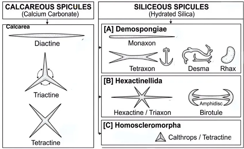
Figure 1: Classification and Morphological Diversity of Sponge Spicules Based on Mineral Composition and Taxonomic Classes.
2.3 Biological Significance and Evolutionary Symmetry
The majority of sponges (Porifera) include skeletal elements called spicules, which vary in size and three-dimensional shape. They are helpful for taxonomic identification due to their unique morphologies. Calcarea (calcareous sponges) produces spicules composed mainly of calcium carbonate, while Demospongiae (common sponges), Hexactinellida (glass sponges), and Homoscleromorpha (a type of marine sponges) produce siliceous spicules.These structures lower the metabolic cost of water exchange, support the sponge's skeleton, help in maintaining its upright position, and may offer protection from predators.Spicule symmetry varies among groups : Demospongiae usually have monaxonic or tetraxonic spicules, Hexactinellida have hexactinic or triaxonic forms, Homoscleromorpha have tetractine spicules (calthrops) and their derivatives, and Calcarea has diactine, triactine, and tetractine spicules [9].
3. MECHANISM OF ACTION
Sponge spicules are microscopic acicular structures that are made of either calcium carbonate or hydrated silica, depending on the biological source. When applied topically, they act as natural microneedles, partially penetrating the stratum corneum and epidermis [5]. Mechanisms include:
3.1 Physical Penetration and Microchannel Formation
The Spicule-Assisted Topical Drug Delivery System (S-ToDDS) uses micro-mechanical poration and surface chemical interactions to form temporary microchannels 30-60 µm deep without permanently damaging the epidermis. Hydrophilic silanol (–Si–OH) groups on the spicule surface stabilize this process by forming temporary hydrogen bonds with skin lipids, which promote the development of regular permeation channels. Also, the microchannels gradually close over a period of up to 120 hours, and the spicules remain in the skin for at least 72 hours. This provides a highly sufficient window for optimal drug diffusion before the skin barrier fully restores its integrity to prevent pathological transepidermal water loss and microbial infection[3].
3.2 Temporary Disruption of The Skin's Lipid and Protein Barriers
By mechanically disorganizing the intercellular lipid lamellae, spicule penetration transiently modulates the stratum corneum barrier, increasing the inter-bilayer distance from approximately 6 nm to 9–10 nm and reducing desmosome compactness. At the same time, intercorneocyte bonds are relaxed by a temporary downregulation of barrier proteins such filaggrin and loricrin, resulting in microfissures that expand the diffusion surface area for carrier vehicles like flexible liposomes. With minimal tissue trauma and no necrosis or long-term structural damage, this highly targeted modulation yields an excellent safety profile. A transient 10–20% increase in transepidermal water loss (TEWL) over the first 12 hours shows the system's functional reversibility. The lipid barrier is then restored in 48–72 hours when the skin's compensatory mechanisms cause lamellar bodies to secrete de novo lipids. Through cellular proliferation and reorganization, the microchannels completely close within 120 hours, returning TEWL and protein expression to baseline and avoiding pathogen entry or chronic irritation[3].
3.3 Enhanced Diffusion and Transport of Active Ingredients
By bypassing the stratum corneum and forming direct pathways that overcome the conventional <500 Da molecular weight limitation, the spicule system enhances topical transport and permits the deep penetration of large hydrophilic macromolecules, peptides, and nanoparticles. Beyond microchannel formation,Spicules modified with mesoporous silica (mSHS) coatings function as local reservoirs , sustaining drug release along a favorable concentration gradient. When paired with ultra-deformable carrier vehicles, like flexible liposomes or ethosomes, which structurally adapt to the channels and protect payloads like siRNA, hyaluronic acid, and stem cell exosomes, this flux is further enhanced.Quantitatively, this approach enhances the transport of high-molecular-weight hyaluronic acid by 19.4-fold (depositing 86.8 ± 4.1% into deep tissue layers), 10 kDa dextran by 33.09-fold, and macromolecular siRNA by 72.95 -fold [3].
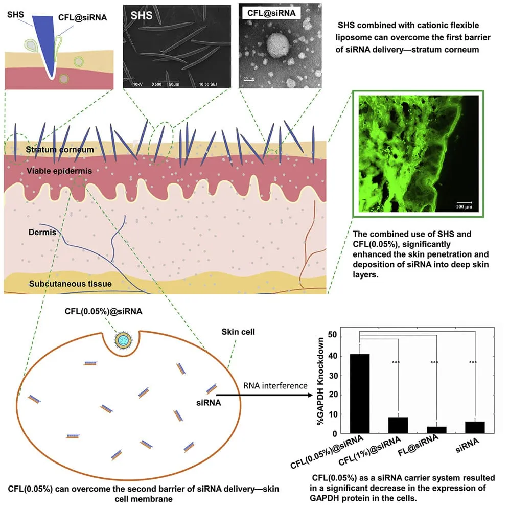
Figure 2: Mechanism of Action of Microspicule (Topical siRNA Delivery via Marine Sponge Spicules & Cationic Liposomes) [10].
3.4 Controlled Drug Retention and Release in The Target Tissue
Spicules embedded within skin tissue for at least 72 hours establish a prolonged physical reservoir, acting as local depots that continuously release payloads or facilitate diffusion straight into the viable dermis. This sustained distribution is controlled by the gradual closure of microchannels over about 120 hours, resulting in a controlled therapeutic window which ensures active components are localized at the target site. This system achieves improved tissue retention and highly sustained release kinetics[3].
4. FORMULATION STRATEGIES
Microspicule-assisted delivery systems are formed by incorporating microspicules into an appropriate topical formulation to allow active ingredients to penetrate into the skin. The formulation strategy used will depend upon the physicochemical properties of the active, nature of the microspicules, site of application and the desired release behaviour. Different strategies have been used to develop microspicule-assisted delivery systems, such as direct drug loading, gels, vesicular systems, polymer systems and hybrid systems.
4.1. Conventional Formulations
Creams:
A cream formulation containing 2% (w/w) microspicules derived from sponges was developed (Tansathien et al., 2019). The cream also contained 0.2% (w/w) deer antler velvet (DAV). Another example is the formulation of a cream containing epidermal growth factor (EGF) encapsulated within the microspicules made from water-soluble hyaluronic acid and suspended in a hydrophobic cosmetic paste [11,12]
Gels:
A gel formulation containing 1% (w/w) microspicules derived from Spongilla was developed to deliver pig placenta extract (PPE). The formulation contained glycerin, disodium EDTA, preservative system, aqueous and gelling agent (SEPIMAX™ ZEN). The gel formulation was also enriched with lipid nanocarriers containing PPE[13]
Overall, this shows that simple cream and gel formulations can be developed with microspicules derived from different materials with active compounds or delivery systems.
4.2. Advanced Carrier Formulations
Microspicules can be incorporated into more advanced delivery systems. This allows encapsulation of the active via nanocarriers and improved skin penetration via spicules. Vesicular systems such as liposomes and niosomes are particularly useful for encapsulating actives within the vesicles.
4.2.1. Niosomes and Liposomes
Liposomes can be incorporated into liposome or niosome systems. The liposomes were developed from phospholipid bilayers whilst niosomes were developed from non-ionic surfactants and often with added cholesterol. The active was encapsulated within the vesicles with addition of microspicules.
A study developed a formulation with PPE encapsulated within liposomes or niosomes developed via thin film hydration and probe sonication. The nanocarriers were added to a gel formulation containing polyacrylate crosspolymer-6 as the gelling agent along with glycerin, disodium EDTA and aqueous and preservative system. 1% w/w spongilla-derived microspicules were then added to form a hybrid formulation. [13]
An example of developing a DPE loaded niosome-microspicule (NS-MS) formulation. A separate niosome formulation containing DPE was developed. Sorbitan laurate and cholesterol were combined in a 5:5 molar ratio and dissolved in methanol/chloroform (2:1 v/v). The solution was evaporated in nitrogen leaving a lipid film. The lipid film was dried in a desiccator for 6 hours and hydrated in distilled water containing 0.2% w/v DPE. The solution was probe sonicated for 30 minutes at 20% amplitude in an ice bath and centrifuged for 15 minutes at 10,000 rpm and 4°C. The niosomes were then added to the gel formulation containing 0.5% w/w polyacrylate crosspolymer-6. Microspicules were then added at 1% w/w.[14]
Another study developed a hybrid system with pectolinarin loaded liposomes attached to sponge-derived spicules. The formulation also contained alginate encapsulation (Kim et al., 2022). The formulation contained calcium alginate, gellan gum, hydrogenated lecithin, glycerin, 1,2-hexanediol and caprylic/capric triglyceride. This system was developed using multiple stages to encapsulate the drug within the liposomes, utilise the spicules for transport and utilise the polymer for encapsulation. [1]
Overall, different vesicular carrier systems have been developed with microspicules. This includes incorporation into gel formulations and encapsulation within polymer capsules. The best carrier and vehicle to use will depend on the active and intended use of the formulation.
4.2.2. Secretome Coated Microspicules
Nano-coated spicules were developed with spicules derived from a marine sponge and the secretome derived from Wharton’s jelly-derived mesenchymal stem cells (WJ-MSC) (Lee et al., 2025). Spray coating was used to coat the secretome onto the surface of spicules. The secretome contained various bioactive factors including EGF, vascular endothelial growth factor (VEGF), fibroblast growth factor 2 (FGF2) and transforming growth factor beta 1 (TGF-β1). Approximately 2.75x10² spicules were coated with secretome containing 200μg total protein. This was then air dried in a sterile environment and added into an oil-in-water cosmetic essence. This allows complex bioactive molecules to be combined with the skin penetration ability of marine sponge derived spicules[15]
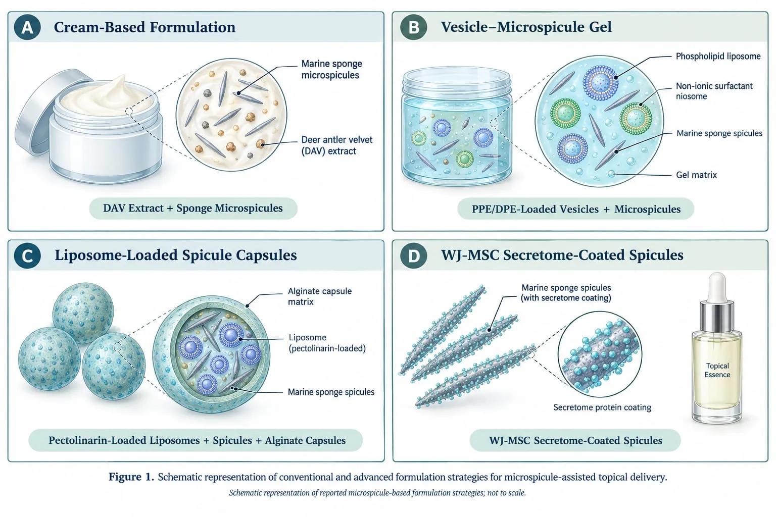
Figure 3: Schematic Representation of Conventional and Advanced Formulation Strategies for Microspicule-Assisted Topical Delivery
5. CRITICAL FORMULATION VARIABLES AND OPTIMIZATION
A number of interconnected factors, including as spicule features, active component qualities, formulation composition, production circumstances, and application settings, affect how well natural microspicule-based topical administration systems work. These variables affect the final formulation's tolerability, active component distribution, skin penetration, and physical stability. Numerous factors have been experimentally examined in the reviewed studies; however, systematic optimization of the relationships between formulation and application parameters is still lacking.
5.1 Microspicule characteristic
Table 1: Critical Formulation Variables Related to Microspicule Characteristics
| MICROSPICULE CHARACTERISTICS | EFFECT ON FORMULATION | REFERENCES |
|---|---|---|
| Natural spicule diversity (type and size) | • S. manconiae: smooth oxeas, spiny microxeas, gemmuloscleres • E. fluviatilis: 250–380 × 12–19 µm; E. fragilis: 165–261 × 10–14 µm • Characterisation only, with no delivery data; diversity alone does not establish differences in delivery performance | [16,17] |
| Morphology and aspect ratio (L/D) | • Five marine species studied (spindle-shaped oxeas, unicuspidate subtylostyles, strongyloxea, spheroxyaster) • Haliclona sp. gave the highest permeability enhancement • Aspect ratio vs enhancement: power-law fit, R² = 0.82 | [18] |
| Spicule size (Haliclona sp.) | • Sharp-edged, rod-shaped; ~120 × 7 µm • 10 kDa FITC-dextran into porcine skin: 33.09 ± 7.16-fold vs control • Optimal dimensions depend on the active ingredient and application conditions | [4] |
| Size and structural integrity | • Intact: 176.77 × 11.89 µm; blending for 30 s → broken; 10 min → fine powder • BSA–FITC enhancement: 2.26-fold (intact), 1.94-fold (broken), 1.15-fold (powder) • Intact spicules caused skin irritation, so broken spicules were selected → trade-off between permeation and tolerability • Effect cannot be attributed to length alone, as blending altered both size and integrity | [12] |
| Natural spicules with liposome loading | • Natural siliceous spicules, ~250 × 12 µm, carried pectolinarin via lecithin-based liposome loading • Porcine skin (Franz cells): dermal absorption 73.4%; percutaneous penetration 2.2% • Spicule sizes not compared, so outcomes cannot be attributed to size alone | [1] |
Note: Fold-enhancement values were obtained with different cargos, skin models and methods and are not directly comparable across studies.
5.2. Drug and Active Ingredient Properties
Molecular weight and physicochemical properties influence how actives are distributed during spicule-assisted delivery. Zhang et al. tested fluorescent dextrans of 4-70 kDa, and Haliclona sp. spicules enhanced penetration of all of them compared with controls [4]However, total skin entry generally fell as molecular weight rose, and larger dextrans showed a lower proportion in the dermis and receptor phase and more in the upper skin layers [4].Molecular weight therefore affects both the extent and the depth of delivery. Insulin reached deeper skin layers with spicules than by passive penetration, whereas the hydrophobic drug cyclosporine A required a carrier-assisted approach to improve epidermal accumulation (see 5.3)[18]. Outcomes thus depend on both the active and the delivery strategy, so drug-specific evaluation is needed.
Carrier loading and measurement limits also matter. In the porcine placenta extract study, liposomes showed higher entrapment efficiency (~40%), whereas niosomes had a higher loading capacity (26.4%) and gave greater protein deposition than the plain gel; these are different attributes and should not be used interchangeably [13]. In the deer placenta study, niosomes were prepared with 0.1-0.4% w/v extract, and 0.2% was selected based on particle size and surface charge, showing that the carrier must be optimized alongside active concentration [4]For liposome-loaded spicules, vacuum loading (25 °C, 10 Torr, 25 min) was reported as 56.4% efficient in the Results but 70.9% in the Discussion, a discrepancy to verify before either value is used [1]. In the spray-coated extracellular-vesicle system, the amount retained after coating was not quantified, so loading efficiency and delivered dose cannot be established, and fluorescence shows distribution but not that the vesicle remains intact and functional [15]. Endogenous skin proteins also interfered with protein quantification in the placenta extract work [14]
5.3. Formulation Composition
Formulation composition defines the environment in which spicules and actives are dispersed and applied. Its variables should be judged together rather than independently.
Spicule concentration. Studies used about 1% w/w in gels and 2% w/w in creams [12–14]while SHS studies used doses per unit area [18] so these values are not directly comparable. In SHS experiments, skin conductivity enhancement rose from 27.00 ± 1.35-fold at 1 mg to 47.50 ± 2.43-fold at 10 mg [4], and insulin delivery was also dose-dependent [9]. However, higher loading is not necessarily better: the 2% cream with intact spicules gave the greatest permeation but caused pain and redness, with redness also at 4% [12], and a later gel study chose 1% partly on tolerability [14]No universal safe concentration can be inferred, so delivery and tolerability should be optimized together.
Carrier, polymer and vehicle. Carrier effectiveness cannot be predicted from its class alone. Ethosomes combined with SHS improved cyclosporine A deposition in the viable epidermis, whereas the tested flexible liposomes did not significantly improve on the control [18]. Niosomes (Span 20 and cholesterol) were also compared with liposomes and modified vesicles [13]Polyacrylate crosspolymer-6 was used in gels at 0.54% w/v and 0.5% w/w, and gel incorporation changed vesicle size and surface charge [13,14]Because units and conditions differed, these are not validated optima, and the reported viscosity unit needs confirming. Vehicles ranged from creams and hydrophobic pastes to hydroethanolic solutions and oil-in-water emulsions [11,15,18], and should be chosen for the active’s solubility and stability, carrier compatibility and intended use. A hydroethanolic vehicle may help lipophilic actives, but its effects on hydration and tolerability must be considered. Vesicular systems were prepared at pH 7.4 [13,14], but no study systematically varied formulation pH.
5.4. Manufacturing and Processing Parameters
Manufacturing affects spicule dimensions, integrity, purity and loading. SHS underwent acid treatment, purification and lyophilization [4].whereas freshwater spicules were purified with alkaline and ascorbic acid treatments and gamma-irradiated at 25 kGy [1]; coated spicules were dry-heat sterilized [15]These methods are not interchangeable and must suit the source material and preserve the properties needed for delivery, especially for biologically active cargo.
Carrier preparation, loading and coating. Short blending of freshwater spicules produced fragments and prolonged blending a fine powder, with corresponding differences in permeation, so the degree of size reduction is a critical process parameter [12]Vesicles were prepared by thin-film hydration, sonication, centrifugation and membrane extrusion, which influence size, distribution and composition [13,14,18]Vacuum loading incorporates liposomes into hollow freshwater spicules [1]whereas spray coating deposits extracellular vesicles on the spicule surface [15]The vacuum loading efficiency needs verification given the conflicting values [1] and for coated systems the retained quantity and cargo integrity after formulation and storage must be characterized [15]
Stability and release. SHS retained ~92% integrity over four weeks [4]and liposomes were stable for six months in one study , while ethosomes and flexible liposomes were tested over shorter periods; these durations are not directly comparable. One hydrophilic capsule formulation broke down within a week, whereas one with more calcium alginate was more stable . Pectolinarin-loaded spicules released about half their cargo in 2-3 hours and nearly all within 30 hours, and release was tested at pH 3.1, 5.0, 7.4 and 9.6, with ~80% release at pH 3.1. This is release data, not proof of stability, and alkaline conditions may affect silica, so pH, carrier stability and spicule integrity need further study. Release profiles depend on the method and medium and should not be generalized.[18]
5.5. Application-Related Factors
In SHS studies, stronger massage gave greater enhancement than weaker massage, and enhancement increased from 30 to 120 seconds with little further gain at 180 seconds, suggesting a plateau [4]. Other studies used about two minutes of massage for creams and gels and a shorter staged rubbing for MS-EGF , which complicates comparison, so force, duration and method should be standardized. Reconstructed human skin and porcine skin responded differently in one freshwater-spicule study, showing that the test substrate can affect results [1]. SHS persistence in skin and barrier recovery time also varied with conditions [4]. MS-EGF caused transient erythema and prickling in six of 20 participants [1]whereas the short secretome essence study reported no adverse reactions [15]this gives preliminary tolerability information but does not establish long-term safety. Exposure times ranged from 4 to 24 hours, so absolute permeation or deposition values should not be compared without accounting for exposure, skin model, application method and endpoint.[11–14]
5.6. Interactions Between Variables and Optimization
Performance depends on interactions among spicule characteristics, active properties, carrier composition and application conditions. Intact spicules gave more permeation but more irritation [12]enhancement rose with dose and time before plateauing [4] and carrier performance and capsule stability varied with the cargo, vehicle and matrix [13,18] Gel incorporation also altered vesicle size and charge, so carriers should be characterized before and after incorporation [14] Spreadability was not systematically quantified in the reviewed studies. Future work should therefore use controlled, multivariable comparisons rather than testing one factor at a time.
6. CHARACTERIZATION AND EVALUATION TECHNIQUES
6.1 Morphological Characterization
The shape and dimensions, along with surface characteristics and structural integrity of the microspicules, will determine their interaction with the skin. These can be easily assessed using optical and scanning electron microscopy (SEM) to confirm if the spicules are suitable for topical delivery [12,19]. Tansathien et al. determined that raw sponge microspicules were needle-shaped with sharp tips measuring approximately 176.77 ± 10.52 µm in length and 11.89 ± 0.11 µm in width. Blending the spicules for 30 seconds produced shorter needles (~85 µm) and cylinders (~62 µm). After 10 minutes of blending, they formed a fine powder. The shape and dimensions, along with surface characteristics and structural integrity of the microspicules, will determine their interaction with the skin. Shortened spicules increased permeation by 1.94-fold with no significant increase. They were selected to continue with [12]. The size and morphology need to be optimised to ensure they provide efficient delivery without causing any issues with the skin.
6.2 Particle Dimensions
The length, diameter and aspect ratio (length: diameter) will vary depending on the biological source. Microscopy techniques are often used to measure these properties, as the sizing will determine their distribution. Zhang et al. have shown that the aspect ratios of spicules from five different marine species had a relationship to the increase in skin permeability [18]. Haliclona spicules had an average length of ~125 µm and diameter of 7.5µm. The sizes did not vary significantly between the three batches, showing that they are reliably sized if you control the initial source [10]. However, processing can change their size significantly. They become shorter when blended and will eventually break down into particles, which could affect performance [12]. Spicules created using an alkaline etch process were longer, and their pores increased in size; however, this made them weaker. The optimal conditions were used to ensure they remained strong [19]. Any data collected should include information about the size of the channels inside the spicules, if applicable and should be considered with the permeation and safety data.
6.3 Drug Content
Drug content analysis determines the quantity of drug present within the formulation and ensures consistent dosing. Validated techniques such as UV-visible spectroscopy, HPLC, protein assays and ELISA are used depending on the nature of the drug. Total protein content within the deer antler velvet extract cream was determined using the Bradford assay (Total protein = ~197.56 ± 18.35 mg/g). Growth factor content (IGF-1, EGF, TGF-β1) was determined using ELISA. The protein content given refers to that within the extract itself and not the cargo retained on the spicules [12]. In a study completed by Zhang et al., fluorescent insulin content and cyclosporine A content were determined using fluorescence and HPLC, respectively. The data obtained refer to the permeation and deposition of the drugs [18]. Another study investigating spicules coated with Wharton’s jelly stem cell secretome highlighted a lack of quantification of the cargo retained on the surface of the spicules. It recommends an ELISA and measurement of extracellular vesicles [20]. Drug content is an important aspect to consider within the study. It should be considered what quantity of drug is present initially, retained following loading and the quantity that has been released or deposited. Loading efficiency and recovery should also be included.
6.4 Release Profile
The release profiles can indicate the rate of availability of a drug. In vitro release profiles can be obtained by determining the cumulative amount released within an appropriate medium at set intervals. Hong et al. attached tripeptide GHK to modified spicules via disulphide bonds (ALTUM) and were able to show sustained release of approximately 60% of the tripeptide over 35 days in non-reducing conditions using dialysis with PBS at 37 °C and UV detection at 220nm. Korsmeyer-Peppas analysis showed that the release was mainly diffusion-controlled. Upon introduction of the reducing agent, reduced glutathione, the disulphide bonds were broken, causing an increased release rate, showing that the system is stimuli-responsive [21]. Tansathien et al. and Zhang et al. performed permeation studies, which indicated how much drug had permeated across the skin and therefore should not be interpreted as release data [12,18].
6.5 Skin Permeation
Permeation studies are typically performed using excised animal or human skin in Franz cells, together with confocal imaging and tape stripping to monitor distribution across layers of skin. In a study by Zhang et al., spicules from different species of sponges were shown to improve delivery of insulin and cyclosporine A, and spicule physicochemical properties influenced their performance [18]. In 2025, a combination of spicules and nanoparticle formulations improved penetration into porcine skin of both hydrophilic and lipophilic compounds [22]. A cream containing spicules provided improved BSA-FITC permeation; however, this may not be the case for all proteins [12]. Hollow spicules provided improved delivery of biomacromolecules [19]. The spicule size, formulation and active agent properties will influence permeation, so it is important to look at deposition and safety data in addition.
6.6 Skin Deposition
Skin deposition refers to the retention of an active within skin layers, distinct from permeation into the receptor medium. It is assessed by separating layers, extracting and quantifying the drug, and by fluorescence or confocal imaging. With Haliclona spicules, the proportion of insulin reaching deeper layers and the receptor compartment rose from 0.6 ± 0.3% (passive) to 54.8 ± 3.9%, and combining spicules with ethosomes improved cyclosporine A accumulation in the viable epidermis [18]. Spicules with cationic flexible liposomes significantly increased siRNA deposition [10], and spiculedelivered Wharton’s jelly secretome reached the viable epidermis and superficial dermis [20]. For hollow spicules, 20 kDa dextran absorption was 7.69 ± 1.72% alone and 35.09 ± 12.00% with iontophoresis, with more uniform deep distribution than subcutaneous injection [19]. Layer-specific quantification and imaging are therefore essential.
6.7 Skin Irritation and Biocompatibility
Toxicity assessments are performed to assess irritation (erythema, oedema, desquamation, inflammation) and histology and healing [18]. Hollow spicules cause little irritation, which is reversible and heals within 72 h. However, the long-term safety has not been confirmed [19]. No significant oedema or erythema was observed on 7 days of testing with a Cutibacterium acnes nanoparticle formulation. There was some mild desquamation and immune cell invasion [22]. No adverse reactions were observed with 21 participants using the secretome-coated spicule formulation. However, there was a small sample size and short duration, so conclusions cannot be drawn [20]. Further studies with larger sample sizes are required using repeated doses and longer durations.
6.8 Skin Barrier Recovery
While barrier disruption can assist drug delivery, it may also cause greater water loss from the skin and irritation. Healing can be assessed using a number of criteria such as Transepidermal Water Loss (TEWL), observable irritation, and tissue changes. Irritation caused by the hollow spicules was reversible within 72 hours. The spicules were slowly removed through desquamation of the epidermis. However, this does not confirm that the barrier has been completely restored [19]. Using traditional microneedles, Ki-67, filaggrin and transglutaminase-1 have all increased after six days of application, which indicates healing of the epidermis; indirect evidence supporting spicule removal [23]. Future studies should assess TEWL, hydration levels and healing at multiple time points.
7. THERAPEUTIC POTENTIAL AND DISEASE-SPECIFIC APPLICATIONS
7.1 Antifungal Delivery
Polymeric microsponges and nanosponges have been explored as porous antifungal carriers; they differ structurally from natural spicules and provide supporting rather than direct evidence. A fluconazole–Eudragit S-100 microsponge gel (mean size ~29.33 µm) released 85.38% of the drug over eight hours versus 83.17% in four hours for conventional gel, with activity against Candida albicans [24]. A butenafine nanosponge gel (543 ± 0.67 nm; encapsulation 71.3 ± 0.34%) showed 89.90 ± 0.87% diffusion over 24 hours [25]. A voriconazole–curcumin nanosponge hydrogel increased skin deposition 2.25- and 3.50-fold versus conventional voriconazole and curcumin formulations [26], and an aripiprazole nanosystem sponge reduced fungal burden in mice [27]. Direct studies of antifungals on natural spicules are still needed.
7.2 Acne and Inflammatory Skin Conditions
By crossing the stratum corneum, spicule-assisted delivery may improve local delivery of antimicrobial and anti-inflammatory agents, though natural spicules differ from polymeric microneedles [28]. In an atopic dermatitis mouse model, spicules with a C. acnes nanoparticle formulation improved epidermal thickening, serum IgE and IL-4 [22]. A freeze-dried secretome–hyaluronic acid sponge reduced mPASI by about 33% and plaque size by 41% in 12 psoriasis patients over 30 days, but it is not a spicule system [29]. Microneedle systems and reviews support the broader approach in acne, psoriasis and atopic dermatitis [28,30]. Direct clinical evidence for natural spicules remains limited.
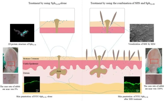
Figure 3: The Topical Application of Sphistin12-38 in Combination with Sponge Spicules for Acne Treatment.
7.3 Wound Healing
Spicule-assisted delivery may transport growth factors and regenerative agents to keratinocytes and fibroblasts, but must be distinguished from polymeric microsponges and wound-dressing scaffolds. Spicules coated with Wharton’s jelly secretome increased fibroblast and keratinocyte viability, keratinocyte migration and procollagen production in vitro, accelerated scratch wound closure, and improved skin penetration in animals; the study focused on rejuvenation rather than open wounds [20]. A resveratrol microsponge gel released about 80.88% over eight hours and improved excision-wound healing in animals [31], and a growth-factor sponge scaffold improved healing and vascularization while reducing scarring [32]. Sponge-based dressings are also under study [33], and biomaterial carriers can protect growth factors and regulate their release [34]. Direct wound models are needed for spicules.
7.4 Hyperpigmentation
The stratum corneum can be a limiting barrier to topical depigmenting agents; therefore, techniques have been developed to increase penetration. Secretome-coated spicules were used in a single-arm study, which decreased pigmentation after 2 weeks [20]. However, a controlled study is required. Microneedle patches containing glabridin, 3-O-ethyl-L-ascorbic acid and tranexamic acid were dissolved to inhibit tyrosinase and decrease melanin production to improve pigmentation [35]. Tranexamic acid combined with microneedling in a split-face study for melasma showed improvement in brown spot score and melanin index when compared to tranexamic acid alone (n=28). There were no obvious adverse effects [36]. Tranexamic acid with vitamin C, both delivered by microneedling, showed similar results [37]. Using microneedling or fractional CO2 laser with tranexamic acid showed better results than topical application; however, there is a risk of postinflammatory hyperpigmentation with the use of laser [38]. These devices cannot be substituted for natural spicules.
7.5 Skin Rejuvenation
Secretome-coated spicules increased fibroblast and keratinocyte proliferation and procollagen secretion in vitro, and improved wrinkles, texture and pigmentation in 21 participants after two weeks, though the trial was short and single-arm [20]. A cream with shortened microspicules and deer antler velvet extract reduced melanin and erythema and improved elasticity and hydration over 14 days; unmodified spicules penetrated more but irritated the skin [12]. A 2026 split-face study of a GHK-conjugated spicule regimen (20 participants, five weekly treatments) showed greater improvements in wrinkle depth, dermal density, roughness and pore volume with a glutathione-containing booster, without adverse effects, but the multi-ingredient booster and small sample limit attribution [21]. Conventional microneedling similarly stimulates collagen and elastin remodelling, providing background support [39]. Larger controlled trials are required.
7.6 Other Applications
Haliclona spicules with cationic flexible liposomes improved dermal siRNA delivery, suggesting potential in gene silencing [10]. Spicules raised insulin flux to 457.0 ± 32.3 ng/cm²/h versus 5.0 ± 2.2 ng/cm²/h for passive delivery, and ethosome combinations improved cyclosporine A accumulation [18]. Hollow spicules improved delivery of proteins, ovalbumin and an anti-PD-L1 antibody fragment, supporting transcutaneous immunization [19], and spicules with C. acnes nanoparticles modulated immune responses in atopic dermatitis [22]. Most evidence remains preclinical; formulation optimization, long-term safety and clinical validation are needed before therapeutic adoption.
8. SAFETY, BIOCOMPATIBILITY AND TRANSLATIONAL CHALLENGE
Irritation and biocompatibility are key factors which must be considered during assessment of microspicule-based drug delivery systems. To promote drug penetration through the stratum corneum, spicules derived from sponges can cause transient skin irritation. Assessment of safety includes assessment of erythema, oedema, desquamation, inflammatory response and histological changes in the skin. It is important to assess how long the irritation lasts and if the skin recovers after treatment[18].
8.1 Skin irritation and inflammation
When assessing the safety of microspicule-assisted drug delivery systems, skin irritation and biocompatibility are crucial factors. Because sponge-derived spicules physically disrupt the stratum corneum to improve drug penetration, their use may cause transient skin irritation or alter skin barrier integrity. Thus, monitoring erythema, swelling, scaling, inflammatory reactions, and histological changes in skin tissue is part of the safety review process. When deciding whether spicules are appropriate for topical and transdermal treatments, the duration of irritation and the skin's capacity to heal after treatment are also crucial factors[22].
8.2 Biocompatibility
The biocompatibility of the spicule-based formulations has also been investigated. A formulation containing nano-encapsulated secretome from Wharton’s jelly-derived mesenchymal stem cells deposited onto marine sponge-derived spicules has been tested. This formulation was tested in pre-clinical studies and in a small clinical trial with 21 participants. During the clinical trial, there were no adverse effects such as swelling, redness, itching, irritation or flaking. However, due to the small number of participants and short duration of the clinical trial, it is difficult to determine how safe the formulation would be if used long term and if the formulation would cause any issues when used multiple times. The results are only applicable to the particular formulation that was used. Overall, the available data suggest that microspicules derived from sponges are well tolerated under the test conditions. Localised irritation, desquamation and inflammation may be observed. As such, full assessments should be performed to evaluate microspicules, including skin irritation scores, histology, inflammatory markers and barrier repair. Studies should be conducted using a larger population, repeated dosing and prolonged follow-up to fully assess the safety and biocompatibility of microspicule systems[20].
8.3 Microbial contamination
Microbial contamination can be a significant problem when working with marine sponge-derived materials. Studies into using primmorphs derived from sponges have shown that penicillin and streptomycin can be used to reduce bacterial contamination in most situations. However, they do not provide effective inhibition of fungal growth. In some cases, antibiotics can also affect primmorph formation[40].
8.4 Reproducibility and translational challenge
A key factor to consider for the development of sponge spicule-based skin delivery systems is reproducibility. Spicule morphology and size can affect the penetration performance, so it is important that the manufacturing process is controlled. Although studies show that the spicules have uniform lengths and diameters within each batch, they will need to follow standardised manufacturing processes and will require quality assurance. The systems must also be validated using human skin models and clinical studies[19].
8.5 Patient acceptability
Spicule-based topical delivery systems have potential to improve patient satisfaction by improving delivery of active ingredients for effective management of acne and pigmentation. Improving appearance of the skin may help contribute to a positive effect on psychological wellbeing and quality of life. Further clinical studies are needed to determine whether patients find these spicule-based formulations acceptable, comfortable and provide long-term satisfaction[41].
8.6 Regulatory consideration
Marine sponge-derived spicules have shown great promise as a vehicle to facilitate the delivery of bioactive compounds. Recently, the secretome of Wharton’s Jelly-derived mesenchymal stem cells (WJ-MSCs) were encapsulated into nanoparticles and applied to the surface of spicules to increase their transdermal penetration. This delivery system demonstrated increased penetration into the skin, enhancing fibroblast and keratinocyte activity, collagen production and regeneration. Observations from clinical trials showed an improvement in skin texture, wrinkles, pore characteristics and pigmentation. This demonstrates the potential of using a spicule-based delivery system for cosmetic and dermatology applications[20].
9. CRITICAL COMPARISON OF EXISTING EVIDENCE
9.1 Conventional topical formulation
Conventional dosage forms in the form of creams and gels are often limited by the permeability of the drug across the skin. The stratum corneum causes a high level of resistance, meaning that drugs may only penetrate slowly through the skin, which will delay the therapeutic effect. This means new methods of delivering drugs topically are needed to increase skin permeability. https://link.springer.com/article/10.1007/s13346 9.2 Spicule-assisted system Spicule-based drug delivery systems have shown great potential in increasing transdermal penetration. Spicules can damage the stratum corneum temporarily and create microchannels which assist in increased penetration of active compounds into deeper skin layers. The literature suggests they may increase skin permeability and increase retention of actives in the epidermis and dermis. The biological activity of spicules may include activation of fibroblasts, collagen production and remodelling. This demonstrates their potential application for skin rejuvenation purposes[42].
9.2 Spicule-assisted system
Spicule-based drug delivery systems have shown great potential in increasing transdermal penetration. Spicules can damage the stratum corneum temporarily and create microchannels which assist in increased penetration of active compounds into deeper skin layers. The literature suggests they may increase skin permeability and increase retention of actives in the epidermis and dermis. The biological activity of spicules may include activation of fibroblasts, collagen production and remodelling. This demonstrates their potential application for skin rejuvenation purposes[43].
9.3 Drug permeation
In vitro skin penetration studies were carried out to investigate permeation characteristics of therapeutic peptides using spicule-based delivery systems. Full-thickness porcine skin was prepared by removing the fatty subcutaneous tissue and trimming the hair. Circular skin samples were created. Electrical conductivity measurements were taken to ensure skin integrity, with only skin samples having values below 10μA being used for experiments[18].
9.4 Skin retention
Spicule-based delivery systems have potential to increase the retention of active ingredients in the epidermis and dermis. The efficiency of spicules at increasing retention can be determined by looking at how much active ingredient is retained in the tissue and how long the spicule persists in the skin. Increased retention of active ingredients could provide constant drug delivery at the site of application. The amount of retention will depend on the characteristics of the spicule, formulation and active ingredient. Spicule-based delivery systems have potential to increase the retention of active ingredients in the epidermis and dermis. The efficiency of spicules at increasing retention can be determined by looking at how much active ingredient is retained in the tissue and how long the spicule persists in the skin. Increased retention of active ingredients could provide constant drug delivery at the site of application. The amount of retention will depend on the characteristics of the spicule, formulation and active ingredient[43].
CONCLUSION
Microspicule-assisted systems represent a highly efficient, minimally invasive physical penetration technology for advanced topical drug delivery. By overcoming the strict penetration limits of the stratum corneum, these natural bio-silica structures establish transient, high-density microchannels that facilitate the localized diffusion of large macromolecules and complex advanced therapies. Compounding these microspicules into advanced carriers—such as liposomes, niosomes, and secretome coatings—significantly increases localized drug retention within target skin tissue while maintaining excellent biocompatibility profiles. Preclinical and clinical evidence confirms their therapeutic efficacy across multiple dermatological applications, including acne management, accelerated wound healing, and skin rejuvenation, without triggering structural trauma. However, successful translation into commercial biomedical applications requires resolving critical variables, including industrial scale-up, strict quality control protocols for batch reproducibility, and standardization of biological purification methodologies. Addressing these translational challenges will establish microspicule formulations as a reliable, patient-acceptable platform for targeted cutaneous therapies
REFERENCES

Sonal Bangar, Soni Suryavanshi, Tripthi Hinger, Khan Zoya Almas, Shravani Shingate, Shrutika Patil, Microspicule-Assisted Skin Delivery: Mechanisms, Formulation Strategies and Therapeutic Potential, Int. J. of Pharm. Sci., 2026, Vol 4, Issue 11, 605-623. https://doi.org/10.5281/zenodo.23938432
 10.5281/zenodo.23938432