
We use cookies to ensure our website works properly and to personalise your experience. Cookies policy
1,4,5,6 L.R Institute of Pharmacy, Jabli Kyar, Solan, Himachal Pradesh, India 173223
2 Himalayan Institute of Pharmacy Kala amb, Sirmaur, Himachal Pradesh, India 173030
3 The School of Pharmaceutical Sciences at Shoolini University, Bajhol, Solan, Himachal Pradesh, India 173223
Microsponges represent a novel and adaptable drug delivery system that has attracted considerable interest in recent years for their potential to achieve controlled and targeted release of therapeutic agents. These sponge-like, porous microspheres, typically ranging between 5 and 300 microns in size, are capable of encapsulating a broad spectrum of active compounds and ensuring their gradual, sustained release.They offer multiple benefits such as enhanced drug stability, reduced side effects, improved aesthetic properties, and better patient compliance. Microsponges have found extensive applications in pharmaceutical, cosmetic, and dermatological formulations, including sunscreens, creams, ointments, gels, and powders. The microsponge delivery system addresses many limitations of conventional topical formulations such as greasiness, odor, and skin irritation. Microsponges have shown promising applications in oral, ophthalmic, and parenteral drug delivery systems. Their drug release can be activated by external factors such as changes in pH, temperature variations, or mechanical pressure. To assess the performance of microsponge formulations, various characterization methods are employed, including evaluation of particle size, porosity, surface morphology, drug loading capacity, and in vitro release behavior. This review outlines the principles, methods of preparation, evaluation parameters, and applications of microsponges. It also highlights recent advancements, and future prospects. Overall, microsponge technology represents a promising platform for the advancement of drug delivery approaches that are safer, more efficient, and convenient for patients across diverse therapeutic fields.
1.1 DEFINITION AND ORIGIN
The Microsponge Delivery System (MDS) is a polymer-based technology consisting of porous microspheres. They are small, sponge-like spherical particles possessing a rigid, non-collapsible structure with numerous interconnected spaces and a porous surface allows for regulated release of active ingredients. Microsponges typically range in size from 5 to 300 μm in diameter, with an average 25 μm particle containing nearly 250,000 pores and an interconnected network extending up to 10 feet in length. This unique architecture provides a pore capacity of approximately 1 ml/g, enabling efficient drug entrapment. Their surface area generally varies between 20 and 500 m²/g, while pore volume lies within 0.1 to 0.3 cm³/g. Owing to this highly porous structure, each microsponge can serve as a reservoir capable of accommodating up to its own weight of active pharmaceutical ingredients.[1,2].
Microsponge technology was first developed by Won in 1987, with the original patents granted to Advanced Polymer Systems, Inc. [3]. The company introduced several modifications of the technique for applications in cosmetics, over-the-counter formulations, and prescription drugs. At present, Cardinal Health, Inc. holds the license for utilizing this advanced technology in topical drug delivery. [4].
1.2 IMPORTANCE OF MICROSPONGES
1.3 CURRENT EVALUATION IN PHARMACEUTICALS
Microsponges have emerged as a promising controlled delivery system in the pharmaceutical industry, particularly for topical, transdermal, and localized drug delivery. Their growing application in dermatology and cosmeceuticals has led to the development of standardized evaluation protocols for assessing their safety, performance, and efficacy.
1.4 OBJECTIVE OF REVIEW
1.5 MICROSPONGE TECHNOLOGY OVERVIEW
1.5.1 STRUCTURE OF MICROSPONGES
The size of microsponges generally ranges between 5 and 300 μm in diameter, depending on surface characteristics [9]. Each microsponge sphere possesses an internal network of nearly 250,000 pores, extending up to 10 feet in length, with an overall pore volume of approximately 1 ml/g. This unique structure provides a large internal reservoir capable of accommodating up to its own weight of active pharmaceutical agents. Importantly, the microsponge particles are too large to penetrate the skin, offering an additional margin of safety. Furthermore, due to their smaller pore diameters, typically less than the size of bacteria (0.007–0.2 μm), microbial penetration into the internal tunnel system is prevented [10]. Unlike conventional topical formulations, which struggle to provide prolonged release of active ingredients, microsponges exhibit a high degree of cross-linking that renders them insoluble and resistant to high shear forces. As a result, they are widely employed in the development of creams, lotions, and powders. Moreover, microsponges ensure uniform and sustained release of active compounds, thereby minimizing irritation while maintaining therapeutic efficacy [11].
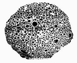
FIGURE 1: STRUTURE OF MICROSPONGE
1.5.2 PROPERTIES OF MICROSPONGES
1.5.3 ADVANTAGES OVER CONVENTIONAL FORMULATIONS
Conventional formulation: Conventional topical pharmaceutical compositions target the skin's outermost layer. Following application, these products release all of their active components. They apply a thick layer of active chemicals to the surface, which are quickly absorbed. It will result in the accumulation of too many components in the dermis and epidermis. The Microsponge system allows for the gradual application of active ingredients to the skin, effectively minimizing adverse effects such as drug-induced irritation while maintaining therapeutic efficacy. A notable example is the Microsponge Delivery System used with benzoyl peroxide formulations, which demonstrates high effectiveness with minimal discomfort [16].
Liposomes and microencapsulation: The MDS may benefit from other techniques like liposomes and microencapsulation. Most of the time, it is impossible to control how quickly active medication releases from microcapsules. The active compounds enclosed within the microcapsule are released immediately when the capsule wall ruptures. Liposomes with small dimensions, seem problematic in preparation, are microbially fragile, and have poor chemical stability [16].
Ointments: Patient adherence to ointments is low because of their sticky, oily, and unsightly nature. Traditional ointments are often inadequate for delivering drugs that require high concentrations of active ingredients, as they can cause irritation and sensitization. Additional limitations of conventional topical formulations include unpleasant odor, rapid evaporation of active components, and potential incompatibility between the drug and the vehicle. In contrast, the microsponge delivery system enhances topical therapy by extending the residence time of active ingredients within the epidermis or on the skin surface, thereby improving efficacy and reducing adverse effects.
2. METHODS FOR THE PREPARATION OF MICROSPONGES
Based on the drug’s physicochemical characteristics, there are two methods for determining the dosage amounts in microsponges. Whether it is inactive or non-polar, the drug will create a porogen, which is a porous structure. Free radicals are absent from porogen, a single-phase medication that neither inhibits nor encourages polymerization [17].
2.1 LIQUID-LIQUID SUSPENSION POLYMERIZATION
Microsponges can be prepared using the liquid–liquid suspension polymerization technique. In this method, polymerization is carried out in a round-bottom flask containing polymers such as polystyrene or methyl methacrylate. Non-polar active ingredients are first dissolved in a suitable solvent along with the monomer and then dispersed into an aqueous phase. The formulation also contains additives, including surfactants and suspending agents, which help maintain the stability of the suspension. Once droplets of the desired size are formed, polymerization is initiated using a monomer initiator, elevated temperature, or radiation [18].
PROCESS
This method involves mixing two phases. One phase consists of the active ingredient dissolved in a nonpolar solution, which is then combined with a second phase containing dispersing agents or surfactants. This mixture is maintained as a suspension, and once the droplets attain the desired particle size, polymerization is initiated by activating the monomer through a catalyst, radiation, or elevated temperature [19].
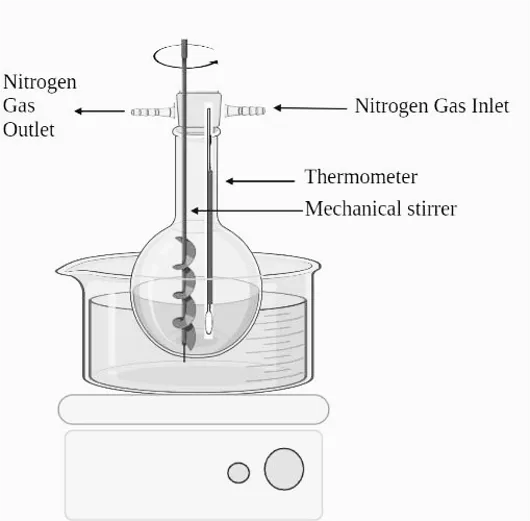
FIGURE 2: LIQUID-LIQUID SUSPENSION POLYMERIZATION METHOD
2.2 QUASI-EMULSION SOLVENT DIFFUSION TECHNIQUE
The quasi-emulsion solvent diffusion technique is the most widely used method for preparing microsponges. This two-step process begins by dissolving an appropriate polymer in solvents such as ethanol, acetone, or dichloromethane, followed by the addition of a dispersible agent (porogen) and a plasticizer. The mixture is continuously stirred for a specified period and, if required, maintained at an elevated temperature. The porogen diffuses into the external phase, forming a highly porous scaffold structure known as a microsponge. The final product is then purified and dried in a vacuum oven at 60°C for 24 hours [21].
PROCESS
All microsponges were prepared using the quasi-emulsion solvent diffusion method. The external phase consisted of 200 mL of distilled water containing 40 mg of polyvinyl alcohol (PVA) [20]. The internal phase was composed of ketoprofen, ethanol, polymer, and triethyl citrate (TEC), added at 20% of the polymer weight to enhance plasticity. The internal phase was initially prepared at 60°C and subsequently introduced into the external phase at room temperature. After emulsification, the mixture was continuously stirred for two hours. The microsponges were then separated by filtration, washed, and dried in a vacuum oven at 40°C for 24 hours [22].
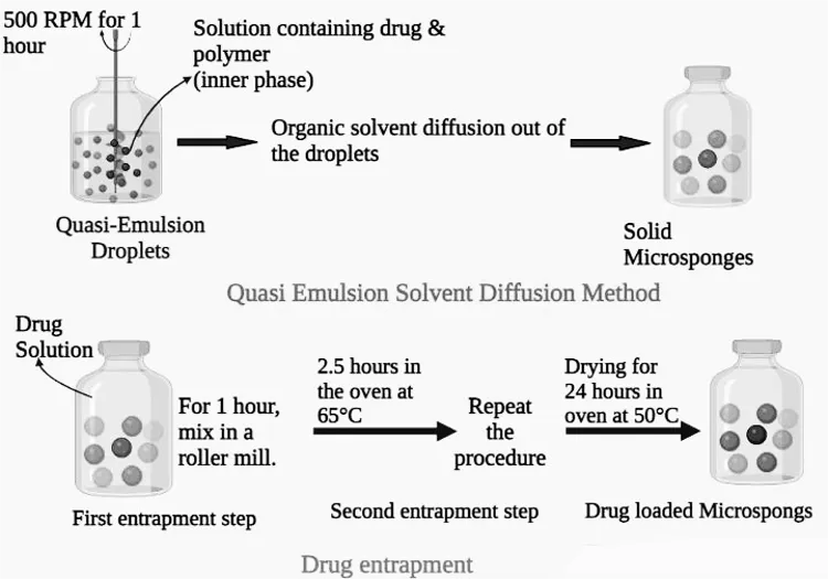
FIGURE 3: QUASI-EMULSION SOLVENT DIFFUSION METHOD
2.3 SOLVENT DIFFUSION METHOD USING WATER-IN-OIL-IN-WATER (W/O/W) EMULSION
This novel strategy resulted in porous microspheres that are biodegradable. Emulsifying compounds are used in this approach such an organic polymeric solution distributed span, polyethyleneimine, and stearyl amine via an inner water phase. To create a double emulsion, The water-in-oil emulsion is added to an external PVA-containing aqueous phase. This approach can catch both aqueous and water-insoluble drugs. They can also entrap thermolabile molecules like proteins [23]. Many researches have identified xanthan gum as an emulsifier capable of stabilizing an interior without emulsion [10].
2.4 ADDITION OF POROGEN
This method entails incorporating a porogen, such as H₂O₂ or NaHCO₃, into the inner aqueous phase of a water-in-oil-in-water (W/O/W) emulsion. The porogen is dispersed within the polymer solution to form a uniform dispersion, which is then emulsified into an external aqueous phase containing PVA. Following the addition of an initiator to the W/O/W emulsion, the organic solvents are allowed to evaporate, leaving behind the microparticles. The use of hydrogen peroxide as a porogen generates uniformly distributed, interconnected pores ranging from 5 to 20 μm [24].
2.5 OIL IN OIL EMULSION SOLVENT DIFFUSION
Unlike the W/O/W method, the oil-in-oil (O/O) emulsion technique employs a volatile organic solvent as the internal phase, which gradually evaporates under continuous stirring. In one study, dichloromethane was used as the internal phase, polylactide-co-glycolic acid (PLGA) as the polymer, and a mixture of fixed oils (corn or mineral) with dichloromethane and sorbitan trioleate as the external phase. Microsponges were formed by adding the internal phase dropwise into the dispersion medium while maintaining continuous stirring [25]. Similarly, hydroxyzine HCl-loaded ERS-100 microsponges were prepared using acetone as the dispersing solvent and liquid paraffin as the continuous phase [26].
2.6 LYOPHILIZATION
The gelation technique was employed to convert the microspheres into porous structures, with lyophilization being a commonly used method. In this process, the microspheres were immersed in a chitosan hydrochloride solution and subsequently subjected to lyophilization [27].The pores in the microspheres were formed by the solvent evaporating too quickly. Although fast and effective, this procedure can result in broken or Collapsed microparticles resulting from the rapid removal of the solvent.
2.7 VIBRATING ORIFICE AEROSOL GENERATOR METHOD
The method was used to synthesize lipid bilayered mesoporous silica particles, as reported previously. Porous particles were synthesized using a VOAG approach by evaporating surfactant in microdroplets. The core particle stock solution of tetraethyl orthosilicate was prepared by refluxing a mixture of water, ethanol, and dilute HCl. To produce monodisperse droplets, this solution was diluted with a surfactant-containing solvent using VOAG and continuously stirred [28]. The resulting microspheres were subsequently incorporated into liposomes, enabling targeted delivery of active compounds to specific sites within the body.
2.8 ULTRASOUND-ASSISTED PRODUCTION
Nanosponges are prepared by modifying the procedure to use β-cyclodextrin (β-CD) as the monomer and diphenyl carbonate as the cross-linker. The mixture is heated and sonicated to achieve the desired particle size. After cooling, the product is crushed into coarse particles and washed with distilled water and ethanol [29]. The resulting cross-linked β-CD microparticles, which are highly porous, can act as drug carriers. However, traces of the cross-linking agent may remain, posing a potential risk.
3. POLYMERS AND MATERIALS USED IN MICROSPONGES
Microsponges are highly porous, polymer-based delivery systems designed for the controlled and sustained release of active pharmaceutical ingredients (APIs). The selection of polymeric materials and excipients directly affects morphology, particle dimensions, loading efficiency, and release kinetics rate, and skin compatibility related to the microsponge system. These materials must be non-toxic, biocompatible, stable, and suitable for the desired route of administration, particularly topical use in the case of antifungal agents like miconazole nitrate.
3.1 POLYMERIC CARRIERS
The polymer forms the backbone of the microsponge system. Various synthetic and natural polymers have been investigated, each offering distinct advantages depending on the drug’s properties, target site, and release profile.
3.1.1 ETHYL CELLULOSE (EC)
Ethyl cellulose is one of the most widely used hydrophobic, water-insoluble cellulose derivatives in microsponge fabrication.
PROPERTIES:
ROLE IN MICROSPONGES:
Ethyl cellulose forms the matrix of the microsponge through quasi-emulsion solvent diffusion or emulsion solvent evaporation techniques. Its hydrophobicity delays drug diffusion, thereby allowing for sustained release [30].
3.1.2 EUDRAGIT RS100 AND RL100
Eudragits are synthetic polymethacrylate polymers, differing primarily in the permeability imparted by quaternary ammonium groups.
PROPERTIES:
Eudragit RS100: Less permeable due to lower ammonium group content (5%)
Eudragit RL100: More permeable with higher ammonium content (10%).
Insoluble in water, but swellable and permeable.
ROLE IN MICROSPONGES:
Used to modulate the rate of drug release by forming semi-permeable, pH-independent matrices that allow controlled diffusion of actives like miconazole.
COMBINATION STRATEGY:
Blending RS and RL in specific ratios allows fine-tuning of release profiles. A higher proportion of RL enhances permeability and faster drug release, whereas RS provides more sustained action [31].
3.1.3 PLA (Polylactic Acid) and PLGA (Poly[lactic-co-glycolic Acid])
FDA-approved biodegradable polyesters often used in injectable and topical microspheres and microsponges.
PROPERTIES:
ROLE IN MICROSPONGES:
These polymers form compact matrices with slow degradation, providing long-term drug release. While commonly used for implants, they are also explored for deep skin fungal infections requiring prolonged action [32].
3.1.4 POLYVINYL ALCOHOL (PVA)
PVA does not serve as a polymer for matrix formation but acts as a stabilizer and emulsifier during microsponge fabrication.
PROPERTIES:
ROLE IN MICROSPONGES:
PVA combined with the external water phase during the Quasi-emulsion technique for solvent diffusion. It prevents coalescence composed of droplets, stabilizes microsponge formation [33].
3.1.5 NATURAL POLYMERS: GELATIN AND CHITOSAN
While synthetic polymers dominate, gelatin and chitosan, which are natural biodegradable polymers are used when biodegradability, mucoadhesion, or biocompatibility is desired.
GELATIN:
CHITOSAN:
USE IN ANTIFUNGAL MICROSPONGES:
Suitable for bioadhesive topical microsponges, particularly where occlusive or prolonged skin contact is needed [34].
3.2. CROSS – LINKING AND SOLVENT AGENT
These materials assist in microsponge formation and polymer hardening, influencing porosity and drug entrapment.
3.2.1 ORGANIC SOLVENTS:
DICHLOROMETHANE (DCM):
Volatile, non-polar solvent used to dissolve hydrophobic polymers like ethyl cellulose or Eudragit.
Upon evaporation, it induces phase separation and pore formation.
ETHANOL:
Often used as co-solvent or in diffusion medium.
3.2.2 CROSS-LINKERS:
GLUTARALDEHYDE:
Used mainly in gelatin/chitosan-based systems.
Induces inter-chain cross-linking, improving mechanical strength [35].
3.3. EMULSIFIERS AND SURFACTANTS
Used during emulsion-based methods to control droplet size and stabilize microsponge dispersion.
COMMON SURFACTANTS:
SPAN 80 (SORBITAN MONOOLEATE): Lipophilic surfactant that helps in reducing interfacial tension [36].
TABLE 2: POLYMERS AND MATERIALS USED IN MICROSPONGES
|
Sr.No. |
Material Type |
Example |
Function |
Suitability |
|
1 |
Synthetic polymer |
Ethyl cellulose, Eudragit rs/rl |
Matrix former, sustained release |
Best for topical antifungal formulations |
|
2 |
Biodegradable polymer |
Pla,plga |
Long-term, controlled release |
Useful in deep fungal infections |
|
3 |
Natural polymer |
Gelatin, Chitosan |
Biodegradable, mucoadhesive |
Suitable for sensitive skin or wounds |
|
4 |
Stabilizer |
Pva |
Emulsion stability |
Needed in quasi-emulsion solvent diffusion |
|
5 |
Solvents |
Dcm,ethanol |
Polymer dissolution, pore formation |
Evaporated post-synthesis |
|
6 |
Cross linker |
Glutaraldehyde matrix former, sustained release |
Structural integrity (for gelatin systems) |
Limited to natural polymer matrices |
4. POLYMERS SELCETION CRITERIA
4.1 POLYMERS WITH TEMPERATURE SENSITIVITY
Thermoresponsive polymers are materials that undergo phase transitions in response to temperature fluctuations, making them suitable for in vivo drug delivery. These polymers exhibit a sol–gel transition at a specific temperature known as the critical solution temperature (CST).
Types of CST:
Lower Critical Solution Temperature (LCST/MinCST): Below this, the polymer remains soluble; above it, the polymer precipitates (e.g., Poly(N-isopropylacrylamide) has an LCST ≈ 32°C).
Upper Critical Solution Temperature (UCST/MaxCST): Above this, the polymer remains soluble; below it, phase separation occurs.
Mechanism:
Below LCST: Hydrogen bonding with water dominates (enthalpy-driven).
Above LCST: Hydrophobic interactions increase, leading to polymer aggregation (entropy-driven).
Examples of Thermosensitive Polymers:
Poly(N-isopropylacrylamide) (PNIPAAm), Poloxamers (Pluronics), Poly(N-vinylcaprolactam), Poly(ethylene oxide)-poly(propylene oxide)-poly(ethylene oxide) triblock copolymers, Chitosan, Xyloglucan, Cellulose derivatives.
These polymers are extensively used for temperature-triggered drug release, injectable gels, and controlled topical systems due to their reversible sol-gel transition at physiologically relevant temperatures [36,37].
4.1.1 MECHANISMS OF ACTION OF THERMALLY RESPONSIVE SMART POLYMERS
Thermally responsive smart polymers function based the lowest critical solution temperature , they become insoluble in water due to the disruption of intermolecular hydrogen bonds between the polymer and water. These polymers, often based on ethylene oxide/ethylene monomers, exhibit temperature-sensitive solubility that can be tuned between 7–70°C by altering their chemical composition. As temperature rises, hydrogen bonding weakens, shifting the hydrophilic-lipophilic balance and triggering a phase transition where the polymer separates into a dilute aqueous phase and a polymer-rich phase. This behavior enables applications in smart drug delivery, cell mapping, and DNA sequencing, with the added benefit of avoiding organic solvents. However, a strong initial burst release may occur due to the rapid volume shrinkage of the polymer network during phase change [37,38].
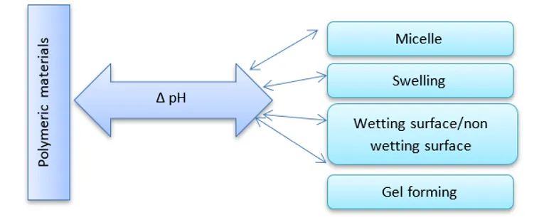
FIGURE 4: VARIOUS POLYMERS AND THEIR RESPONSES TO CHANGES IN PH
4.2 PH-RESPONSIVE POLYMERS
Polymers that respond to pH changes are classified as smart polymers that respond to changes in the surrounding pH by altering their structural and physical properties such as solubility, chain conformation, self-assembly, and surface behavior. These polymers contain ionizable acidic or basic groups (like carboxyl, sulfonic, phosphate, or amine groups) that exhibit protonation or deprotonation in response to changes in pH., leading to transitions such as swelling, deswelling, micelle formation, or strand collapse. They can be linear, branched, or networked in structure, with their sensitivity and response varying accordingly. pH-reactive polymers are widely used in drug delivery, gene therapy, responsive membranes, and chromatography. Both synthetic and natural polymers have been explored, with biopolymers being especially favored for their biodegradability, biocompatibility, and modifiability in biomedical applications [39,40,41,42,43].
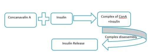
FIGURE 5: VARIOUS POLYMERS AND THEIR RESPONSES TO GLUCOSE
4.3 POLYMERS RESPONSIVE TO DUAL STIMULI (PH AND TEMPERATURE)
These polymers are designed to respond to both temperature and pH changes, and they are typically formed via the integration of ionizable components having lipophilic characteristics groups in a simple and direct manner [44]. Examples of such dual-responsive polymers include chitosan, acrylic acid, and N,N-dimethylaminoethyl methacrylate. The creation of these systems generally involves one of three strategies: copolymerizing monomers with these functional groups, blending thermosensitive polymers with polyelectrolytes, or synthesizing new monomers capable of simultaneously responding to both stimuli [45, 46].
4.4 GLUCOSE-RESPONSIVE POLYMERS
Glucose-responsive polymers are capable of mimicking physiological insulin release, thereby assisting in diabetes management and facilitating targeted drug delivery. These react to glucose within various mechanisms and are useful for both monitoring and insulin delivery. However, challenges include fast reaction times and potential biocompatibility issues. Common methods to create these systems involve glucose oxidation used (GOx), interaction with glucose via lectin binding, or through the formation of reversible bonds with phenylboronic acid. For example, in poly(acrylic acid) combined with GOx, high glucose levels trigger gluconic acid formation, lowering pH and releasing insulin faster—closely resembling natural insulin release [47, 48].
Another approach exploits the specific carbohydrate-binding ability of lectins to develop glucose-responsive systems. Lectins are proteins with dual functionality, and their glucose-binding property enables the formation of glucose-sensitive materials. These systems selectively respond to glucose and mannose, while showing minimal interaction with other sugars. Concanavalin A, a lectin with four binding sites, is frequently employed in insulin delivery. In this method, insulin is chemically modified by attaching functional groups or glucose molecules and subsequently linked to a carrier through glucose-sensitive interactions. The system leverages the competitive binding of Concanavalin A to glucose and glycosylated insulin: when free glucose concentrations rise, glucose displaces the glycosylated Concanavalin A–insulin complex, triggering insulin release. Other studies have reported the synthesis of a mono-substituted glucosyl PEG–insulin complex covalently attached to Concanavalin A within a PEG–poly(vinylpyrrolidone-co-acrylic acid) network. As glucose levels increase, glucose competes with the complex for Concanavalin A binding, resulting in the controlled release of the insulin complex [49].
4.5 PHOTORESPONSIVE SMART POLYMERS
Photoreactive Polymers offer significant potential for delivering bioactive compounds in response to light stimuli. These materials enable highly precise and nearly instantaneous drug release, facilitated by light-triggered structural changes within nanocarriers [50]. Typically, three main strategies are employed for this light-responsive drug delivery. This non-invasive approach relies on exposure to specific wavelengths of light and can operate through either single or repeated on–off release mechanisms [51].
The three main mechanisms through which photo-sensitive drug delivery is achieved include: (1) a light-triggered transformation from hydrophobic to hydrophilic properties, (2) photo-cleavage reactions, and (3) photo-induced thermal effects. Electromagnetic radiation in the ultraviolet (UV) range (250–380 nm) and near-infrared (NIR) range (700–900 nm) is commonly employed to activate these photo-responsive systems. However, light with wavelengths exceeding 900 nm is ineffective for targeting certain regions of the body, such as the posterior segment of the eye, due to its inability to penetrate ocular soft tissues. Although several polymers have been studied for ocular applications, many have been excluded because of chromophore instability and the potential for tissue damage caused by light exposure [52].To regulate the osmolality of ocular gel systems, UV-sensitive polymers have been utilized to initiate ionization upon UV exposure, leading to drug release via solvent influx [53]. Viger et al. [54] demonstrated thermally triggered drug release by using light to activate aqueous nano-platforms within a hydrated poly(lactic-co-glycolic acid) (PLGA) microparticle system. When exposed to NIR light at 980 nm, the absorbed light was rapidly converted into heat, softening the PLGA matrix. This thermal effect enabled easier release of dyes. Compared to non-irradiated particles, a significantly enhanced release was observed, as confirmed in vitro [54].
Macromers are designed to contain at least one water-soluble segment, one biodegradable component, and a minimum of two sites that can undergo free radical polymerization. These macromers are polymerized through the action of free radical initiators, which are activated by UV light, visible light, or thermal energy. Commonly used polymerizable units include acrylates, methacrylates, and diacrylates, which are generally considered biocompatible. Promoters such as ethyl eosin, camphorquinone, and acetophenone derivatives are often utilized to initiate the formation of free radicals for the polymerization process [55].
4.6 ENZYME SENSITIVE POLYMERIC MATERIAL
When designing enzyme-sensitive polymers for biomedical applications, certain essential principles must be followed. Enzymes require specific conditions to function effectively— usually an aqueous medium containing multiple ions, with a pH around 7.4, occasionally slightly acidic or alkaline. Enzyme-responsive polymers must remain stable under these physiological conditions. In addition to the presence of a target substrate or a substrate-mimicking molecule, the enzymatic activity must induce changes in the polymer’s properties to trigger the desired response. This transformation may occur either simultaneously with enzyme activity or through a sequential mechanism.
For example, proteins have been used to crosslink DNA nanoparticles, and upon exposure to proteases, these proteins are rapidly degraded, resulting in the disintegration of the nanoparticles [56, 57].
Natural polymers such as chitosan, alginate, dextran, (PEG), polyacrylamide, polyethylene oxide, and butyl methacrylate have all been studied as matrices for fabricating enzyme-sensitive delivery systems [58–59].
4.7 OXIDATION AND REDUCTION SENSITIVE POLYMERIC MATERIAL
Redox-responsive polymeric materials are generally categorized into two types: reduction-responsive systems and oxidation-responsive systems, based on the type of redox stimulus involved. In reduction-sensitive systems, disulfide and diselenide bonds are commonly used. These bonds can be cleaved when there is an increase in reducing agents such as glutathione (GSH) in the surrounding environment. There are two primary methods to introduce disulfide bonds into polymers: one involves directly forming disulfide linkages, while the other uses disulfide-containing crosslinkers to connect polymer chains.Disulfide bonds can be incorporated into polymers through controlled or living polymerization methods, such as reversible addition–fragmentation chain transfer (RAFT) or atom transfer radical polymerization (ATRP) [60]. Another milder method, often conducted at room temperature, is the thiol–disulfide exchange reaction, which is widely employed in the preparation of reduction-sensitive prodrugs and gene delivery systems [61].To prevent premature drug release, polymeric micelles can be stabilized using covalent crosslinkers like bis(2,2′-hydroxyethyl)disulfide or dithiodipropionic acid. These crosslinked structures remain stable until they reach the target site, where the disulfide bonds are cleaved, triggering the release of the encapsulated drug [62].Because the bond dissociation energies of C–Se (244 kJ/mol) and Se–Se (172 kJ/mol) are lower than those of C–S (272 kJ/mol) and S–S (251 kJ/mol), substituting disulfide bonds with diselenide bonds can enhance the sensitivity of redox-responsive systems. However, incorporating diselenide bonds into polymer structures is more complex than using disulfide bonds, and further research is needed to develop efficient synthetic methods for this purpose [63].
TABLE 3 ADVANTAGES AND DISADVANTAGES OF CHOOSING A POLYMERIC SYSTEM [64]
|
SR.NO. |
RESPONSIVENESS |
ADVANTAGES |
DISADVANTAGES |
|
1 |
Thermal |
Introducing active moieties is simple. Manufacturing and composition are simple. |
Issues with injectability during the application process. Issues with mechanical sturdiness, biocompatibility, and stability of thermolabeled medicines. |
|
2 |
pH |
hermolabile drugs will profit from this. |
There is a lack of information about toxicity. The mechanical strength is low. |
|
3 |
Light
|
Managing the trigger procedure is straightforward. Controlling stimuli with accuracy. |
Gel's low mechanical strength increases the possibility of noncovalently bound chromophores leaking out. |
|
4 |
Electric field |
Electrical charge fluctuations lead to pulsative release. |
Unpredictable behaviors are brought to light. Implantation requires surgery. External stimulation requires additional equipment. It is difficult to achieve the ideal size of an electric charge. |
|
5 |
Ultrasound
|
Protein release can be controlled. |
Controlling the release with specialist equipment. Non-biodegradable delivery systems require surgical implantation. |
|
6 |
Mechanical abrasion |
Possibility to obtain medicine release |
Managing the release pattern is challenging. |
5. EVALUATION PARAMETERS OF MICROSPONGES
5.1 PARTICLE SIZE DISTRIBUTION
The distribution of particle sizes is assessed using either an optical or electron microscope [65]. Particle size plays a crucial role in determining the formulation's uniformity and overall stability. Particles larger than 30 µm may create a gritty sensation, making them unsuitable for topical use. Therefore, a particle size range of 10 to 25 µm is generally considered ideal for such formulations [66].In this analysis, optical microscopy was utilized at 10x and 40x magnification to evaluate the particle size. The microsponges were placed on a glass slide and examined under the microscope, with measurements taken from around 50 to 100 microsponges to determine the size distribution [67].
5.2 MORPHOLOGY AND SURFACE TOPOGRAPHY OF MICROSPONGES
The prepared microsponges can be coated with a thin layer of gold-palladium under an argon atmosphere at room temperature to facilitate morphological and surface topography analysis. Scanning Electron Microscopy (SEM) is then employed to examine the surface features of the microsponges. Additionally, SEM can be utilized to study the ultrastructural characteristics of broken or fragmented microsponge particles [68, 69].
5.3 PRODUCTION YIELD OF MICROSPONGES
The production yield of microsponges is determined by precisely measuring the initial weight of the raw materials and the final weight of the dried microsponges. Each batch of dried microsponges is weighed individually, and the percentage yield is calculated using the following formula [70]
Percentage Yield = (Initial weight of raw materials/Weight of dried microsponges)×100
5.4 ENTRAPMENT EFFICIENCY
Entrapment efficiency refers to the proportion of the core drug material that is successfully encapsulated within a formulation. Several factors influence entrapment efficiency, including the drug-to-polymer ratio and the quantity of pore-forming agents used [70]. An increase in the drug-to-polymer ratio can reduce the diffusion rate of the drug solution from the concentrated polymeric phase into the external phase [71]. This leads to a longer time for droplet formation, which improves both the microsponge yield and entrapment efficiency. This effect is likely because a higher amount of drug is present per unit of polymer [72].
Entrapment efficiency can be determined indirectly by centrifuging the microsponge suspension at 2000 rpm for 10 minutes. The supernatant is then diluted appropriately with a suitable solvent, and the amount of free (unentrapped) drug in the supernatant is measured using UV-Visible spectroscopy or High-Performance Liquid Chromatography (HPLC). The drug entrapment efficiency is calculated using the following formula [70]:
Entrapment Efficiency (%) = (Total drug added - Total drug added/Free drug in supernatant)×100
5.5 TOTAL DRUG CONTENT (TDC)
Total Drug Content (TDC)is calculated using the formula:
TDC=Mact/Mms×100
Where:
Mact = Actual amount of drug present in the weighed quantity of microsponges
Mms = Weighed quantity of microsponges
This calculation helps determine the percentage of drug loaded into the microsponge formulation [73].
5.6 CHARACTERIZATION OF PORE STRUCTURE
The duration and strength of an active ingredient’s effect depend largely on the volume and size of the pores in the microsponge system. Specifically, the pore diameter controls how the active compounds migrate from the microsponges into the surrounding medium. To study how pore size and volume impact the rate and extent of drug release, mercury intrusion porosimetry is commonly employed.
This technique can measure various porosity characteristics of microsponges, including pore size distribution, bulk and apparent density, percentage of porosity filled, total pore surface area, interstitial void volume, percent porosity, as well as the shape and morphology of the pores. Mercury intrusion also generates intrusion-extrusion isotherms and provides data on the average pore diameter.
PORE DIAMETER
The Washburn equation is commonly used to calculate the pore diameter:
D=−4γcoθ/P
Where:
D = pore diameter (µm)
γ = surface tension of mercury (485 dyne/cm)
θ = contact angle between mercury and the material surface (130°)
P = applied pressure (psi)
TOTAL PORE AREA
The total pore surface area (Atot can be calculated using the following equation:
Atot=1/γcosθ 0∫Vtot P dV
Where:
P = pressure (psi)
V = intrusion volume (ml/g)
Vtot = total specific intrusion volume (ml/g)
AVERAGE PORE DIAMETER
The average pore diameter (Dm) is calculated by:
Dm=4Vtot/Atot_
Vtot= total specific intrusion volume (ml/g)
Atot= total pore surface area
These equations help in quantitatively characterizing the pore structure of microsponges by relating mercury intrusion data to pore size and surface area [74].
5.7 SCANNING ELECTRON MICROSCOPY (SEM)
SEM is employed to examine the morphology, surface texture, and particle size of the microsponges. This technique provides high-resolution images that reveal the structural features of the surface, aiding in the evaluation of formulation quality [74].
5.8 FOURIER TRANSFORM INFRARED SPECTROSCOPY (FTIR)
FTIR spectroscopy is used to analyze the chemical compatibility between the drug and polymer (Eudragit RS-100). Spectra are recorded for the pure drug, a physical mixture of the drug and polymer, and the final formulation using the potassium bromide (KBr) disc method. Compatibility is confirmed by identifying characteristic peaks using an FTIR spectrophotometer [74].
5.9 DIFFERENTIAL SCANNING CALORIMETRY (DSC)
DSC is conducted to investigate the thermal behavior of the physical mixture of the drug and Eudragit RS-100. Accurately weighed samples are sealed in aluminum pans and subjected to heating at a constant rate of 20°C/min, across a temperature range of 40°C to 430°C. This analysis helps detect any thermal interactions or changes in crystallinity that may occur during formulation [75].
6. APPLICATION OF MICROSPONGES
Microsponges have diverse applications, primarily for topical use, although recent innovations have extended their use to oral drug delivery. Their high drug-loading capacity and controlled release behavior make them suitable as pharmaceutical excipients, as highlighted in various patents [76].
6.1 ORAL DRUG DELIVERY
Microsponges improve the solubility of poorly water-soluble drugs by entrapping them within their porous structure. Controlled oral formulations of Ketoprofen and Flurbiprofen have been developed using Eudragit RS 100 via the quasi-emulsion solvent diffusion method [77–80].
6.2 TOPICAL DELIVERY
Microsponges have been employed to deliver benzoyl peroxide with sustained release, which reduces irritation and enhances skin penetration. Their incorporation into gel-based formulations has demonstrated improved therapeutic efficacy while minimizing adverse effects on sensitive skin [77, 81, 82].
6.3 BONE AND TISSUE ENGINEERING
Collagen-based microsponges have shown promise in bone regeneration. When implanted into mice subcutis, these scaffolds promoted angiogenesis in a dose-dependent manner, indicating their potential as a reservoir for Basic Fibroblast Growth Factor (bFGF) and for tissue healing [83].
6.4 CARDIOVASCULAR TISSUE ENGINEERING
In cardiovascular surgery, collagen microsponges blended with poly(lactic-co-glycolic acid) have been utilized to create vascular patches. These biodegradable constructs encourage autologous cell growth and tissue regeneration, enhancing healing post-implantation [86].
6.5 VASCULAR WALL RECONSTRUCTION
A biodegradable scaffold composed of polyglycolic acid mesh and collagen microsponges, reinforced with polylactic acid, was designed for vascular repair. These tissue-engineered grafts were used without prior cell seeding, yet showed excellent in situ cell proliferation without thrombosis. Histological and genetic analysis confirmed the presence of endothelial and smooth muscle cells two months post-implantation [84, 86].
6.6 ANTI-ULCER THERAPY
Microsponges can be employed for targeted delivery of anti-ulcer agents to the intestinal lining. Their high drug encapsulation capacity and compatibility with capsule formulations offer sustained release and enhanced therapeutic action for conditions like peptic ulcers.
6.7 ANTIFUNGAL TREATMENTS
Topical antifungal agents like fluconazole have been incorporated into microsponge-based gels, providing controlled and prolonged drug release. These formulations demonstrate high drug yield and improved therapeutic efficiency for severe fungal skin infections.
6.8 ANTICANCER APPLICATIONS
Traditional cancer therapies often harm healthy tissues. Microsponge systems are being explored for targeted delivery of anticancer agents in cancers such as colorectal, pancreatic, gastric, and breast cancers. These delivery systems may reduce side effects while maintaining or enhancing drug efficacy.
6.9. ANTI-ARTHRITIC USE
Diclofenac sodium microsponges have been formulated for rheumatoid arthritis treatment, offering localized, sustained release to reduce inflammation and pain with fewer systemic side effects.
6.10 ANTIEPILEPTIC DRUG DELIVERY
Microsponges of Carbamazepine (CBZ), used for conditions such as epilepsy, trigeminal neuralgia, and bipolar disorder, have been synthesized using ethyl cellulose (EC) and polyvinyl alcohol (PVA) through a quasi-emulsion solvent diffusion method. These were analyzed using FTIR, DSC, and XRD techniques to confirm their structural integrity and drug loading [85].
7. RECENT ADVANCES IN MICROSPONGE DRUG DELIVERY SYSTEM
Significant progress has been made by refining the fabrication techniques to develop nanosponges, nanoferrosponges, and porous microbeads. One major advancement includes the formulation of β-cyclodextrin (β-CD) based nanosponges, which are suitable for encapsulating both hydrophilic and hydrophobic drugs—unlike traditional polymeric microsponge systems. These β-CD nanosponges are synthesized by cross-linking β-CD with diphenyl carbonate, and have been evaluated for the oral delivery of various drugs, including dexamethasone, flurbiprofen, doxorubicin hydrochloride, itraconazole, and serum albumin.
Research also highlights the potential of nanosponges as effective carriers for gaseous drug delivery. Furthermore, incorporating cytotoxic agents into nanosponge systems has shown to significantly enhance drug potency, suggesting their potential use in targeted cancer therapy.
The concept of nanoferrosponges introduces self-guiding drug carriers that can penetrate deeper tissues using external magnetic fields. Once delivered to the target site, the magnetic core is removed, leaving behind a porous delivery system for sustained drug release.
Additionally, porous microbeads have been developed using a high internal phase emulsion (HIPE) method, which involves a monomer dispersed in a continuous oil phase, along with a cross-linking agent and an aqueous internal phase. This technique has demonstrated improved RNA stability and efficient siRNA encapsulation, opening new possibilities for siRNA-based therapeutic delivery [87,88,89].
8. FUTURE PROSPECTS
Microsponge Drug Delivery Systems (MDS) exhibit a range of exceptional characteristics, including enhanced extended-release, improved drug release profiles, reduced skin and mucosal irritation, and superior physical, chemical, and thermal stability. These features make microsponges highly adaptable for the development of novel pharmaceutical formulations.
Beyond conventional drug delivery, microsponges hold promise in cell culture technologies, where their porous architecture allows them to serve as scaffolds for stem cell cultivation and tissue regeneration. This opens avenues in regenerative medicine and biomedical research.
Their aesthetic and functional qualities have also led to increasing use in cosmetic products, where controlled ingredient release and product elegance are critical.
Looking forward, one of the key challenges lies in developing oral peptide delivery systems using optimized polymer ratios, ensuring both protection and bioavailability of sensitive biomolecules. Additionally, research into biodegradable and bioerodible polymers is expanding the safe and effective delivery of therapeutic agents.
Microsponge systems have also been explored for pulmonary drug delivery, demonstrating effective release even in environments with limited dissolution media, making them suitable for colon-targeted therapies. The flexibility of these carriers suggests they could also be engineered for parenteral routes, expanding their utility in systemic drug administration.
Altogether, these innovative formulation strategies not only broaden the scope of microsponge applications across pharmaceutical, biomedical, and cosmetic fields, but also pave the way for advanced, patient-friendly, and targeted drug delivery solutions [90,91,92].
9. CONCLUSION
The microsponge drug delivery system is an innovative and versatile technology offering controlled, sustained release of a wide range of drugs. It improves therapeutic efficacy, reduces side effects, and enhances patient compliance. With applications in both topical and oral delivery, microsponges are non-toxic, non-irritant, and highly compatible with various active ingredients. Their potential extends to pharmaceuticals, cosmetics, and even tissue engineering. Given their numerous advantages over conventional systems, microsponges represent a promising field that warrants further research and development for future therapeutic applications.
REFERENCES


Payal, Monika, Rishabh Sharma, Priya Thakur, Avneet Gupta, Diwan Chand, Microsponges: Comprehensive Review of Formulation Techniques, Evaluation Parameters, and Future Prospects, Int. J. of Pharm. Sci., 2026, Vol 4, Issue 7, 390-413. https://doi.org/10.5281/zenodo.21140368
 10.5281/zenodo.21140368