

We use cookies to ensure our website works properly and to personalise your experience. Cookies policy
Department of Pharmacology, Caritas College of Pharmacy, Kottayam, Kerala, India.
Neurodegenerative diseases are a group of disorders characterized by progressive neuronal loss leading to cognitive and memory impairment. Alzheimer’s disease, the most predominant neurodegenerative condition, involves neuroinflammation, oxidative stress, tau accumulation, and ?-amyloid–induced neurotoxicity. Current therapies offer only symptomatic relief, highlighting the need for novel neuroprotective agents. This study evaluated the in-vitro neuroprotective activity of the ethanolic leaf extract of Gliricidia sepium using SH-SY5Y human neuroblastoma cell lines with ?-amyloid to induce neurotoxicity. Cell viability and morphological changes were assessed using the MTT assay and microscopic analysis across five concentrations of the extract. ?-amyloid exposure significantly reduced cell viability, whereas treatment with Gliricidia sepium extract resulted in a dose-dependent increase in cell viability and improved cellular morphology. Statistical analysis confirmed a highly significant dose-dependent neuroprotective effect (P<0>0.05) effect was observed on comparison with the ?-amyloid group. The findings of our study suggest that the ethanolic extract of Gliricidia sepium leaves possesses neuroprotective potential, possibly mediated through the presence of neuroprotective phytochemicals, antioxidant and anti-inflammatory mechanisms, and the ability to overcome ?-amyloid-induced toxicity, which warrants further investigation for its clinical applicability.
Neurodegenerative disorders (NDs) belong to a broad category of conditions characterised by progressive death of neurons, leading to movement dysfunction, hippocampal atrophy, cognitive decline, neuroinflammation, metabolic issues and other neurological symptoms. The neurodegenerative diseases encompass a broad array of conditions, such as Alzheimer's disease (AD), Parkinson's disease (PD), amyotrophic lateral sclerosis (ALS), Huntington’s disease (HD), along with several other disorders[1]. Neurodegeneration is acknowledged as the key pathophysiological alteration in most of the brain-related conditions[2]. In such a scenario, neuroprotection contributes significantly. Neuroprotection provides preservation, recovery, or regeneration of the cells, structure, and function of the nervous system[3]. As per WHO reports, approximately 3 billion individuals globally - about one in three suffer from various types of neurological disorders. By the year 2050, it is projected that there will be around 139 million dementia cases globally, with the occurrence of neurodegenerative diseases anticipated to rise even more[4]. Alzheimer's disease is the most prevalent of all the neurodegenerative diseases, affecting over 55 million individuals globally[5].
ALZHEIMER’S DISEASE
Alzheimer’s disease (AD) is a complex condition influenced by multiple genes and factors, marked by the accumulation of beta-amyloid peptides and hyperphosphorylated tau protein in the brain, causing plaques and neurofibrillary tangles (NFTs), which ultimately lead to dendritic dysfunction, neuronal death, memory impairment, behavioural alterations, and organ failure[6].
Aetiology of Alzheimer’s disease
In addition to the crucial involvement of Aβ and tau, various other elements may play a role in the pathology of AD, including acetylcholine scarcity, neuroinflammation, oxidative damage, bio-metal imbalances, glutamate disruption, insulin insensitivity, gut microbiome irregularities, cholesterol homeostasis issues, Down syndrome, mitochondrial impairment, and autophagy dysfunctions[7]. Acetylcholine is a key neurotransmitter that influences cognitive function across different regions of the brain. Genetic factors have been linked to both early and late onset AD. Mutations in APP, PSEN1 or PSEN2 lead to overproduction of pathological Aβ fragments and consequently to amyloid pathology with increased plaque formation. Alterations to chromosomes 1, 14, and 21 are associated with early-onset AD, whereas the presence of APOE- ε4 allele a form of the apolipoprotein E gene on chromosome 19 increases risk of developing late-onset AD.[8]
Pathogenesis of Alzheimer’s disease
Two pathological features of AD are present, including senile amyloid plaques that contain the amyloid-beta (Aβ) peptide, and neurofibrillary tangles formed by hyperphosphorylated tau protein. A further crucial discovery is brain atrophy, particularly in the hippocampus[9]. The Aβ hypothesis suggests that Aβ accumulation is critical to AD, leading to Aβ plaques, cell death, and dementia. Aβ peptide results from the improper cleavage of a larger Amyloid precursor protein (APP). APP is a membrane-spanning protein that is typically cut by enzymes known as secretases to generate different peptide fragments vital for neuronal activities. Long species (Aβ42) have a higher tendency to aggregate compared to short species. Aβ oligomers, being smaller clusters of Aβ peptides, are regarded as especially harmful to neurons and synapses. In addition to the accumulation of amyloid-beta plaques, another hallmark pathology of AD is the presence of neurofibrillary tangles (NFTs) composed of abnormally hyperphosphorylated aggregates of tau protein[10]. Phosphorylated Tau aggregates to form oligomers that later mature into paired helical and straight filaments, which are the major components of neurofibrillary tangles[11]. The formation and accumulation of NFTs disrupt neuronal function and contribute to neurodegeneration.
Clinical manifestations
AD is progressive and develops over the years, resulting in the gradual emergence of cognitive impairments[12]. It is marked by early memory difficulties and mental deterioration that may eventually impact behaviour, language, visual-spatial awareness, along with the presence of the following impairments: aphasia, apraxia, agnosia, or cognitive, behavioural and emotional problems and the motor system, and it is the most prevalent type of dementia[13]. These conditions lead to difficulties in daily activities and indicate a cognitive and functional deterioration[12]. Along with these symptoms, like psychotic features, aggression, depression, sleep disturbances, and apathy are also significant[12].
Complications of Alzheimer’s disease
Complications that correlate to AD can be broadly put into 2 categories: mental/behavioural and physical complications. Mental/behavioural complications include depression, difficulty concentrating, social withdrawal, mood changes, sleep disturbances, wandering, agitation, delirium, and sundowning – common in more advanced stages[14]. Physical complications include difficulty swallowing and breathing, aspiration pneumonia, loss of appetite, incontinence and urinary tract infection[15].
Management of Alzheimer’s disease
Alzheimer's disease presents one of the most significant challenges in medical care this century and is the leading cause of dementia, yet it remains incurable. A multifactorial approach, involving both pharmacological and non-pharmacological intervention, is required for the management of Alzheimer’s disease[16]. Pharmacological intervention involves the use of various categories of drugs for the treatment and management of AD and its related symptoms. Acetylcholinesterase inhibitors (AChEIs) and N-methyl-d-aspartate receptor (NMDAR) antagonists are the FDA-approved classes of drugs for the treatment of AD[17]. AChEIs inhibit the action of acetylcholinesterase and maintain the ACh levels at the synaptic cleft. Three medications have been approved by the FDA in this category: donepezil, galantamine, and rivastigmine. These agents prevent the degradation of acetylcholine by binding to the cholinesterase enzyme. NMDA receptor antagonists drug, Memantine works by inhibiting NMDA receptors, which are voltage-dependent cation channels, thus stopping their excessive activation and lowering calcium entry into the cells, since calcium triggers excitotoxicity that results in neuronal death. It is recommended for the treatment of moderate to severe AD[8]. It helps in memory and learning, neuroplasticity, and neuronal control. Nootropics are a class of agents that are widely used in the management of Alzheimer’s disease. Also known as smart drugs, brain boosters or memory-enhancing agents, nootropics are substances that can enhance mental abilities such as memory, attention and concentration[21]. Nootropics can be classified as synthetic agents, vasodilators or cerebral metabolism enhancers, and natural compounds[22]. Dietary supplements that include vitamins, nutraceuticals and other plant-derived bioactive constituents are gaining wide popularity in the management of Alzheimer’s disease. Vitamin E is an antioxidant that is prescribed as an adjuvant therapy for AD patients[23].
Newer Therapeutic Interventions
Although conventional medications like acetylcholinesterase inhibitors and NMDA receptor blockers are accessible for treating AD-related dementia, limited drug penetration through the blood-brain barrier (BBB) and disease relapse from ageing continue to pose difficulties[6]. Many advanced and site-specific therapies have evolved for the treatment of AD. Monoclonal antibodies (MABs) that target amyloid have marked the beginning of a new age in therapeutics for Alzheimer's disease with targeted therapy. Three agents—aducanumab, lecanemab, and donanemab have received FDA approval, while additional ones are undergoing development. These agents are given intravenously, and upon reaching the brain, they stimulate microglia to absorb amyloid-beta protein fibrillar plaques[24]. Genetic therapies, in general, refer to any techniques that alter genes or their expression, while gene therapies specifically focus on the genes themselves. RNA-based or gene silencing treatments can lower the synthesis of proteins like amyloid or tau by modifying the amount of mRNA that serves as the template for protein translation[25]. Owing to an increase in neurodegenerative diseases, the challenge of delivering and releasing drugs into the brain is attracting much attention. Soft nanoparticles, such as liposomes and exosomes, are nanovesicles that have the potential to deliver drugs and genes across the BBB. The role of apolipoprotein E (ApoE) as a pivotal factor related to Aβ level and amyloid deposition in the progression of AD. Liposomes can be used to deliver plasmid-encoded ApoE2 (pApoE2) for the effective treatment of AD. The exosomes contain non-coding RNAs, microRNAs, mRNAs, lipids, and proteins. There have been successful attempts where the exosomes loaded with siRNA are used for effective brain delivery. [26] Stem cell therapy is another area of intensive research aiming at the treatment of Alzheimer’s disease. Stem cell therapies, including human embryonic stem cells or neural cells derived from induced pluripotent stem cells[27].
Importance of Herbal Medicines in Alzheimer’s Diseases
The rising interest in herbal remedies stems from the therapeutic capabilities and enduring advantages of phytochemicals. Numerous traditional plants are utilized to treat cognitive and neurodegenerative conditions involved in Alzheimer’s disease[28]. Herbal medications contain bioactive substances like flavonoids, alkaloids, glycosides, terpenes, and tannins, which are usually regarded as effective with few side effects[29]. In traditional Indian medicine, various plants such as Allium sativum, Bacopa monniera, Centella asiatica, Withania somnifera, Ginkgo biloba, Curcuma longa, Celastrus paniculatus, Terminalia chebula, and Crocus sativus, among others, exhibit promising potential against Alzheimer's disease[30].
MATERIALS AND METHODS
Plant Profile
Gliricidia sepium (Jacq.) Kunth. ex. Walp is a medium-sized, semi-deciduous tree that typically grows in the tropical and subtropical areas[31]. Belonging to the family of Fabaceae, the plant is widely available in the Western Ghats of India, and studies have reported with mainly anti-inflammatory, antioxidant, anthelmintic, antimicrobial and wound healing properties. Fig. No.1 shows the leaves of Gliricidia sepium used in the present study.

Fig. No.1 Gliricidia sepium leaf
Various parts of Gliricidia sepium have several ethnomedical uses. The leaves of the plant are used to prepare teas to treat cough and asthma. A decoction of the leaf is utilized to cure urticaria, rash, and burns. The whole plant is used to treat headache, fever, burns, bruises, ulcers, wounds, and rheumatism, as an expectorant and sedative[32]. The phytochemical examination of the plant revealed numerous bioactive phytoconstituents, with key components being flavonoids such as isoquercetin and isorhamnetin, along with isoflavans, 3-hydroxy-9-methoxy pterocarpan, kaempferol -O-glycosides, triterpene saponins like gliricidoside A and B, and coumarin[33]. Each of these agents exhibits notable neuroprotective properties.
Chemicals and Reagents
Ethanol (95%), SH-SY5Y Human neuroblastoma cell line (NCCS Pune), Dulbecco’s Modified Eagle's Medium (DMEM) (Sigma Aldrich, USA D5648), β-amyloid45-55 (10µM), 0.25% Trypsin (Invitrogen, USA 25200-056), 3-(4,5-dimethylthiazol-2-yl)-2,5-diphenyltetrazolium bromide (MTT) (Sigma Aldrich M5655), Foetal Bovine Serum (Gibco, US origin), Phosphate-buffered saline (PBS), Dimethyl sulfoxide (DMSO), L-glutamine, Sodium bicarbonate, Penicillin (100 U/ml), Streptomycin (100 µg/ml), Amphotericin B (2.5 µg/ml).
Collection of Raw Material
Fresh Gliricidia sepium leaves were identified and then collected in October from the local areas of Kottayam, Kerala. The leaves were shade-dried for five weeks until crispy dry, ground into fine powder using a mechanical grinder, and stored in airtight glass containers for subsequent extraction and in-vitro analysis.
Preparation of Extract
The powdered leaves were extracted by simple maceration using 95% ethanol (1:6 ratio), where 66.66 g of powder was soaked in 400 mL of ethanol. The mixture was shaken continuously for 30 min and macerated for seven days with intermittent shaking. On day seven, it was double-filtered using Whatman filter paper to separate the liquid extract from the marc. The filtrate was then evaporated at 40 °C to remove ethanol. The crude ethanolic extract was stored below 4 °C for subsequent in-vitro studies[34].
The percentage yield was calculated for the Gliricidia sepium leaf extract based on the crude material used, using the formula:
Percentage Yield (%) = Weight of Plant Material Taken (g) × 100
Weight of Plant Extract (g)?
In-Vitro Neuroprotective Activity Determination by MTT Assay
Cell Culture
The cell line SHSY-5Y (Neuroblastoma cells) was obtained from the National Centre for Cell Sciences (NCCS) in Pune, India, and was kept in Dulbecco’s Modified Eagle's medium (DMEM) (Sigma Aldrich, USA). The cell line was cultured using DMEM enriched with 10% FBS, L-glutamine, sodium bicarbonate, and an antibiotic solution that included: Penicillin (100 U/ml), Streptomycin (100 µg/ml), and Amphotericin B (2.5 µg/ml). Cell lines were maintained at 37ºC in a 5% CO2 humidified incubator (NBS Eppendorf, Germany)[35].
Cell Seeding and Compound Stock Preparation
Confluent monolayers of cells that were two days old were trypsinised, and then the cells were resuspended in 10% growth medium. 100 µl suspension of the cell (5x10³ cells/well) was placed in a 96-well tissue culture plate and incubated at 37ºC in a humidified incubator with 5% CO2. Then, 1mg of the sample was measured and fully dissolved in 1mL DMEM utilizing a cyclomixer. The extract solution was passed through a 0.22 µm Millipore syringe filter to ensure it remained sterile.
Morphological analysis for Neuroprotective activity
Once the cells achieved adequate growth, Beta Amyloid45-55 (10µM) was introduced to provoke toxicity and was then incubated for one hour[36]. The 96-well plate contained wells for the different concentrations of sample, control and beta amyloid groups. Following the incubation period, samples of Gliricidia sepium (100, 50, 25, 12.5, and 6.25 µg/mL in DMEM) were applied in triplicate across the respective wells and incubated for 24 hours at 37 °C with 5% CO?. Post-treatment, cells were examined under an inverted phase-contrast microscope (Olympus CKX41 with Optika Pro5 CCD camera), and images were captured for analysis.
Cell viability assay by MTT method
15mg of MTT (Sigma, M-5655) was dissolved in 3 ml PBS, and then sterilized using filter sterilization. Following the 24-hour incubation post-treatment, the contents of the wells were discarded, and 30µl of reconstituted MTT solution was introduced into the wells. The plate was gently agitated and then incubated for 4 hours at 37°C in a humidified 5% CO2 incubator. After the incubation, the supernatant was discarded, and 100µl DMSO was introduced; the wells were then gently shaken to dissolve the formazan crystals produced by the viable cells. The absorbance measurements were taken with a microplate reader at a wavelength of 540 nm[37]. The percentage cell viability was calculated by the formula:
Percentage cell viability (%) = Mean optical density of sample × 100
Mean optical density of control
Statistical Analysis
The values obtained after the experiments were expressed as mean ± Standard error mean, where n = 3. The results were analyzed by one-way analysis of variance (ANOVA) followed by Dunnett’s post hoc multiple comparison test. The difference between groups was considered significant at P<0.05*, P<0.01**, P<0.001***, P<0.0001****, and non-significant (NS) at P>0.05 when compared to the β-amyloid group. The statistical analysis was carried out using SPSS software Version 22.
RESULTS
Preparation of Extract and Calculation of the Percentage Yield
The Gliricidia sepium leaves were extracted by the technique of simple maceration using 95% ethanol as solvent. The final percentage yield of the plant extract was found to be 10.2%w/w.
Neuroprotective Activity Studies
The study was carried out to evaluate the in-vitro neuroprotective activity of the ethanolic extract of Gliricidia sepium leaves in the SH-SY5Y human neuroblastoma cell line using β-amyloid 45-55 to induce neurotoxicity. The Aβ-induced cells were treated with five different concentrations of the plant extract (6.25, 12.5, 25, 50, and 100 µg/ml) to assess its neuroprotective effect. The control and β-amyloid wells were also maintained for comparative studies. Cell viability was measured using the MTT colorimetric assay, and microscopical examination was conducted to study the changes in cell morphology. Statistical analysis was conducted using One-way ANOVA followed by Dunnett’s post hoc test.
Effect of Gliricidia sepium Leaf Extract on Cell Morphology
The morphological changes in SH-SY5Y cell lines following Aβ exposure and treatment with different concentrations of the ethanolic extract of Gliricidia sepium leaves were observed through microscopical examination. Control cells displayed normal neuronal morphology with intact and elongated structure with strong adherence to the cell surface, whereas the Aβ group showed shrinkage of cells, dissolution of the cell content, reduced cell density and loss of adherence, exhibiting severe cytotoxicity. Treatment with the plant extract showed a dose-dependent advancement in cell morphology. Partial protection was observed in a lower concentration (12.5 µg/ml). Reduced distorted features of cells were observed in intermediate concentrations (25 and 50 µg/ml). Advanced neuroprotective activity with maximum protection was observed for 100 µg/ml, where the cell morphology was almost similar to that of the control group. Fig. No. 2 illustrates the microscopic images showing the effect of various concentrations of the ethanolic extract of Gliricidia sepium leaves on β-amyloid–induced cytotoxicity in SH-SY5Y cells.
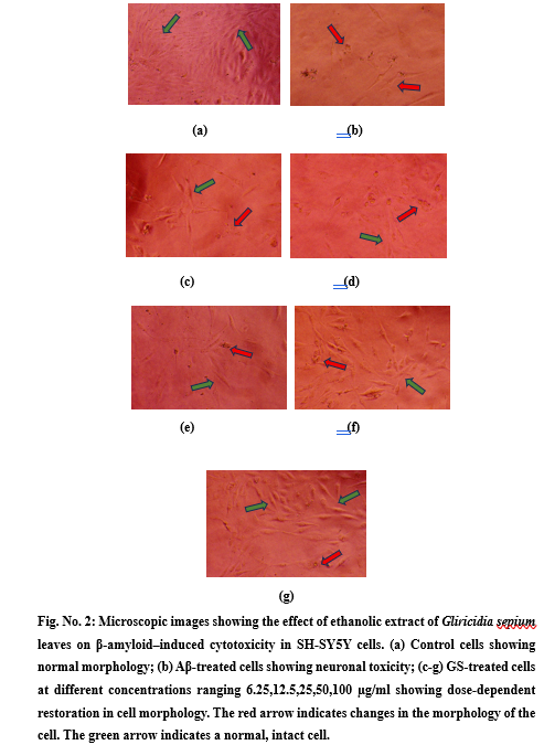
Fig. No. 2: Microscopic images showing the effect of ethanolic extract of Gliricidia sepium leaves on β-amyloid–induced cytotoxicity in SH-SY5Y cells. (a) Control cells showing normal morphology; (b) Aβ-treated cells showing neuronal toxicity; (c-g) GS-treated cells at different concentrations ranging 6.25,12.5,25,50,100 µg/ml showing dose-dependent restoration in cell morphology. The red arrow indicates changes in the morphology of the cell. The green arrow indicates a normal, intact cell.
Effect of Gliricidia sepium Leaf Extract on Cell Viability by MTT Assay
Cell viability of control, β-Amyloid and extracts at various concentrations was assessed using the MTT assay by estimating absorbance at 540nm. The neuroprotective activity of Gliricidia sepium leaf extract is shown in Table 1.
Table 1: Neuroprotective activity of Gliricidia sepium leaf extract
|
Sample Concentration (µg/ml) |
Average Absorbance @ 540nm |
Cell Viability (%) |
|
|
Control |
0.4132 |
100.00 ± 0.0000**** |
|
|
β-Amyloid |
0.2033 |
49.19 ± 0.2194 |
|
|
GS + β-amyloid |
|||
|
6.25 |
0.2172 |
52.57 ± 0.0740NS |
|
|
12.5 |
0.2429 |
58.79 ± 1.1329**** |
|
|
25 |
0.2641 |
63.92 ± 1.3244**** |
|
|
50 |
0.2811 |
68.02 ± 0.5071**** |
|
|
100 |
0.2972 |
71.93 ± 1.2090**** |
|
At concentrations 12.5, 25, 50 and 100 µg/ml, Gliricidia sepium extract showed a highly significant (P value < 0.0001****) increase in cell viability to 58.79 ± 1.1329, 63.92 ± 1.3244, 68.02 ± 0.5071 and 71.93 ± 1.2090 %, respectively, when compared to the β-amyloid group, with a cell viability of 49.19 ± 0.2194 %. The concentration 6.25 µg/ml showed a non-significant increase in cell viability (52.57 ± 0.0740 %) with the P value > 0.05. Fig. No. 3 shows the graph obtained after the statistical analysis of the data attained from the experiment.
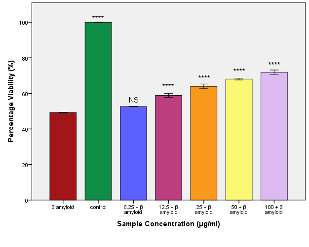
Fig. No. 3: Effect of ethanolic extract of Gliricidia sepium leaves on percentage cell viability in β-amyloid–treated SH-SY5Y cells by MTT assay. All experiments were performed in triplicate, and the data are expressed as mean ± SEM (n = 3). Statistical analysis was carried out using one-way ANOVA followed by Dunnett’s post hoc test. **** p< 0.0001 compared to β-amyloid–treated group; NS: non-significant (p > 0.05) compared to β-amyloid–treated group.
DISCUSSION
Alzheimer’s disease is the most prevalent neurodegenerative disorder, characterized by progressive neuronal loss leading to memory, cognitive, and behavioral impairments. Its pathology involves β-amyloid plaque deposition and neurofibrillary tangles, resulting in synaptic dysfunction, neuronal loss, and cerebral atrophy. Despite extensive research, current therapies mainly offer symptomatic relief. Owing to its global impact, Alzheimer’s disease was chosen as the focus of this study. Gliricidia sepium was selected based on literature evidence highlighting its bioactive constituents (flavonoids, saponins, and essential oils) and established antioxidant and anti-inflammatory properties. However, its neuroprotective potential lacks experimental validation, forming the research gap that provided the rationale for the present investigation.SH-SY5Y human neuroblastoma cells are commonly utilized in neuronal studies due to their neuron-like characteristics. Exposure to β-amyloid peptides leads to Alzheimer’s disease–related pathological characteristics, such as decreased cell viability, morphological alterations, and cell death, establishing SH-SY5Y as a classic in-vitro model for investigating β-amyloid–induced neurotoxicity and evaluating neuroprotective compounds. Since β-amyloid plaque buildup is a key feature of Alzheimer’s disease, it is frequently utilized to trigger neurotoxicity. In this research, β-Amyloid 45–55 was selected for its greater cytotoxicity and enhanced cellular penetration noted in earlier studies. The ethanolic extract of Gliricidia sepium demonstrated a neuroprotective effect through consistent MTT absorbance and microscopic findings. Cell viability was significantly decreased by β-Amyloid, and morphology was altered, whereas treatment with the extract led to a dose-dependent improvement. At 6.25 µg/ml, no protection was noted; slight improvement occurred at 12.25 µg/ml, and significant recovery of cell density, adherence, and morphology was seen at 25–100 µg/ml, with optimal restoration at greater concentrations
The MTT assay was utilized to measure the neuroprotective effects of the ethanolic extract of Gliricidia sepium. Control cells demonstrated 100% viability, whereas the β-amyloid group decreased the viability to 49.19%, confirming its neurotoxic effects. Extract treatment resulted in a dose-dependent rise in viability ranging from 12.5 to 100 µg/ml. No notable protection was detected at 6.25 µg/ml with a viability of 49.19%. Viability increased to 58.79%, 63.92%, and 68.02% at concentrations of 12.5, 25, and 50 µg/ml, respectively, peaking at 100 µg/ml with 71.93% viability. These quantitative outcomes matched the microscopic observations, showing the maintenance of neuronal integrity and survival. Pre-treatment of the ethanolic extract of Gliricidia sepium resulted in a remarkable improvement in the β-Amyloid- exposed SH-SY5Y cell lines. A concentration-dependent increase in cell viability was observed, pointing to the neuroprotective effect of the extract against β-Amyloid induced toxicity. Modest protective effects were observed at the lowest concentrations. The dose-dependent protective effect observed clearly states that the phytochemical constituents present in Gliricidia sepium have the efficacy to effectively counteract the cellular toxicity. The extract preserved cell viability, suggesting a role in supporting mitochondrial function and neuronal metabolism. The phytoconstituents of Gliricidia sepium, along with its antioxidant and anti-inflammatory properties, likely contribute to this neuroprotective effect. Overall, the ethanolic leaf extract showed promising protection against β-amyloid–induced toxicity in SH-SY5Y cells. Further in-vivo studies are needed to confirm their therapeutic efficacy and safety. Moreover, research studies should aim to isolate and characterise the specific bioactive constituents responsible for neuroprotective action. The mode of action of the extract can be clearly studied once the mechanistic studies evaluating the neuroprotective action, such as oxidative stress markers, inflammatory mediators and apoptotic pathway, are identified exclusively.
CONCLUSION
The present study was conducted to evaluate the neuroprotective properties of the ethanolic extract of Gliricidia sepium leaves against β–amyloid–induced neurotoxicity using SH-SY5Y human neuroblastoma cell lines. Significant reduction in cell viability and pronounced morphological alterations were identified upon exposure of the SH-SY5Y cell line to β-amyloid, confirming the neurotoxicity. Treatment with the ethanolic extract of Gliricidia sepium leaves showed a dose-dependent increase in cell viability, pointing to its ability to counteract β-amyloid–induced neurotoxicity. The protective action of the extract was further confirmed by the improvement in the cellular morphology of the treated group. The results of the study suggest that the neuroprotective activity of Gliricidia sepium is likely derived from its active phytoconstituents, which are known to possess anti-inflammatory and antioxidant properties along with the plant’s ability to overcome β-amyloid–induced neurotoxicity. Although the study portrays the neuroprotective effect of Gliricidia sepium leaves, it is limited by its in-vitro nature. The underlying mechanisms, the active neuroprotective phytoconstituents, need to be explored further to confirm the neuroprotective potential of the plant extract. The results of the present study add to the existing knowledge and emphasizes the potential role of medicinal plants as valuable candidates in the development of neuroprotective interventions.
ACKNOWLEDGEMENT
The authors would like to thank the Principal, Management and Department of Pharmacology, Caritas College of Pharmacy, Kottayam for their institutional support and co-operation.
CONFLICTS OF INTEREST
The authors declared no potential conflicts of interest with respect to the research, authorship, and/or publication of this article.
REFERENCES

Anjana George, Athira Anilkumar, Maria Joseph, Meenakshy Rajeev, Neuroprotective Activity of Gliricidia Sepium Leaf Extract Against Beta-Amyloid Induced Neurotoxicity in SH-SY5Y Cell Line, Int. J. of Pharm. Sci., 2026, Vol 4, Issue 3, 788-799. https://doi.org/10.5281/zenodo.18920380
 10.5281/zenodo.18920380