

We use cookies to ensure our website works properly and to personalise your experience. Cookies policy
1Student of Vikash College of Pharmacy, Vissannapeta, Andhra Pradesh, India.
2Principal and HOD department of pharmaceutical chemistry, Vikash College of Pharmacy, Vissannapeta, Andhra Pradesh, India.
Nitrogen-containing fused heterocyclic compounds are important scaffolds in medicinal chemistry because of their structural versatility and potential biological activities. The present study aimed to synthesise, characterise, and computationally evaluate substituted hexahydroquinoline-3-carbonitrile derivatives as potential anticancer agents targeting the anti-apoptotic protein myeloid cell leukaemia-1 (Mcl-1). The compounds were synthesized through a two-step procedure and characterized using physicochemical parameters, FT-IR, ¹H NMR, ¹³C NMR, and mass spectrometry. Molecular docking was performed using MOE software (version 2009.10) with the Mcl-1 protein (PDB ID: 2PQK), while SwissADME and ADMETlab were employed to predict drug-likeness, pharmacokinetic properties, and toxicity profiles. Among the synthesized derivatives, F8 exhibited the most favorable docking score (?16.73 kcal/mol), followed by F7 (?15.09 kcal/mol), compared with gambogic acid (?16.67 kcal/mol). The ADMET predictions indicated that the compounds generally satisfied Lipinski’s rule of five and Veber’s criteria, with favorable predicted gastrointestinal absorption and preliminary safety profiles. Overall, F7 and F8 emerged as promising computational leads for further investigation of Mcl-1-targeting anticancer activity. Experimental validation through biochemical inhibition assays, cytotoxicity studies, and apoptosis evaluation is required to establish their biological efficacy and therapeutic potential.
Cancer remains a major global health challenge, highlighting the need for novel small molecules with promising anticancer potential and improved selectivity. Myeloid cell leukemia-1 (Mcl-1), an anti-apoptotic member of the B-cell lymphoma-2 (Bcl-2) protein family, represents an important molecular target because its overexpression contributes to cancer cell survival and resistance to apoptosis (Tantawy et al., 2023) . Consequently, the development of structurally optimized small molecules capable of interacting with Mcl-1 has attracted considerable interest in medicinal chemistry (Kump & Nikolovska-Coleska, 2020) . Quinoline and hexahydroquinoline derivatives have demonstrated diverse pharmacological activities, including antimicrobial, antifungal, and anticancer effects, as well as the ability to modulate multidrug resistance (Abo Al-Hamd et al., 2023; Jain et al., 2019; Oduselu et al., 2026) . Previous studies have reported anticancer activities associated with Mcl-1 inhibition, caspase-mediated apoptosis, cell-cycle arrest, carbonic anhydrase inhibition, DNA interactions, and modulation of P-glycoprotein-mediated drug efflux (Abo Al-Hamd et al., 2023; Jain et al., 2019) . These findings highlight the versatility of quinoline-based heterocyclic scaffolds for medicinal chemistry applications and support further structural optimization to explore their potential against cancer-associated molecular targets.
Structure–activity relationship studies indicate that substituent modifications can substantially influence the biological activity and physicochemical properties of quinoline derivatives. In particular, the incorporation of electron-withdrawing groups, such as fluorine, chlorine, and bromine, has been associated with enhanced activity in selected compounds (Alizamir et al., 2026). Fluorinated substituents may also modify lipophilicity, electronic distribution, metabolic stability, and ligand–protein interactions, while trifluoromethoxy groups offer an additional strategy for tuning molecular properties (Vishwakarma et al., 2026) . These observations provide a chemical rationale for investigating substituted hexahydroquinoline derivatives as potential Mcl-1-targeting candidates.
Accordingly, the present study focuses on the design and synthesis of substituted 2-amino-7,7-dimethyl-5-oxo-1,4,5,6,7,8-hexahydroquinoline-3-carbonitrile derivatives incorporating selected fluoro, trifluoromethoxy, and other aromatic substituents. The synthesized compounds were characterized using FT-IR, ¹H NMR, ¹³C NMR, and mass spectrometry to establish their chemical structures. Molecular docking was performed to investigate their predicted binding interactions with Mcl-1, while in silico ADMET and drug-likeness analyses were conducted to assess their pharmacokinetic suitability and potential toxicity. This integrated approach aims to identify promising computational leads for further investigation as potential anticancer agents.
MATERIALS AND METHODS
2.1 Chemistry
All chemicals and solvents were purchased from SD Fine-Chem. Melting points were determined using a digital melting-point apparatus (Scientific Equipment) and are uncorrected. FT-IR spectra were recorded using a Shimadzu FTIR-8400 spectrophotometer by the KBr pellet method. The ¹H NMR and ¹³C NMR spectra were recorded using a Bruker Avance III spectrometer at 400 MHz in DMSO-d₆. Mass spectra were obtained using LC–MS.
2.2 Synthesis of Enaminone Intermediates (3a–3d)
The enaminone intermediates 3a–3d were synthesized by reacting 5,5-dimethylcyclohexane-1,3-dione (dimedone, 1) with the corresponding substituted aromatic amines (2): 4-fluoroaniline, 3,4-dimethoxyaniline, 2,4-difluoroaniline, and 4-(trifluoromethoxy)aniline, respectively. Equimolar quantities (0.01 mol each) were refluxed in methanol (30 mL) containing 2–3 drops of acetic acid for 6 h. After cooling, the reaction mixture was poured into cold water, and the precipitated products were filtered, washed with water, air-dried, and recrystallized from ethanol (El-Gazzar et al., 2008; Karad et al., 2015).
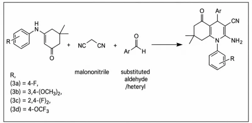
Scheme 1: Synthesis of Enaminone Intermediates 3a–3D. Reaction Conditions: Methanol, Catalytic Acetic Acid, Reflux for 6 H.
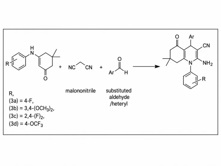
Figure 1: Synthesis of Substituted 2-Amino-7,7-Dimethyl-5-Oxo-1,4,5,6,7,8-Hexahydroquinoline-3-Carbonitrile Derivatives via a One-Pot Three-Component Reaction of Substituted Enaminone Intermediates (3a–3D), Malononitrile, and Aromatic or Heteroaromatic Aldehydes.
2.2.1 Synthesis of Hexahydroquinoline Derivatives (F1–F8)
The final hexahydroquinoline derivatives were synthesized by reacting the appropriate enaminone intermediate (3a–3d), an aromatic or heteroaromatic aldehyde, and malononitrile (0.003 mol each) in boiling methanol (35 mL). Triethylamine (2–3 drops) was added, and the mixture was refluxed for 6–8 h. Reaction progress was monitored by thin-layer chromatography (TLC). The precipitated products were filtered, washed with methanol, and recrystallized from 1,4-dioxane (Ahmed et al., 2017). The synthesized compounds comprised three series: F1–F3, containing a 4-fluorophenyl substituent; F4–F5, containing a 3,4-dimethoxyphenyl substituent; and F6–F8, containing a 2,4-difluorophenyl substituent at the N-1 position. The aromatic or heteroaromatic substituents at the C-4 position varied among the compounds.
Scheme 2: Synthesis of Substituted Hexahydroquinoline-3-Carbonitrile Derivatives F1–F8. Reaction Conditions: Methanol, Catalytic Triethylamine, Reflux for 6–8 H.
2.2.2 2.1.4. Chemical Structures of the Synthesized Compounds
The chemical structures and corresponding C-4 aryl or heteroaryl substituents (Ar) of the synthesized compounds are presented in Tables 1–3.
Table 1: Chemical Structures of the First Series (F1–F3).
| Core structure | Compound (F1) Ar |
Compound (F2) Ar |
Compound (F3) Ar |
|---|---|---|---|
Table 2: Chemical Structures of the Second Series (F4–F5).
| Core structure | Compound (F4) Ar |
Compound (F5) Ar |
|---|---|---|
Table 3: Chemical Structures of the Third Series (F6–F8).
| Core structure | Compound (F6) Ar |
Compound (F7) Ar |
Compound(F8) Ar |
|---|---|---|---|
2.3 Molecular Docking
Molecular docking was performed using MOE software (version 2009.10). The Mcl-1 crystal structure (PDB ID: 2PQK) was retrieved from the Protein Data Bank, and the BIM peptide, ions, and water molecules were removed, followed by hydrogen addition using the 3D-protonation tool. Ligands were drawn in ChemDraw (version 10), saved in MOL format, and prepared by energy minimization and partial charge assignment. Docking was performed using the Triangle Matcher placement method and London dG scoring function, with other parameters set to default. Docking scores, binding poses, and molecular interactions, including hydrogen-bond distances, were analyzed using MOE tools, and the final images were exported at 600 dpi resolution (Shaker et al., 2020).
2.4 In Silico ADMET study
The pharmacokinetic and toxicity profiles of all designed compounds were predicted using the online tools SwissADME and ADMETlab. Parameters evaluated included Lipinski’s and Veber’s rules, blood–brain barrier permeability, gastrointestinal absorption, topological polar surface area (TPSA), P-glycoprotein inhibition, CYP1A2 inhibition, and Ames mutagenicity. SwissADME tools, including BOILED-Egg, iLOGP, and the Bioavailability Radar, were used to assess drug-likeness and predicted absorption properties. The results were analyzed to identify compounds with favorable pharmacokinetic characteristics and potential toxicity profiles (Daina et al., 2017; Xiong et al., 2021) .
RESULTS AND DISCUSSION
3.1 Synthesis and Chemistry
The hexahydroquinoline-3-carbonitrile derivatives were synthesized via a two-step procedure, as illustrated in Schemes 1 and 2. In the first step, 5,5-dimethyl-1,3-cyclohexanedione (1) was refluxed with substituted anilines (2) in equimolar amounts in methanol, using catalytic glacial acetic acid for 6 h to obtain enaminone intermediates (3a–3d), whose synthesis and spectral data were previously reported (Ghozlan et al., 2016). In the second step, the enaminones were refluxed with malononitrile and substituted aryl/heteroaryl aldehydes in equimolar amounts in methanol, using catalytic triethylamine for 6–8 h. The reaction proceeds through Knoevenagel condensation between the aldehyde and malononitrile, followed by Michael addition and cyclization to yield the target hexahydroquinoline derivatives (Othman et al., 2019).
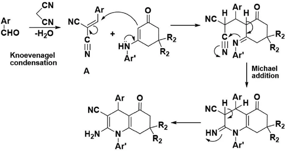
Figure 2: Mechanistic Scheme for the Synthesis of Hexahydroquinoline Derivatives.
3.2 General Characterization of Hexahydroquinoline Derivatives (F1–F8)
The synthesized hexahydroquinoline-3-carbonitrile derivatives (F1–F8) were characterized by melting point, molecular weight, percentage yield, and thin-layer chromatography (TLC). The molecular weights ranged from 233.03 to 578.39 Da, with yields of 54–90%. TLC was performed using ethyl acetate: chloroform (0.2:1.8, v/v), and the \(R_f\) values were calculated based on the distance travelled by each compound relative to the solvent front. The structures of the synthesized compounds were confirmed using FT-IR, ¹H NMR, ¹³C NMR, and mass spectrometry. The FT-IR spectra showed characteristic bands for NH₂ stretching (3219–3479 cm⁻¹), aromatic C–H stretching (3047–3188 cm⁻¹), aliphatic C–H stretching (2704–2972 cm⁻¹), nitrile (C≡N) stretching (2168–2226 cm⁻¹), and carbonyl (C=O) stretching (1635–1662 cm⁻¹). The ¹H NMR spectra exhibited signals corresponding to methyl, methylene, methine, amino, methoxy (where present), and aromatic protons. The ¹³C NMR spectra showed characteristic signals for aliphatic, aromatic, and carbonyl carbons, with the carbonyl signal at δ 194.15–195.55 ppm. Mass spectrometry further supported the proposed structures through molecular-ion, protonated molecular-ion, and isotope peaks, where applicable. The spectral characterisation data are provided in Table 4.
Table 4: Characterization of Hexahydroquinoline Derivatives (F1–F8)
| Compound | Spectral data (cm−1) |
|---|---|
| F1 | Pale yellow solid (76.24%, ), m.p.215–217 °C; Rf value 0.38, FORMULA: C27H28FN3O4 FTIR (KBr, max/cm-1): 3462, 3323 (NH2), 3123 (CH-Ar), 2947, 2883 (CH aliph.), 2177 (CN), 1651 (CO); 1H NMR (400MHz, DMSO) δ ( ppm): 0.82 (s, 3H, CH3), 0.93 (s,3H, CH3) 1.73 (d, 1H, J= 17.6 Hz, CH), 2.04 (d, 1H, J= 16 Hz, CH), 2.22 (d, 1H, J= 16.4 Hz, CH), 2.27 (d, 1H, J= 18.4 Hz, CH), 3.47 (s,3H, OCH3),3.76 (s, 6H, 2OCH3) 5.48 (s, 2H, NH2, D2O exchangeable), 6.45-6.49 (m, 2H, Ar-H), 7.39-7.44 (m, 4H, Ar-H). MS (ESI): [M+] 477.04 |
| F2 | Pale yellow solid (67.07%) m.p 205–207 °C, Rf value 0.41. FORMULA: C26H26FN3O3 FTIR (KBr, max/cm-1): 3423, 3360 (NH2), 3049 (CH-Ar), 2958, 2868 (CH aliph.), 2173 (CN), 1641 (CO), 1H NMR (400MHz, DMSO) δ ( ppm): 0.82 (s, 3H, CH3), 0.93 (s,3H, CH3) 1.73 (d, 1H, J= 17.6 Hz, CH), 2.04 (d, 1H, J= 16 Hz, CH), 2.22 (d, 1H, J= 16.4 Hz, CH), 2.27 (d, 1H, J= 18.4 Hz, CH), 3.47 (s,3H,OCH3),3.76 (s, 6H, 2OCH3) 5.48 (s, 2H, NH2, D2O exchangeable), 6.45-6.49 (m, 2H, Ar-H), 7.39-7.44 (m, 4H, Ar-H) 13C NMR (400MHz, DMSO) δ ( ppm): 26.17 (CH3), 29.35 (CH3), 30.26 (C(CH3)2), 31.80 (CH), 41.08 (CH2), 49.30 (CH2CO), 55.07 (OCH3), 56.36 (OCH3), 60.22 (C-CN), 110.59 (Ar-C), 111.35 (CN), 112.65, 113.78, 116.97, 117.19, 121.33, 135.55, 150.56, 151.11, 151.32, 153.22, 160.93, 163.38 (12C, Ar-C), 194.62 (CO) MS (ESI): [M+] 477.04 |
| F3 | White solid (72.18%), m.p.195–197 °C; Rf value 0.54 FORMULA: C24H20Cl2FN3O FTIR (KBr, max/cm-1): 3462, 3327 (NH2), 3074 (CH-Ar), 2956, 2868 (CH aliph.), 2281 (CN), 1651 (CO). 1H NMR (400MHz, DMSO) δ ( ppm): 0.80 ( s, 3H, CH3), 0.89 (s, 3H, CH3), 1.76 ( d, 1H, J = 17.6 Hz, CH), 1.96 (d, 1H, J = 16 Hz, CH), 2.14-2.19 (m, 2H, CH2), 4.97 (s, 1H, CH), 5.46 (s, 2H, NH2, D2O exchangeable), 7.39-7.43 (m, 4H, Ar-H), 7.49- 7.52 (m, 3H, Ar-H) MS (ESI): [M+] 455.81 |
| F4 | White solid (62.47%), m.p. 189–191 °C; Rf value 0.59 FORMULA: FTIR (KBr, max/cm-1): 3464, 3367 (NH2), 3068 (CH-Ar), 2956, 2820 (CH aliph.), 2171 (CN), 1635 (CO), 1519, 1346 (NO2) 1H NMR (400MHz, DMSO) δ ( ppm): 0.75 (s, 3H, CH3), 0.90 ( s, 3H, CH3), 1.95 (d, 1H, J = 16.8 Hz, CH), 1.85 (d, 1H, J = 17.6 Hz, CH), 2.18 (d, 1H, J = 16 Hz, CH), 2.30 (d, 1H, J = 16.4 Hz, CH), 3.84 (s, 6H, OCH3), 4.60 (s, 1H, CH), 5.59 (s 2H, NH2, D2O exchangeable), 6.96 (s, 2H, Ar-H), 7.07 (d, 1H, J = 8.8 Hz, Ar-H), 7.55 (s, 2H, Ar-H), 8.21 (dd, 2H, J = 8 Hz, Ar-H) MS (ESI): [M+] 474.35 |
| F5 | White solid (67.53%), m.p. 203–205 °C; Rf value 0.53 FORMULA: C26H26N4O5 FTIR (KBr, max/cm-1): 3471, 3373 (NH2), 3064 (CH-Ar), 2960, 2850 (CH aliph.), 2171 (CN), 1633 (CO). 1H NMR (400MHz, DMSO) δ ( ppm): 0.75 (s, 3H, CH3), 0.89 ( s, 3H, CH3), 1.82 (d, 1H, J = 17.6 Hz, CH), 1.98 (d, 1H, J = 16 Hz, CH), 2.14-2.23 (m, 2H, CH2),3.81-3.83 (s, 6H, 3OCH3), 4.47 (s, 1H, CH), 5.39 (s, 2H, NH2), 6.87-7.01 (m, 2H, Ar- H), 7.03-7.15 (m, 3H, Ar-H), 7.29 (s, 2H, Ar-H). 13C NMR (400MHz, DMSO) δ ( ppm):26.21 (CH3), 29.10 (CH3), 31.83 (C(CH3)2), 35.81 (CH), 40.64 (CH2), 49.30 (CH2CO), 55.56 (OCH3), 55.73 (OCH3), 62.01 (C-CN), 111.86 (CN), 112.99, 152.90, 107.54, 117.24, 129.85, 135.24, 135.87, 142.16, 152.78, 159.37, 163.43 (11C, Ar-C), 194.71 (CO). MS (ESI): [M+] 447.70 |
| F6 | White solid (69.52%), m.p. 265–267 °C; Rf value 0.67 FORMULA: C24H20F3N3O FTIR (KBr, max/cm-1): 3466, 3336 (NH2), 3061 (CH-Ar), 2968, 2889 (CH aliph.), 2181 (CN), 1653 (CO) 1H NMR (400MHz, DMSO) δ ( ppm): 0.75 (s, 3H, CH3), 0.90 (s, 3H, CH3), 1.66 (d, 1H, J = 17.2 Hz, CH), 1.86 (d, 1H, J = 17.2 Hz, CH), 2.19 (d, 1H, J = 15.6 Hz, CH), 2.27 (d, 1H, J= 18 Hz, CH), 4.45 (s, 1H, CH), 5.67 (s, 2H, NH2, D2O exchangeable), 7.12 (t, 2H, J = 8.8 Hz, Ar-H), 7.26-7.31 (m, 3H, Ar- H), 7.54-7.59 (m, 1H, Ar-H),7.64-7.70 (m, 1H, Ar-H). 13C NMR (400MHz, DMSO) δ (ppm):25.86 (CH3), 29.19 (CH3), 31.88 (C(CH3)2), 38.83 (CH), 40.51 (CH2), 49.15 (CH2 CO), 60.58 (C-CN), 112.90 (CN),105.33, 112.03, 112.69, 114.97, 115.05, 128.56, 133.24, 142.66, 149.77, 150.87, 151.01, 159.59, 161.99 (13C, Ar-C), 194.85 (CO) MS (ESI): [M+] 423.80 |
| F7 | White solid (60.20%), m.p. 263-264 °C; Rf value 0.58 FORMULA: C27H27F2N3O4 FTIR (KBr, max/cm-1): 3421, 3335 (NH2), 3074 (CH-Ar), 2958, 2829 (CH aliph.), 2179 (CN), 1653 (CO). 1H NMR (400MHz, DMSO) δ ( ppm): 0.82 (s, 3H, CH3), 0.93 (s,3H, CH3) 1.73 (d, 1H, J= 17.6 Hz, CH), 2.04 (d, 1H, J= 16 Hz, CH), 2.22 (d, 1H, J= 16.4 Hz, CH), 2.27 (d, 1H, J= 18.4 Hz, CH), 3.47 (s,3H,OCH3),3.76 (s, 6H, 2OCH3) 5.48 (s, 2H, NH2, D2O exchangeable), 6.45-6.49 (m, 2H, Ar-H), 7.39-7.44 (m, 4H, Ar-H). 13C NMR (400MHz, DMSO) δ (ppm): 25.71 (CH3), 29.44 (CH3), 32.87 (C(CH3)2), 36.85 (CH), 40.65 (CH2), 49.19 (CH2CO), 55.45 (OCH3), 55.64 (OCH3), 59.89 (C-CN), 60.13 (OCH3), 112.77 (CN), 105.64, 106.43, 111.62, 112.96, 121.30, 133.85, 133.96, 136.2, 150.57, 150.93, 152.69, 152.78, 161.50 (13C, Ar-C), 194.96 (CO) MS (ESI): [M+] 495.20, 496.8 |
| F8 | White solid (67.50%), m.p. 245-246 °C; Rf value 0.49 FORMULA: C25H23F2N3O2 FTIR (KBr, max/cm-1): 3439, 3348 (NH2), 3188 (CH-Ar), 2821, 2704 (CH aliph.), 2212 (CN), 1639 (CO) 1H NMR (400MHz, DMSO) δ ( ppm): 0.84 (s, 3H, CH3), 0.91 (s, 3H, CH3), 1.64 (d, 1H, J = 17.2 Hz, CH), 2.04-2.08 (m,1H, CH), 2.18 (d, 1H, J = 16 Hz, CH), 2.44 (d, 1H, J = 16 Hz, CH), 3.39 (s, 3H, OCH3), 4.83 (s, 1H, CH), 5.68 (s, 2H, NH2), 7.05 -7.27 (m, 3H, Ar-H),7.30-7.43 (m, 2H, Ar-H), 7.61-7.78 (m, 2H, Ar-H). 13C NMR (400MHz, DMSO) δ (ppm): 26.83(CH3), 29.31(CH3), 32.13 (C(CH3)2, 36.02 (CH), 41.03 (CH2), 49.80 (CH2CO), 61.17 (OCH3), 62 (C-CN), 113.25 (CN), 104.42, 112.99, 114.82, 115.80, 118.06, 122.07, 134.59, 138.12, 144.25, 145.14, 145.40, 149.50, 149.59, 150.97, 151.32 (15C, Ar-C), 195.37 (CO)MS (ESI): [M+] 434.10 |
3.3 Molecular Docking
Molecular docking was performed to evaluate the binding interactions of the synthesized hexahydroquinoline-3-carbonitrile derivatives with the anti-apoptotic Mcl-1 protein (Shaker et al., 2020) . The docking scores ranged from −14.29 to −4.67 kcal/mol for the first series, −13.35 to −14.67 kcal/mol for the second series, and −13.79 to −16.73 kcal/mol for the third series, compared with −16.67 kcal/mol for gambogic acid. Among the evaluated compounds, F8 (−16.73 kcal/mol) exhibited the most favorable docking score, followed by F7 (−15.09 kcal/mol). Interaction analysis revealed hydrophobic and arene–cation interactions within the Mcl-1 binding pocket, while gambogic acid formed two hydrogen bonds with Arg263 at distances of 1.7 and 2.0 Å. The favorable docking scores and binding interactions, particularly for F7 and F8, suggest potential Mcl-1-targeting activity that requires experimental validation. The docking scores are presented in Table 5, and the binding poses are illustrated in Figure 3.
Table 5: Docking Scores of Compounds.
| a) | b) |
| c) | d) |
Figure 3: Docking Pose of Active Compounds (F6, F7, and F8) in the Active Site of Mcl-1: A) F6; B) F7; C) F8, and (D) Gambogic Acid. Hydrogen-bond and Arene-Cation Interactions Are Illustrated as Dotted Lines in Pink and Green, Respectively.
| Compounds | Docking Score (kcal/mol) |
|---|---|
| F1 | -14.29 |
| F2 | -13.78 |
| F3 | -14.67 |
| F4 | -13.34 |
| F5 | -14.37 |
| F6 | -13.79 |
| F7 | -15.09 |
| F8 | -16.73 |
| Gambogic acid | -16.67 |
3.4 In Silico ADMET Studies
The designed compounds were evaluated for their predicted ADMET properties using SwissADME and ADMETlab, and the results are presented in Table 6. All compounds satisfied Lipinski’s rule of five and Veber’s criteria, with TPSA values ranging from 67.34 to 134.40 Ų, indicating favorable preliminary drug-likeness characteristics. The compounds also exhibited predicted high gastrointestinal absorption, P-glycoprotein inhibition, no CYP1A2 inhibition, negative Ames mutagenicity, and no predicted blood–brain barrier penetration. These findings suggest potentially favorable pharmacokinetic profiles; however, the predicted P-glycoprotein inhibition and safety characteristics require experimental validation. Comparison with gambogic acid and available database information provided an additional reference for interpreting the computational results. Overall, the ADMET findings support further investigation of the designed compounds as potential anticancer candidates.
Table 6: In Silico ADMET Study
| Code | Lipinski rule | Veber | P-gp Inhibition | CYP1A2 Inhibitors |
Ames Mutagenicity | Skin Sensitivity | GI Absorption |
BBB | TPSA (Å2) |
|---|---|---|---|---|---|---|---|---|---|
| F1 | Yes | Yes | + | No | No | No | High | No | 97.81 |
| F2 | Yes | Yes | + | No | No | No | High | No | 80.43 |
| F3 | Yes | Yes | + | No | No | No | High | No | 70.12 |
| F4 | Yes | Yes | + | No | No | No | High | No | 134.40 |
| F5 | Yes | Yes | + | No | No | No | High | No | 88.58 |
| F6 | Yes | Yes | + | No | No | No | High | No | 70.12 |
| F7 | Yes | Yes | + | No | No | No | High | No | 97.81 |
| F8 | Yes | Yes | + | No | No | No | High | No | 115.94 |
| Std | Yes | Yes | + | No | No | Yes | Low | No | 119.36 |
“–’’ negative effects; “+’’positive effects, std- gambogic acid (Pubchem CID- 3451, Zinc 15 Id-3933041)
SUMMARY AND CONCLUSION
The present study focused on the design, synthesis, characterization, and computational evaluation of substituted hexahydroquinoline-3-carbonitrile derivatives as potential Mcl-1-targeting anticancer agents. Fluoro, difluoro, trifluoromethoxy, and methoxy substituents were incorporated to explore their influence on molecular interactions and drug-like properties. The study combined molecular docking using MOE software (PDB ID: 2PQK) with SwissADME and ADMETlab analyses to assess predicted binding interactions, pharmacokinetic properties, and toxicity profiles. The target compounds were synthesized through a two-step procedure involving enaminone formation followed by cyclization with malononitrile and aromatic or heteroaromatic aldehydes. The structures were confirmed using FT-IR, ¹H NMR, ¹³C NMR, and mass spectrometry. Among the synthesized derivatives, F8 exhibited the most favorable docking score (−16.73 kcal/mol), followed by F7 (−15.09 kcal/mol), compared with gambogic acid (−16.67 kcal/mol). The ADMET predictions indicated generally favorable drug-likeness characteristics. These findings identify F7 and F8 as promising candidates for further experimental evaluation of Mcl-1 inhibition, cytotoxicity, and apoptosis-inducing activity.
CONCLUSION
The present study successfully synthesized and characterized substituted hexahydroquinoline-3-carbonitrile derivatives using a two-step synthetic approach. Molecular docking identified F8 (−16.73 kcal/mol) and F7 (−15.09 kcal/mol) as promising candidates for interaction with Mcl-1, while in-silico ADMET analysis indicated generally favorable predicted drug-like properties. These findings support further investigation of fluorinated hexahydroquinoline derivatives as potential anticancer agents; however, experimental validation is essential to establish their biological efficacy and safety.
Future studies should prioritize F7 and F8 for direct Mcl-1 inhibition assays, cancer-cell cytotoxicity, normal-cell selectivity, and apoptosis evaluation. Further structural optimization and experimental pharmacokinetic, toxicity, and in-vivo studies are required to validate their potential for anticancer drug development.
REFERENCES

Dirsipam Param Geetha, Dr. N. Srinivasa Rao, Nitrogen-Based Fused Heterocyclic Compounds: Synthesis, Characterization, and Computational Study Profiling, Int. J. of Pharm. Sci., 2026, Vol 4, Issue 10, 5057-5065. https://doi.org/10.5281/zenodo.23831330
 10.5281/zenodo.23831330