

We use cookies to ensure our website works properly and to personalise your experience. Cookies policy
Department of Pharmacy, Lokmanya Tilak Institute of Pharmacy, Kharghar.
Hyperpigmentation is a common skin disorder in which melanin is overproduced and accumulated leading to development of dark spots on the skin. It can be caused due to various reasons like exposure to uv rays, changes in hormonal levels-inflammatory conditions, genetic factors and skin damage. Various topical drugs like hydroquinone, kojic acid, arbutin, azelaic acid, glycolic acid, niacinamide and vitamin-C have been widely employed in the treatment of hyperpigmentation. However, they suffer from several disadvantages including low solubility, instability, low permeability and causes adverse effects like irritation and allergy. Various advancements have been made in the field of topical drug delivery systems that focus on providing stability, controlled release and permeability of the active drug. Emulgel formulation is gaining popularity as a topical formulation as it combines the advantages of both emulsions and gels, thus improving drug loading, easy spreading properties, stability and patient compliance. Advanced drug delivery systems like liposomes, nanoemulsions, niosomes, solid lipid nanoparticles and nanostructured lipid carriers have shown potential to overcome the shortcomings of conventional drug delivery systems. The present review describes the mechanism of melanogenesis, widely used depigmenting agents and its various drawbacks. Various formulations, permeation enhancers and recent advancement in topical drug delivery have been discussed in this review. Limitations and future perspectives for stable, effective and patient friendly topical formulations for the treatment of hyperpigmentation.
The pigmentation of skin is mainly controlled by melanin which is a natural pigment produced by melanocytes present in the Basal Layer of the Epidermis. The two main types of melanin found are eumelanin and pheomelanin. Eumelanin produces browns and blacks in the skin whilst pheomelanin produce yellows and reds. Melanin plays a key role in protecting the skin from UV rays however if there is a change in melanin production, distribution or deposition this can cause pigmentation disorders such as Hyperpigmentation[1]. Hyperpigmentation is a common skin disorder in which patches of skin become darker than normal. This most commonly occurs due to increased melanin production in the epidermis and/or dermis. However, in some cases pigmentation can occur from deposition of other pigments in the skin, such as hemosiderin, iron and some heavy metals. Pigmentation can be affected by several factors such as carotenoids, blood vessels and skin thickness. Pigments can appear brown/black/gray or even red/pink depending on the cause [2] . Hyperpigmentation occurs due to a variety of different factors. Excessive sun exposure, fluctuating hormones, inflammation, skin damage and some medications can all cause hyperpigmentation. Common types of hyperpigmentation include melasma, solar lentigines and post-inflammatory hyperpigmentation (PIH). This can occur in any skin type however is very common particularly in those with darker skin phototypes. Individuals with darker skin phototypes are often more likely to suffer from PIH following inflammatory skin conditions. UV can aggravate melanogenesis and therefore worsen pigmentation, so is important to protect against this [3]. Hyperpigmentation can cause people great psychological distress due to the visible nature of this condition, particularly if it occurs on the face. This can lead to low selfesteem, feelings of sadness and poor quality of life. There are many ways to treat hyperpigmentation however due to the variety of causes, it can be difficult to manage. Hyperpigmentation is often recurrent, and can cause irritation from treatments. Darker skin types can be especially problematic. Treatments include sun protection, topical depigmenting agents, chemical peels and some treatments. Treatment will depend on the cause of hyperpigmentation and skin type [4]. Emulgel is an emulsion that has been gelled. They can be either an o/w or w/o emulsion. Emulgel is a stable and superior delivery system for poorly water soluble drugs. An emulgel can be described as an emulsion in a gel. Gels have many benefits however a major con is delivering hydrophobic drugs. Due to this, an emulsion is being used to overcome this issue. Even hydrophobic drugs can benefit from the properties of a gel. Emulgels can deliver both hydrophilic and lipophilic drugs as they have both aqueous and non-aqueous phases. In recent years they have been used as a controlled release formulation. Emulgels are biphasic with higher drug loading and stability. Emulgels have many positive properties, they are easily spreadable, non-greasy, thixotropic, long shelf-life, odourless and aesthetically pleasing compared to traditional topical formulations. Emulgels have properties of both a gel and an emulsion. Therefore, they act as a dual controlled release system[5].
1.1 .FACTORS AFFECTING HYPERPIGMENTATION:
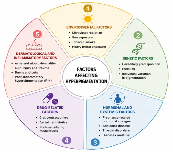
Figure 1: Factors Affecting Hyperpigmentation
1.2.TYPES OF HYPERPIGMENTATION
Table 1: Types of Hyperpigmentation
| SR NO. |
TYPE | CHARACTERISTICS | COMMON CAUSES OR ASSOCIATED FACTORS |
|---|---|---|---|
| 1) | Freckles (Ephelides) | Small tan or brown spots on sun-exposed skin; may fade in winter | Genetics and sun exposure. |
| 2) | Solar lentigines (Age spots | Tan to dark brown spots on the face, hands, forearms, and chest. | Chronic sun exposure and sunburn |
| 3) | Melasma | Irregular, dark patches, usually on the face | Hormonal changes and sun exposure. |
| 4) | Maturational hyperpigmentation | Gradual darkening, particularly on the sides of the face | Chronic sun exposure; sometimes associated with metabolic disorders |
| 5) | Periorbital hyperpigmentation | Darkening of the skin around the eyes (dark circles). | Genetic, vascular, structural, and pigmentary factors |
| 6) | Acanthosis nigricans | Dark, thickened, velvety skin in body folds, such as the neck, armpits, and groin | Insulin resistance and type 2 diabetes. |
| 7) | Post-inflammatory hyperpigmentation (PIH | Dark spots or patches that remain after skin inflammation or injury | Acne, burns, wounds, and skin rashes. |
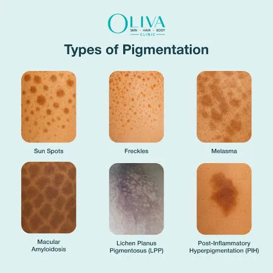
Figure 1.2: Types of Hyperpigmentation
2. SKIN PHYSIOLOGY :
The skin is the primary organ for administration of topical drug delivery systems (TDDS). Drugs can penetrate the skin through three routes, namely stratum corneum, sweat ducts and sebaceous hair follicles. The adult human skin has a surface area of about 2 m² and receives about one third of total blood volume within the body. In human skin there are approx. 40-70 hair follicles and 200-300 sweat ducts per cm². The pH on the surface of the skin is acidic, normally between 4.0 and 5.6. This pH helps to maintain the skin barrier functionality.
2.1.Skin Layers:
Skin can be divided into the following layers.
1. Epidermis
2. Dermis
3. Subcutaneous layer
2.1.1.EPIDERMIS LAYER: This is the top layer of skin. The epidermis has a thickness of approx. 150 µm.
2.1.2. DERMIS LAYER : This layer is a connective tissue made up of mainly proteins. It is made up of collagen, reticular fibres and elastic fibres. The dermis is approx. 2000-3000 µm in thickness and provides a structural connective tissue matrix. It provides structural support to the epidermis and also connects the skin to muscles and bones.
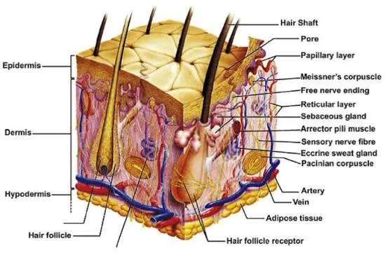
Figure 2: Skin Layers
2.1.3.SUBCUTANEOUS LAYER: This layer is also known as the hypodermis. It is made up of areolar connective tissue rich in fat. This layer is also known as superficial fascia and connects the dermis to the tissues beneath. This layer contains adipose tissue and larger blood vessels such as arteries and veins[6]
2.2.SKIN BARRIER
Table 2: = The Stratum Corneum and Simultaneous Multitasking: Individual Ep Epidermal Barrrier Functions[7].
| BARRIER FUNCTION | MAJOR COMPONENTS | |
|---|---|---|
| Permeability Barrier | Formation of stratum corneum lipids in specific ratio from precursor lipids | |
| Formation of natural moisturizing factor from filaggrin (converted from profillagrin) | ||
| Maintenance of water gradient, calcium gradient, acid mantle (acidic pH) | ||
| Response of primary pro-inflammatory cytokines to impairment of permeability barrier | ||
| Antimicrobial Barrier | Maintenance of an acidic skin pH decreases skin colonization by pathogenic bacteria | |
| Antibacterial activity of stratum corneum lipids (eg, free fatty acids, sphingosine, other) | ||
| Antioxidant Barrier | Network of enzymatic and non-enzymatic antioxidant systems to counter oxidative stress | |
| Antioxidants present in epidermis (stratum corneum, skin surface lipids) and dermis | ||
| Gradients in stratum corneum for ascorbic acid and tocopherol (lowest near surface) | ||
| Immune Response Barrier | Dendritic cells involved in immune surveillance and antigen recognition (eg, plasmacytoid dendritic cells, myeloid dendritic cells, Langerhans cells) | |
| Toll-like receptors involved in recognition of microbial pathogens and other agonists | ||
| Innate and acquired immune response pathways and balance with T regulatory cell system |
3. CLASSIFICATION OF TOPICAL DOSAGE FORMS
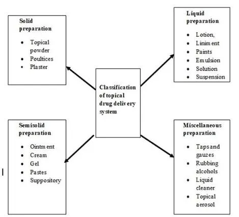
Figure 3: Classification of Topical Dosage Forms
Top of Form
Bottom of Form
Top of Form
Bottom of Form
4. CLASSES OF EMULGEL
1) Microemulsion :- These are thermodynamically stable, optically clear, isotropic mixtures of two phase O/W system with surfactants. The droplet sizes range between 10-100nm which prevents coalescence. These consist of a fixed amount of oil, co-surfactant, surfactant and water.
2) Nanoemulgel :- These are optically clear (transparent) dispersions of oil in water and are thermodynamically stable due to the presence of surfactant and cosurfactant molecules. The globule sizes range from 1-100nm.
3) Macroemulsion gel :- An emulgel with emulsion droplet particle sizes larger than 400nm. These cannot be seen with the naked eye but can be viewed under the microscope. They are not thermodynamically stable although can be stabilised by surface active agents.
4.1.Advantages of emulgel:
4.2. Disadvantages of emulgel :
5. MELANOGENESIS:
Melanin is formed from amino acids by melanocytes via a series of enzymatic reactions. Tyrosinase is used as the substrate. Melanin has a close relationship to skin and hair colour in humans. Knowing the mechanisms and proteins involved in melanin formation is important. The type, amount and distribution of melanin are the main factors that affect human skin colour. Melanin is present in high amounts in the skin, hair, mucous membranes and other tissues. Melanin is formed from melanocytes (MC) [8].
These are derived from NCCs (Neural crest cells) and are mainly located in the basal layer of mammalian epidermis and hair follicles. Melanin is formed in melanosomes. Three enzymes; TYR, TYRP-1 and TYRP-2 play key roles in melanin formation. Melanogenesis begins with the formation of dopaquinone, an o-quinone. Pulse radiolysis is a powerful technique to study the fate of these reactive melanin precursors
There are three stages of melanogenesis:
1) Cysteinyldopas are formed by fast addition of cysteine to dopaquinone. This will continue until there is no more cysteine (1μM).
2) Cysteinyldopas are oxidized to produce pheomelanin. This will continue until there are no more cysteinyldopas (10μM).
3) Eumelanin is produced. This doesn’t begin until most of the cysteinyldopas have been used up.
This suggests that eumelanin is added onto the pheomelanin.
The ratio of eu to pheomelanin depends on the tyrosinase activity and concentration of cysteine. During eumelanogenesis the dopachrome is a relatively stable species. It decays spontaneously to produce mainly 5,6-dihydroxyindole. Dopachrome tautomerase (Dct) catalyses the tautomerisation of dopachrome to mainly produce 5,6-dihydroxyindole-2-carboxylic acid (DHICA). We have shown that Dct increases the DHICA ratio in eumelanin which increases the amount of eumelanin produced. The cytotoxicity of o-quinone melanin precursors was found to be related to protein binding through cysteine residues. The regulation of cysteine availability within the melanosome is unknown[9].
Top of Form
Bottom of Form
Top of Form
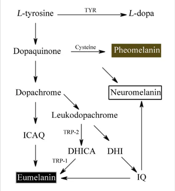
Figure 5: Melanogenesis
5.1.FACTORS INFLUENCING MELANOGENESIS:
Melanin production is affected by genetic, environmental, and hormonal factors,as well as inflammatory and biochemical ones. Through specific signaling pathways, these factors control melanocyte activity and the synthesis of melanin[10].
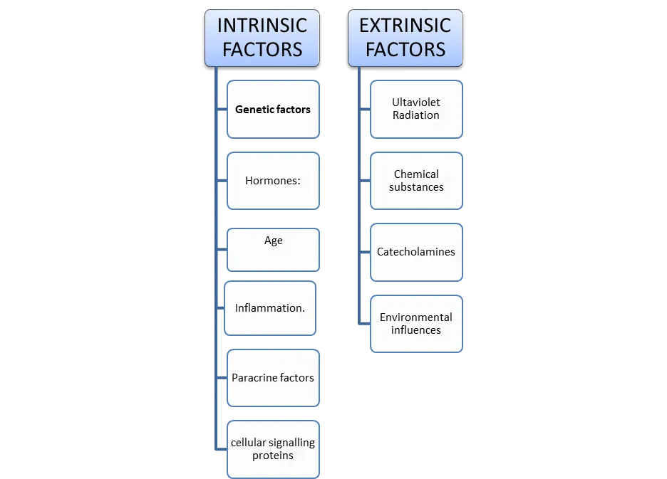
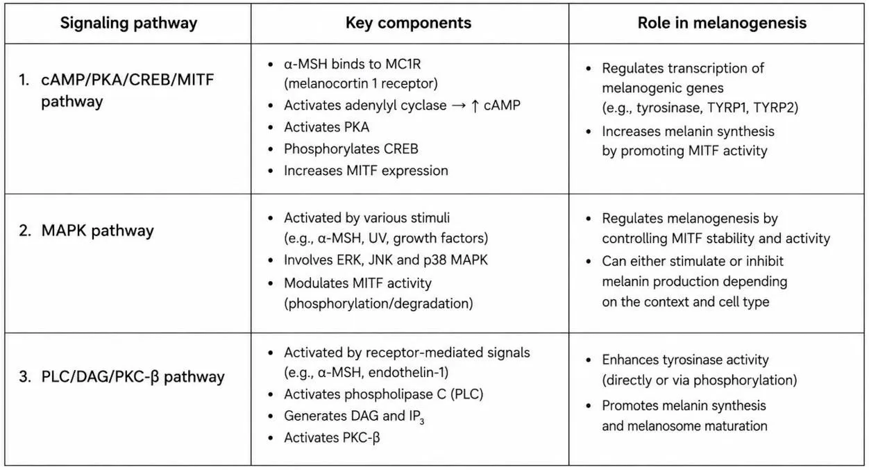
Figure 5.1: Major Signaling Pathway
6.WIDELY USED TECHNIQUES FOR EMULGEL FORMULATION:
6.1. Emulsion incorporation technique:
The oil phase and the aqueous phase are prepared separately, heated when needed, and then blended with continuous stirring to create an emulsion. This prepared emulsion is subsequently incorporated into a gel base that has been prepared separately[11]
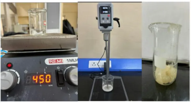
Figure 6.1: Emulsion Incorporation Technique
6.2. Method of high-shear homogenization
The oil and aqueous phases are combined with a homogenizer running at high speed, which reduces droplet size and enhances the uniformity of the emulsion. The emulsion obtained is then blended with the gel base[12].
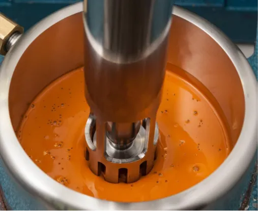
Figure 6.2: High Shear Homogenization Method
6.3. Method using ultrasonication:
Ultrasound energy is used to shrink the size of dispersed oil droplets and enhance how they are distributed. It can be applied following the initial emulsification, with the temperature kept under control so that heat-sensitive ingredients do not degrade[13]
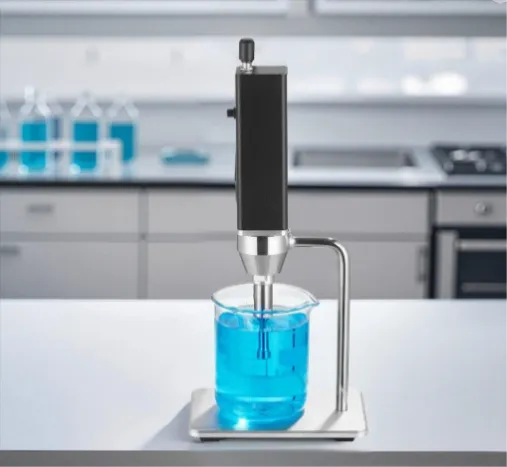
Figure 6.3: Ultrasonification Method
6.4. Method of magnetic stirring:
The phases are combined with a magnetic stirrer, frequently under controlled heating. This method works well for small-scale laboratory preparation, especially when the ingredients do not need intensive homogenization.[14]
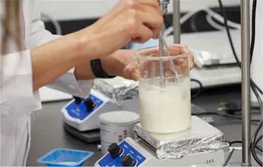
Figure 6.4: Magnetic Stirring Method
6.5. Method of mechanical stirring
An overhead stirrer or mechanical mixer is employed to prepare the emulsion and to blend it with the gel. It is well suited to viscous formulations and to scaling up laboratory procedures.[15]
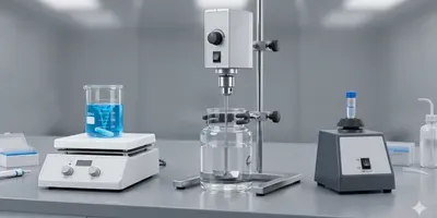
Figure 6.5: Mechanical Stirring Method
Method dependent on preparation:
Other methods include; Carbopol gel method: Carbopol 934 or 940 is dispersed within water and allowed to swell. An appropriate neutralising agent (such as triethanolamine) is added and mixed until neutral.
The emulsion is then added gradually into the gel base. Hydrogel polymer method: Polymers such as HPMC, sodium CMC, or xanthan gum are swelled or dispersed to make a gel. The emulsion can then be added.
In situ gelation: A formulation must be created to make or strengthen a gel when applied under the correct conditions. This is a specialist method and is not normally required for conventional emulgels. Design of Experiments: Design of Experiments (DoE), factorial design or response surface methodology may be used to optimise oil, surfactant and gelling agent concentrations, and mixing speed.[16]
7. EMULGEL PREPARATION:
1. Emulsions formulation (O/W or W/O): The first part involves dissolution of the water soluble ingredients (tween 80 in sterile water) and the oil soluble ingredients (span 20 in liquid paraffin). In order to ensure dispersion of both phases into small droplets the phases are brought together under turbulent conditions. In a laboratory setting, mechanical stirrers are used to prepare emulsions. However, in an industrial setting, ultrasonicators, homogenisers, colloid mills and mechanical stirrers are commonly used.
2. Gel base formulation: The water soluble ingredients (excipients) are dissolved using mechanical stirring in a mixing vessel. The hydrophilic polymer is then added slowly to the mixture to prevent aggregation. The stirring continues until the polymer has fully dissolved in the correct pH range. Over-stirring can cause entrapped air in pharmaceutical gels so a moderate mixing speed should be used.
3. Addition of Emulsion to gel base with consistent mixing: The emulsion and gel are mixed together in a 1:1 ratio to form the emulgel [17].
8. CONVENTIONAL TREATMENT:
Topical treatments are often prescribed to treat localised hyperpigmentation. Drugs used to treat hyperpigmentation include;
Hydroquinone: The gold standard in the treatment of hyperpigmentation, this drug has been used topically since the 1960s. This drug works by inhibiting tyrosinase.
Arbutin: This is a derivative of hydroquinone with much less melanotoxic activity. Arbutin causes hypopigmentation due to its inhibitory effect on tyrosinase and melanosome maturation. It has a dose dependent effect on tyrosinase, but if higher concentrations are used caution may need to be taken as paradoxical hyperpigmentation may occur.
Glycolic acid: This is a white crystalline alpha hydroxy acid derived from sugarcane. Depending on the concentration used this will cause different effects. At low concentrations it will cause desquametion of keratinocytes while at high concentrations it will cause epidermolysis.
Kojic acid: This is commonly used to treat hyperpigmentation disorders due to its many mechanisms of action. Kojic acid works mainly by inhibiting the catecholase of tyrosinase
Retinoids: These are derivatives of Vitamin A/Retinol. This work by several mechanisms leading to hypopigmentation. Retinoids will inhibit melanogenesis by MSH or L-tyrosine but have no effect on melanocyte growth and morphology, tyrosinase and dopachrome tautomerize.
Tretinoin: A first generation retinoid which is a natural derivative of retinol. It has been proposed to be effective for photoageing induced hyperpigmentation.
Azelaic acid: Inhibits tyrosinase and has a direct inhibitory effect on melanogenesis pathway. Has no effect on normal melanocytes and will not cause ochronosis after long term use.
Niacinamide: An active form of vitamin B3. This inhibits transfer of melanosomes to surrounding keratinocytes and also interrupts the signalling pathway between melanocytes and keratinocytes.
Triple combination: Hydroquinone is the most commonly used agent to combine with several other agents including kojic acid, glycolic acid, azelaic acid or corticosteroids. This has been found to be more effective in treatment than hydroquinone alone. (Ferreira Cestari et al, 2007). Triple combination of 5% hydroquinone with 0.1% tretinoin and 0.1% dexamethasone has been proposed to be effective for PIH, melasma and ephelides.
Vitamin C: Ascorbic acid (vitamin C) is known to inhibit melanin formation by decreasing o-quinone. The antioxidant property prevents free radical formation which causes melanogenesis,22 and also decreases oxidative melanin so pigmentation becomes tan instead of black[18].
Chemical peels: Chemical peels or microdermabrasion can also be used as an adjuvant treatment. These work best when used in a course of treatment. Usually 3-5 sessions would be required every month[18,19]
Combination therapy: New combination therapies have been introduced which have been proven to be as effective as hydroquinone. A combination of glycolic acid, antioxidants and licorice root was found to be as effective as 4% hydroquinone in an 8 week blind split face trial.31 Another combination of kojic acid, alpha hydroxyl acids and emblica were as effective as 4% hydroquinone after 12 weeks. Combination therapy may be a new way of treating hyperpigmentation with significant results[20,21].
8.1. LIMITATIONS:-
Table 8.1: Topical Drug and Their Limitation
| SR. NO. | TOPICAL DRUG | LIMITATION |
|---|---|---|
| Hydroquione | May cause skin irritation and other adverse effects. Prolonged use can be associated with exogenous ochronosis | |
| Arbutin | Higher concentrations may cause paradoxical hyperpigmentation; concentration-dependent effects require careful monitoring | |
| Glycolic acid | Its effects depend on concentration. Higher concentrations may cause excessive epidermal exfoliation or epidermolysis, increasing the risk of irritation | |
| Kojic acid | May cause contact dermatitis, limiting its tolerability in some individuals. | |
| Niacinamide | Does not directly inhibit tyrosinase or melanin synthesis; its main effects involve reducing melanosome transfer, which may limit its usefulness when used alone for some types of hyperpigmentation. | |
| Hydroquinone-based combinations | May cause irritation and other adverse effects depending on the ingredients and their concentrations. Combining multiple agents may increase the risk of irritation | |
| Triple-combination therapy | The combination containing higher tretinoin concentrations may cause skin irritation. Prolonged or inappropriate corticosteroid use can also cause adverse skin effects. | |
| Vitamin C | Lower efficacy than hydroquinone and a delayed onset of visible skin-lightening effects. |
9. PENETRATION ENHANCERS:
Excipients often contain penetration enhancers to improve drug absorption. These may work by temporarily disrupting the skin barrier, dissolving the lipid pathways between corneocytes, improving the distribution of drug within skin structures or improving delivery to the skin.
9.1.Characteristics of Penetration Enhancers
9.2. Mechanism of Penetration Enhancement:
Penetration enhancers can work by one or more of the following mechanisms: Disrupt the highly organised lipid structure of the SC. Interact with extracellular proteins. Increase the penetration of drug/activator/solvent into stratum corneum[22].
9.3.Reasons why penetration enhancement is required:
10. ADVANCED FORMULATION APPROACHES:
Advanced topical drug delivery systems have been considered as a promising strategy to overcome these drawbacks. These drug delivery systems can improve drug solubility, protect bioactive compounds from degradation, control drug delivery and improve drug penetration into the epidermis, which is the site where melanocytes and melanogenesis process mainly occur. Liposomes, niosomes, nanoemulsions, solid lipid nanoparticles (SLNs) and nanostructured lipid carriers (NLCs) have attracted much attention for formulation of anti-hyperpigmenting agents.
10.1 Liposomal Formulations:
Liposomes are spherical vesicles comprised of a single or multiple phospholipid bilayer surrounding an aqueous core. Due to their amphiphilic nature, liposomes are able to form complexes with both hydrophilic compounds (in the aqueous phase) and lipophilic compounds (in the bilayer). This makes liposomes an ideal carrier for depigmenting agents to be delivered topically.
Liposomal encapsulation can improve the stability of sensitive compounds, increase affinity to the skin barrier and change the release profile of the compound. Certain lipophilic compounds including hydroquinone, kojic acid derivatives, and some antioxidants/pigment modulators have been investigated using lipid based delivery systems. The extent of improvement will depend on the active ingredient used, the composition of the liposome, encapsulation efficiency and formulation characteristics.
Method of preparation: Thin-film hydration method
Thin Film Hydration is a commonly used lab technique to produce liposomes:
1. Prepare the lipid phase by dissolving phospholipids and cholesterol if necessary in a suitable volatile organic solvent.
2. A thin film is formed by removing solvent using a rotary evaporator.
3. The lipid film is hydrated using an aqueous solution containing the hydrophilic active ingredient. Alternatively another suitable solvent could be used. This should be compatible with the lipid.
4. The dispersion may be further processed using sonication, extrusion or homogenisation to obtain the desired size distribution of vesicles.
5. Characterise the formulation to determine particle size, polydispersity index, zeta potential, encapsulation efficiency, drug content, release profile and physical stability. Optimisation of conditions during preparation is required, depending on the specific active ingredient. If the drug is sensitive then care needs to be taken during preparation to avoid damage. Any leftover solvent and suitability of the final topical vehicle should be considered.
Top of Form
Bottom of Form
Top of Form
Bottom of Form
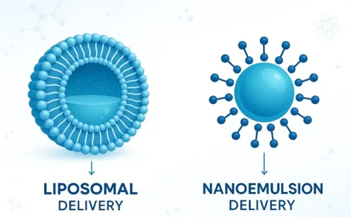
Figure 10: A) Liposomal and Nanoemulsion Delivery
10.2 Formulations Based on Nanoemulsions:
Nanoemulsions are oil in water dispersions stabilized by surfactants, which have nanoscale droplet sizes. Depending on the desired application and nature of the active substance they can be either oil in water or water in oil.
Nanoemulsions are advantageous for poorly soluble compounds as the oil phase can increase the solubility of the drug. They have a relatively large surface area therefore could result in increased interaction with the skin and impact the release and partitioning of the active. Kojic acid derivatives and other depigmenting agents have been investigated using nanoemulsions to improve topical delivery.
However, nanoemulsions should not be confused with thermodynamically stable microemulsions. They are generally a kinetically stable system and can suffer from droplet growth, coalescence and phase separation during storage. Excessive amounts of surfactant can cause irritation so choosing appropriate excipients is key.
Preparation method: High-energy emulsification
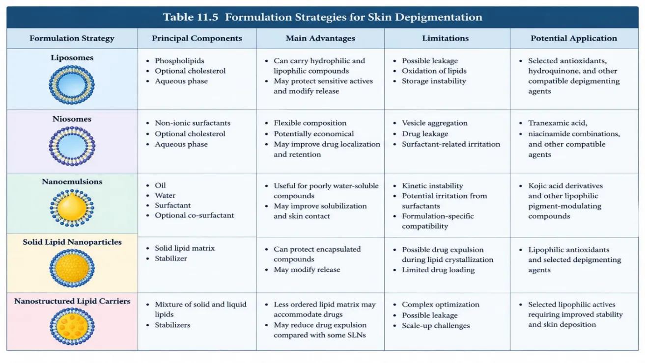
Figure 10: B) Formulation Strategies for Skin Depigmentation
Bottom of Form
11. THERAPEUTIC EFFECTIVENESS
Adverse Drug Reactions, Toxicity, Effectiveness, and Drug–Drug Interactions of Kojic Acid Emulgel for Hyperpigmentation
11.1. Adverse Drug Reactions (ADRs)
Kojic acid is a topical depigmenting agent that is used in cosmetic preparations to manage hyperpigmentation. When applied topically, it may lead to local adverse reactions, especially in people with sensitive skin or a tendency toward allergic contact dermatitis.
The adverse reactions that have been reported include:
Skin irritation – redness, burning, stinging and discomfort. Allergic contact dermatitis – in sensitive individuals kojic acid can cause delayed hypersensitivity reactions.
Erythema and pruritus – may cause localised redness and itch.
Sensitivity of damaged skin – applying to damaged or irritated skin may increase the risk of adverse reactions. Investigated contact sensitisation to kojic acid in patients with suspected cosmetic related dermatitis showed that 5 out of 8 patients who had used skincare products containing kojic acid were positive to kojic acid. This indicates that allergic contact dermatitis may occur but due to a small sample size the prevalence of kojic acid allergy in all users cannot be determined. The rate of adverse reactions will depend on the concentration of active ingredients, excipients, formulation characteristics, frequency of application and individual skin sensitivity for the kojic acid emulgel [25].
11.2. Toxicity of Kojic Acid:
The toxicity of Kojic acid has been investigated in studies. Potential issues of concern are allergic reaction, over bleaching and systemic toxicity. The Cosmetic Ingredient Review Expert Panel considered the safety of using Kojic acid in cosmetics. It took into consideration animal toxicity, genotoxicity, reproductive toxicity and allergic reactions in humans. They concluded Kojic acid can be used safely in cosmetics up to 1% concentration [26].
The Scientific Committee on Consumer Safety (SCCS) of the European Commission also concluded Kojic acid can be considered safe to use as a skin lightening agent in cosmetics up to 1%. However, they expressed concern that absorption may increase when the skin barrier is compromised such as with peeling agents[27].
These conclusions are for the use in cosmetics only. This does not mean all formulations and concentrations will be safe. The final emulgel formulation needs to be investigated for toxicity and tolerance, especially if it contains enhancers or other ingredients which may affect the skin barrier.
11.3. Effectiveness of Kojic Acid Emulgel in Treating Hyperpigmentation
Kojic acid primarily works by inhibiting the enzyme tyrosinase within melanin synthesis. This can lead to reduced melanin formation and therefore may help hyperpigmentation issues like Melasma. A randomised controlled trial was performed using topical Kojic Acid, Hydroquinone and Betamethasone Valerate. It was shown that Melasma improved when Kojic Acid was used. The combination of Kojic Acid and Hydroquinone showed the best clinical efficacy of the treatment groups[28].
A study was performed looking at the effects of adding Kojic Acid to a hydroquinone/glycolic acid gel in patients with Melasma. The results showed that more people responded to treatment with >50% clearance on the side of application with the Kojic Acid containing gel[29].
An emulgel formulation containing 1% Kojic Acid was developed and compared to a simple gel. When stress conditions were applied, the optimised emulgel had a higher stability with 86% of the original concentration remaining compared to the optimised simple gel which had 76%. This shows that there is greater retention of the active ingredient however this does not prove it has a greater depigmenting effect
There is evidence to suggest that Kojic Acid has depigmenting properties. It must be proven that a specific emulgel formulation will have an effective depigmenting effect by performing appropriate biological and clinical testing. This could involve looking at inhibition of tyrosinase, melanin content and validated clinical pigmentation scores[30].
11.4. Drug–Drug Interactions
There are a few considerations to take into account: Other depigmenting agents: Hydroquinone and Kojic acid have been combined into formulations. This can improve pigmentation results however you should consider whether the formulation will interact well, cause irritation and if it is safe.
Exfoliants/Chemical peels: Glycolic acid and other exfoliating agents can cause damage to the skin barrier. Using this alongside can cause increased irritation and increase the absorption into the dermis of kojic acid. Topical retinoids or irritating products: These can cause increased dryness, redness, flaking or irritation.
Excipients: Ingredients such as antioxidants, solvents, surfactants and penetration enhancers can affect the stability, release and delivery of kojic acid. This would be considered an interaction between the formulation components rather than a drug interaction. It was shown that the stability of kojic acid in the emulgel depended upon the formulation and storage conditions.
This study also looked at using sodium metabisulfite as an antioxidant. Therefore it is important to consider whether there are interactions with other topical agents when developing a formulation. Accordingly, potential interactions with other topical agents should be evaluated based on the available evidence and the intended formulation. The absence of reported interactions does not prove that every combination is safe[30]
Challenges and future perspectives of emulgel for hyperpigmentation
The following version is suitable for your dissertation and addresses emulgel as a topical delivery system overall, including various depigmenting agents such as azelaic acid, kojic acid, arbutin, niacinamide, tranexamic acid, and plant-derived ingredients
12.CHALLENGES:
1) Active Ingredient Stability: Active ingredients can oxidise and degrade therefore causing changes in colour. The optimisation of this can be affected by many factors including pH, temperature, light exposure and excipient used within the formulation. Therefore stability studies are needed[31,32].
2) Poor Drug Penetration: The stratum corneum can act a barrier to drug penetration. This will depend on the solubility of active ingredient, molecular characteristics and partitioning between the emulsion and gel[32].
3) Skin Irritation: Some active ingredients may cause skin irritation leading to redness, dryness, burning sensations, flaking and allergic reactions. Skin can become irritated and lead to post-inflammatory hyperpigmentation depending on skin type[31,33].
4) Not enough clinical evidence: Just because an ingredient can lighten pigmentation does not mean it will be suitable for use in an emulgel. There is a large amount of research looking at the active ingredient in a different dosage form or using lab analysis[32,34].
5) Hyperpigmentation can be complex: Melasma, post inflammatory hyperpigmentation and sun induced pigmentation have different causes. This means a formulation may not be as effective for a different type of pigmentation[33,34].
6) Risk of pigmentation returning: Pigmentation can come back after treatment. Improvement may not be seen quickly. This can cause issues with patient compliance due to irritation, long treatments and irregular sunscreen application[33,34].
7) Formulation issues: Emulgels need to use suitable emulsifiers, gelling agents and correct ratios between phases. This will keep the formulation stable with the correct consistency and drug concentration. Changing the formulation can affect the release of drug into the skin[32].
13. FUTURE PERSPECTIVES
1. Advanced drug delivery systems: The development of nanoemulgels, liposomal systems, transfersomes and other lipid-based systems may be investigated in future research. These may improve the drug delivery of different skin-lightening agents and have many benefits however these need to be proven experimentally[32].
2. Multi-component formulations: Formulating products containing different complementary components such as tyrosinase inhibitors and antioxidants may provide a more comprehensive approach. However it is important to investigate if they will be compatible, cause irritation and provide a clinical benefit[33,34].
3. Improved stability: Research should focus on incorporating the correct antioxidant, optimum pH, protective packaging and storage conditions to ensure the amount of active ingredients remains in the formulation and the emulgel remains in good condition[31,32].
4. Quality by Design (QbD): Using Quality by Design principals and experimental design can help determine the key variables within the formulation such as emulsifier level, oil/water ratio, gelling agent level and pH. The formulation can be optimised to improve consistency, stability and drug delivery[32].
5. Tailored formulations: Future research should consider what type of pigmentation, skin phototype, any underlying inflammation and tolerability the active ingredients would be most suitable for. This can help improve treatment outcomes and reduce pigmentation caused by irritation[33,34].
6. Clinical studies: Large well designed clinical studies should be undertaken to compare emulgels with traditional topical treatments. The pigmentation should be measured using validated clinical scoring, melanin readings, standardised images and long term follow up[33,34].
7. Improve safety and patient compliance: Future formulations should aim to have good skin tolerance, easy to apply and consistent. Clinical studies should include patient compliance, reoccurrence of pigmentation and long term safety[33,34].
CONCLUSION:
Hyperpigmentation is a common skin condition that requires effective and suitable treatment strategies due to its complex etiology and high recurrence rate. Conventional depigmenting agents include hydroquinone, kojic acid, arbutin, azelaic acid, niacinamide and vitamin C. These have been shown to be essential in controlling overproduction of melanin. However, these agents may also cause issues in application as they have poor solubility, instability, poor penetration into the skin and skin irritation.Emulgel delivery systems have emerged as an effective method of delivering drugs topically. This is due to having properties of both emulsions and gels. Emulgel delivery systems offer benefits such as improved incorporation of lipophilic drugs, improved spreadability, increased stability and increased patient acceptability. Advanced delivery systems such as liposomes, nanoemulsions, niosomes, solid lipid nanoparticles and nanostructured lipid carriers have potential to improve drug stability, sustained release and penetration.However, challenges do exist when using these delivery systems such as drug instability, formulation compatibility, skin irritation, poor drug penetration and lack of clinical data. Future research should focus on optimising formulation, QbD, increasing stability, correct combination of agents and designing suitable clinical studies.To conclude, formulation of new topical delivery systems including emulgels and advanced nano-carrier based systems offer a promising strategy for managing hyperpigmentation. Further experimentation and clinical investigations are required to prove their long term safety, efficacy and suitability for various skin typesTop of Form
REFERENCES


Christina Viju, Shreeya Shelar, Saniya Nadhavale, Anushka More, Nadim Khan, Shrutika Patil, Novel Topical Formulations for Hyperpigmentation: Recent Advances and Therapeutic Perspectives, Int. J. of Pharm. Sci., 2026, Vol 4, Issue 11, 649-666. https://doi.org/10.5281/zenodo.23938826
 10.5281/zenodo.23938826