

We use cookies to ensure our website works properly and to personalise your experience. Cookies policy
Department of pharmacology, RBVRR Women’s college of Pharmacy (Affiliated to Osmania university) Barkatpura, Hyderabad-500027.
Hinokitiol (?-thujaplicin) is a naturally occurring monoterpenoid tropolone derivative present mainly in the heartwood of some species of the Cupressaceae family . It features a unique seven-membered tropolone ring with hydroxyl and carbonyl functional groups, which confer strong metal-chelating properties and diverse biological activities. Extensive experimental studies have shown that hinokitiol has broad-spectrum antimicrobial, antibiofilm, antioxidant, anti-inflammatory, anticancer, antiviral, neuroprotective, dermatological, and emerging metabolic activities. These effects are mediated by several molecular mechanisms including modulation of metal homeostasis, oxidative stress, NF-?B, MAPK, Nrf2-ARE, apoptosis, autophagy and cell-cycle regulatory pathways. Hinokitiol has shown promising activity in various in vitro and animal models including infectious diseases, cancer, inflammatory conditions, neurological disorders and metabolic abnormalities . However, its therapeutic development is hindered by poor aqueous solubility, low oral bioavailability, rapid systemic elimination and limited pharmacokinetic information. Its bioavailability and therapeutic potential have been studied using several formulation approaches, including phytosomal systems, nanocapsules, cyclodextrin complexes, and other advanced delivery systems. Toxicological studies that are available indicate dose- and route-dependent effects, highlighting the need for careful evaluation of long-term systemic exposure and organ-specific toxicity. Despite extensive preclinical evidence, there is limited clinical research on hinokitiol. Most available human evidence is limited to topical and oral-health applications. Hence, further studies on pharmacokinetics, formulation optimisation, safety assessment and well-designed clinical trials are required to determine its therapeutic efficacy and translational potentials. To summarise, hinokitiol is a promising natural compound with a wide range of pharmacological activities, but there are significant translational challenges to overcome before it can be more widely used as a therapeutic
Hinokitiol (β-thujaplicin) was first found in the heartwood of Chamaecyparis species. It is a tropolone that mainly comes from plants in the Cupressaceae family like Chamaecyparis obtusa and Thujopsis dolabrata. It’s also present in Chamaecyparis taiwanensis, Thuja plicata and other conifer trees .[1]
In terms of structure hinokitiol has a seven-membered ring known as a tropolone. This kind of ring is rare but important. It’s found in powerful natural compounds like colchicine and stipitatic acid. These compounds form through four pathways—using polyketide, terpene, alkaloid and shikimate building blocks. [2] For hinokitiol the tropolone ring gives it an ability to bind with metal ions. This chelating action is a reason, behind its biological effects and medical potential .[1]
Hinokitiol has attracted interest for more than twenty years because it works in many ways. Hinokitiol shows pharmacological properties. Hinokitiol has anti‑inflammatory effects that work by modulating NF‑κB, MAPK and related signaling pathways. Hinokitiol has broad‑spectrum activity that kills bacteria, fungi and viruses. Hinokitiol has anticancer effects in cancer cell lines and animal models, including breast, lung, endometrial and liver cancer. These anticancer effects come from hinokitiol inducing cell death stopping the cell cycle and altering autophagy .[3]Antimicrobial data are also extensive: hinokitiol shows activity, with MIC values from about 0.1 to 164 µg/mL for many pathogens [4]. Hinokitiol also has documented antifungal, antiparasitic and even neuroprotective activity having been shown for the time to reduce brain damage and improve outcomes in a rodent model of stroke caused by blood clots .[5]Even though there is a lot of evidence moving hinokitiol into clinical use has been slow. Hinokitiol is hard to use in clinics because it does not dissolve well in water it breaks down easily. It does not reach good levels in the body. [6]Even recent thorough reviews say that earlier articles about hinokitiol chemistry and how it works mainly looked at how to synthesize it and its structure but they did not give focus to the challenges of making it a drug, how to formulate it and the barriers that stop it from being used clinically. [1] So there is a gap: hinokitiol has mechanistic and preclinical evidence but lacks good data on how it behaves in the body and, in patients. That is why a full current review that brings together the plant chemistry, how it works on a level the drug actions, how to deliver it and safety information in one place is needed.
2.Botanical Sources and Phytochemical profile of hinokitiol
2.1 Natural sources and distribution
Hinokitiol was first isolated in 1936 from the Formosan cypress by Nozoe and his team making it one of the tropolones ever identified from a natural source .[4] Since that discovery it has been found in other genera, within the Cupressaceae family. These include Chamaecyparis obtusa, C. Taiwanensis, C. Lawsoniana, Thujopsis dolabrata, Cupressus lusitanica and Thuja plicata .In all of these trees hinokitiol is mostly found in the heartwood. The leaves and bark usually have smaller amounts. The yield of hinokitiol also changes depending on where the plant grows, which part of the plant is used and the species involved.This natural variation makes it very difficult to get batches of the compound especially when doing pharmacological research. Because of this having extraction methods and carefully selecting the raw material is extremely important. [1]
2.2 Chemical structure and physicochemical properties
Hinokitiol is another name for β-thujaplicin. Its full IUPAC name is 2-hydroxy-4-(propan-2-yl)cyclohepta-2,4,6-trien-1-one. It is a tropolone. A tropolone is a seven-membered ring that is not made of benzene. This ring has a hydroxyl group and a ketone that is connected. It also has an isopropyl group attached. [7] The key properties of hinokitiol taken from PubChem, Wikipedia and a new detailed review are as follows:
TABLE NO:01 PHYSICOCHEMICAL PROPERTIES OF HINOKITIOL
|
Property |
Value |
|
Molecular formula |
C₁₀H₁₂O₂ [7] |
|
Molecular weight |
164.20 g/mol [7] |
|
Appearance |
Colorless to yellow crystalline solid [7] |
|
Melting point |
50–52.5 °C [7] |
|
Boiling point |
140 °C (at 10 mmHg) [7] |
|
Water solubility |
~1.2 g/L (0–25 °C) [7] |
|
Solubility in DMSO/ethanol |
~20–30 g/L [7] |
|
pKa |
~7.0–7.3 [7] |
|
TPSA |
37.3 Ų [7] |
Low water solubility and high ability to pass through cell membranes. Hinokitiol is usually put in BCS Class II [1]. Chemically like tropolones, hinokitiol can bind to metal ions. These metals include Zn²⁺, Fe²⁺, Cu²⁺, Co²⁺, Mn²⁺ and Ag²⁺. When it binds it makes salts. The salt with iron is called "hinokitin".[8]
2.3 Biosynthesis and occurrence in medicinal plants
Tropolones are a class of natural products that include colchicine, stipitatic acid and hinokitiol. They are biosynthesised via four parallel routes from polyketide, terpene, alkaloid or shikimate precursors, but all converge on oxidative ring-expansion of a six-membered precursor ring to give the seven-membered tropolone core.[9] In plants, the pathway derived from terpenes is thought to be in use; hinokitiol is considered to be a monoterpenoid, which is in agreement with biosynthesis via a terpenoid pathway different from the polyketide route used by fungi (e.g. Talaromyces stipitatus) or the shikimate-derived route used by some bacteria (e.g. Streptomyces spp.).[10]The specific enzymatic steps of ring expansion and aromatisation in Cupressaceae species are still only partially characterised and recent authors have noted that later steps may involve other cytochrome P450s, dehydrogenases or peroxidases whose identities are speculative at this point.[1]
3.Extraction, Isolation and Analytical Characterization
3.1 Extraction and isolation methods
Hinokitiol was first isolated in 1936 by Tetsuo Nozoe from the heartwood of Chamaecyparis obtusa. Since then, hinokitiol has been identified in several other Cupressaceae species such as C. taiwanensis, Thujopsis dolabrata, Thuja plicata, Thuja occidentalis, and Cupressus lusitanica. However, the reported yields of hinokitiol vary widely depending on the species, tissue type and extraction conditions.
Over the years different extraction protocols have been developed for these species, generally consisting of an initial solvent partitioning step, followed by column chromatography and HPLC-purification. Since then, a common three-step protocol has been established: (1) initial extraction with a polar solvent, (2) liquid–liquid partitioning with a non-polar solvent and (3) chromatographic purification. [1]
The literature compares a number of extraction techniques:
Although conventional solvent extraction (methanol, dichloromethane) can be replicated in a lab setting, it usually delivers poor yields (usually less than 1%, depending on the plant source) and presents environmental and solvent-toxicity issues.
Improved yield and reduced solvent toxicity are provided by ionic liquids and deep eutectic solvents, but their industrial application is limited by cost and difficulties with downstream purification.
Supercritical fluid extraction (especially CO₂-based) delivers enhanced yield, less solvent residues, and better selectivity, but requires specialist high-pressure equipment and has higher operational expenses.[1]
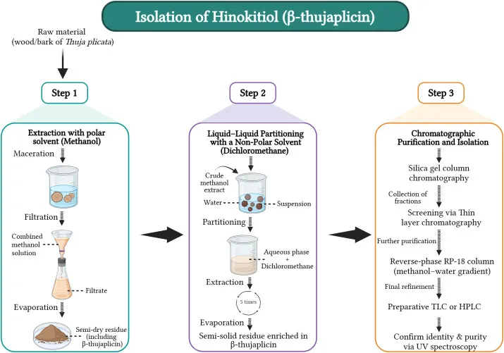
Figure No:01 Stepwise process for the isolation of hinokitiol (β-thujaplicin) from Thuja plicata, including extraction, fractionation, and purification steps.
Synthetic and biosynthetic (biocatalytic) production routes are becoming more popular as alternatives to plant-based isolation because traditional isolation techniques are constrained by low yield, hazardous large-volume solvents, and extensive purification requirements, which limit their economic feasibility and regulatory acceptance for industrial-scale production.[1]
3.2 Chromatographic and spectroscopic characterization
UV-visible and NMR spectroscopy confirmed the structure of hinokitiol. Hojo et al. employed combined 1 H and 13 C NMR, UV-visible spectroscopy and theoretical calculations to characterise the 4-isopropyltropolonate anion, neutral hinokitiol and its protonated form in acetonitrile to directly resolve the compound's tautomeric and ionisation behaviour .[11]Higashi et al [45] developed and validated a reversed-phase HPLC method with dual-wavelength UV detection (240 and 345 nm) using a C4 column for the chromatographic quantification of hinokitiol, with a retention time of 7.1 min and successfully applied to hair restorer, skin lotion and body soap samples. [12]
3.3 Quantification and standardization
Previous methods for determining hinokitiol include formation of difluoroborane derivatives (detection limit ~ 40 pg) and capillary zone electrophoresis (detection limit 0.21 µM), both cited as reference points in Higashi et al.'s HPLC validation paper, which itself specifically developed their dual-wavelength UV method as a simpler, less laborious alternative to earlier copper(II)-complex-based HPLC detection methods.[12]
4.Molecular Mechanisms of Action
4.1 Metal-Chelating and Metal-Homeostasis Mechanisms
Krenn et al. showed that hinokitiol is a zinc ionophore, which leads to a three-fold increase of intracellular zinc within minutes and inhibits the replication of picornavirus (rhinovirus, coxsackievirus, mengovirus) by inhibiting viral polyprotein processing by inhibiting eIF4GI cleavage. [13]This ionophore mechanism was later extended to SARS-CoV-2: hinokitiol in combination with zinc gluconate increased intracellular Zn2+ to levels that were able to disrupt viral papain-like protease (PLpro) and main protease (3CLpro) function in Vero-E6 cells without significant cytotoxicity. [14]
4.2 Oxidative Stress and Redox Regulation
The tropolone ring of hinokitiol is a bidentate enol-ketone coordination motif that chelates transition metals (Fe2+, Cu2+, Zn2+) directly correlating its structure to redox and metal-homeostasis regulation [1].In endometrial cancer cell lines (Ishikawa, HEC-1A, KLE), hinokitiol induced ROS-mediated apoptosis together with p53-driven cell-cycle arrest increasing cleaved-PARP and active caspase-3, and shifting the Bax/Bcl-2 ratio toward apoptosis.[15
4.3 Inflammatory Signaling Pathways
The seminal mechanistic study here is Byeon et al. (Planta Medica, 2008) who demonstrated that hinokitiol suppresses TNF-α production in LPS-activated RAW264.7 macrophages by down-regulating phosphorylation of PDK1, Akt/PKB and ERK, which blocks NF-κB nuclear translocation and activation.[16]This anti-inflammatory mechanism has been recapitulated in other tissue contexts. For example, in human corneal epithelial cells, hinokitiol preserved IκBα levels and prevented nuclear translocation of NF-κB p65, leading to reductions in IL-8, IL-6 and IL-1β.[17]
4.4 Apoptosis, Autophagy and Cell-Cycle Regulation
Hinokitiol induced autophagy via LC3B accumulation and Beclin-1 induction, and activated caspase-3 in a dose-dependent manner and downregulated cyclin B1, cyclin D1 and CDK1 in oral squamous cell carcinoma (SCC4, SCC25).[18] Hinokitiol increased cleaved-PARP and phospho-p53 in breast cancer cell lines (MCF-7, T47D, MDA-MB-231) to induce apoptosis, and also modulated autophagic flux separately. Disruption of autophagosome-lysosome fusion resulted in p62 accumulation, activation of caspase-8 and decrease of cancer stem-like cell populations, whereas blockade of autophagy was shown to enhance the apoptotic effect .[19]
4.5 Other Molecular Targets and Signaling Pathways
Hinokitiol, besides its role in metal binding and various cellular processes, also affects other signaling pathways outside of cancer and infections.
Antiplatelet signaling: In terms of blood platelets, hinokitiol helps prevent their activation. It does this by dampening the PLCγ2-PKC pathway and reducing the formation of certain reactive molecules. As a result, there is less thrombus, or blood clot, formation in living organisms. Interestingly, hinokitiol doesn’t block the binding of fibrinogen to its receptor but works through earlier signaling steps instead. This means it affects the process before the receptor interactions happen.[20]
Vascular smooth muscle cell (VSMC) proliferation:
In smooth muscle cells stimulated by PDGF-BB, hinokitiol showed selective effects by inhibiting the phosphorylation of certain proteins—specifically JNK1/2 and PLC-γ1—without affecting others like Akt, ERK1/2, or p38. It also lowered the levels of the proliferation marker PCNA while increasing the cell-cycle inhibitor p27^kip1^. This caused a halt in the cell cycle at the G0/G1 phase. These actions are different from how hinokitiol works in cancer cells, but they are important for conditions like restenosis and atherosclerosis.[21]
5. Pharmacological Activities of Hinokitiol
5.1 Antimicrobial and antibiofilm activity
A 2026 study showed that hinokitiol had antibacterial and antibiofilm activity against biofilm-producing Staphylococcus epidermidis isolates, and transcriptomic and RT-qPCR analysis revealed significant down-regulation of biofilm-formation genes (icaA, fnbA, aap), quorum-sensing genes (agrA, luxS), and global regulators (sarA, sigB), and up-regulation of the biofilm repressor icaR, via membrane damage and oxidative stress induction .The compound also exhibited a notable synergistic effect with the antibiotic ceftiofur sodium.[23] Hinokitiol was effective against fungal pathogens preventing biofilm formation in fluconazole-susceptible and -resistant Candida strains with MIC values of 3.1–12.5 µg/mL for biofilm inhibition, while pre-formed mature biofilms required 2–4 times higher concentrations.[24]
5.2 Anti-inflammatory activity
Byeon et al. reported that hinokitiol down-regulates PDK1/Akt/ERK phosphorylation and inhibits NF-κB activation, and thus inhibits TNF-α production in LPS-activated macrophages. [16]The mechanistic review further states that hinokitiol additionally inhibits the binding of LPS to TLR4 and suppresses COX-2 expression. COX-2 was reduced by 0.5–1% hinokitiol in osteoblast-like (MG-63) cells and by 1% in gingival fibroblast (HGF) cells, with decreased HIF-1α levels in both cell types.[3]
5.3 Anticancer activity
Hinokitiol induces apoptosis and modulates autophagy in different cancer models. It induces ROS-mediated apoptosis and p53-dependent cell-cycle arrest in endometrial cancer cell lines.[15] In oral squamous cell carcinoma it activates caspase-3, downregulates cyclin B1/D1 and CDK1, and induces autophagy via LC3B/Beclin.[18] The treatment increased cleaved-PARP and phospho-p53 in breast cancer lines and inhibits autophagosome-lysosome fusion, reducing the cancer stem-like cell populations.[19]
5.4 Antioxidant activity
A 2025 in vivo study demonstrated that hinokitiol protects from radiation-induced haematological, hepatic and inflammatory injury through Nrf2-ARE pathway activation, with significant upregulation of Ho-1, Nqo1 and Txnrd1 and suppression of Nf-κB and Tnf-α expression.[25] Mechanistic reviews have described this antioxidant activity as complementary to hinokitiol’s direct reactive-oxygen-species scavenging capacity and anti-inflammatory NF-κB suppression.[3]
5.5 Neuroprotective activity
In this study, we first demonstrated that hinokitiol reduced infarct volume and improved neurobehavioral outcomes in a rodent model of thromboembolic stroke, implying hinokitiol as a candidate neuroprotectant.[5]
5.6 Dermatological and wound-healing effects
Byeon et al. contextualised their results of TNF-α suppression specifically in terms of skin and hair biology: considering that TNF-α drives hair-follicle apoptosis, the authors suggested that hinokitiol might be useful in hair loss treatment by suppressing follicular apoptosis in addition to stimulating new hair growth.[16]This was confirmed by in vivo studies: mice treated with shampoo and hair-tonic formulations containing hinokitiol-loaded poly(ε-caprolactone) nanocapsules showed hair-growth-promoting effects comparable to those achieved with a 3% minoxidil solution.[26]
5.7 Metabolic and other emerging pharmacological activities
Recent research has looked into hinokitiol as a way to help treat metabolic diseases. A specific compound known as bis(hinokitiolato)zinc(II), or Zn(hkt)₂, has demonstrated potential benefits for type 2 diabetes in a study involving mice. This compound not only improved blood sugar levels and insulin resistance more effectively than similar zinc compounds, but it also helped reduce fat buildup in the liver and kidneys. These findings hint at its possible use in managing dyslipidemia related to obesity[.27]
6. Structure–Activity Relationship and Hinokitiol Derivatives
6.1 Structural determinants of activity
A study on different antimalarial compounds revealed that the way the tropolone ring is oxidized significantly affects how effective they are. For instance, while basic forms like tropone, unsubstituted tropolone, and hinokitiol didn't show much strength against malaria, a variant called 7-hydroxytropolone was found to be more than 18 times more potent. This finding highlighted that the number of oxygen atoms next to each other on the ring really influences its effectiveness.[28]It also suggests a bigger pattern in how hinokitiol works: the arrangement of hydroxyl and carbonyl groups around the seven-membered ring is crucial for its ability to combat diseases, including its known ability to bind with iron, alongside its antimicrobial and antifungal properties.[29]
6.2 Synthetic derivatives and metal complexes
The most studied class of hinokitiol derivatives are its metal-chelate complexes, formed due to the coordination of the hinokitiol monoanion (hino⁻) through two non-equivalent oxygen donor atoms. Nomiya and colleagues synthesised and structurally characterised 15 different hinokitiol metal complexes belonging to eight structural classes, and found that antimicrobial activity varied markedly by metal center — silver(I), iron(III) and copper(II) complexes showed significantly different, generally enhanced, antimicrobial activity compared to free hinokitiol itself.[30] In addition, the ternary inclusion complex of copper(II)-hinokitiol with γ-cyclodextrin exhibited higher antibacterial activity than hinokitiol alone.[31]
6.3 Therapeutic implications of structural modification
The structural modifications of hinokitiol have led to derivatives with different, sometimes enhanced, therapeutic profiles compared with the parent compound. Metal complexation may uncover antiviral activity not detected by basic antimicrobial screening of hinokitiol alone. Thujaplicin–copper chelates were able to inhibit apoptosis caused by influenza A (H1N1, H2N2, H3N2) and influenza B virus infection in MDCK cells at micromolar concentrations.[30] In a separate line of work, synthetic derivatives of tropolones related to hinokitiol in structure have been explored for neuroprotection against oxidative-stress-induced neuronal damage, an application not previously explored for tropolones as a class before that work. These studies underscore that the core scaffold of hinokitiol provides a viable synthetic starting point for the development of analogues targeting activities beyond that of the established antimicrobial/anticancer profile of the parent molecule.[29]
7.Pharmacokinetics, Bioavailability and Drug-Delivery Strategies
7.1 Absorption, distribution, metabolism and elimination
A study by Fung et al. (PNAS, 2022) provides the main information about the pharmacokinetics of hinokitiol (HKT). They have administrated HKT intraperitoneally to C57BL/6J mice at a dose of 30 mg/kg and 100 mg/kg. The investigators found out that 30 mg/kg is the lowest dose permitting a reliable quantitative measurement by HPLC. Moreover, they have detected that the plasma concentration of HKT reached approximately 150 µM and 500 µM at 15 minutes post-injection in the mice which have been treated with 30 and 100 mg/kg, respectively. Additionally, the half-life of elimination was about 0.5 hours for the lowest dose and 1 hour in the highest dose, meaning that it is rapidly cleared from the circulation with a short mean residence time. This rapid elimination profile is characteristic for independently reported oral pharmacokinetics: a 3D-printed hinokitiol tablet orally administered to male Wistar rats at 600 µg/kg resulted in a C max of 399.67 ± 62.56 ng/mL at T max ~3 h with an AUC 0–∞ of 2285.67 ± 499.29 ng/mL·h, reflecting hinokitiol’s poor aqueous solubility.[30]
7.2 Solubility and bioavailability limitations
The aqueous solubility of Hinokitiol is about 1.2 mg per mL, and since it can easily pass through cell membranes, it is classified as BCS(Biopharmaceutics Classification System ) class II molecule. This means that dissolution, rather than membrane permeability, is the rate-limiting step in its oral absorption.This is the exact reason why in the rat experiment with the non-lipid-complexed tablet, the oral exposure was so low, and this is the exact limitation that phytosomal and similar technologies were developed to address.[6]
7.3 Nanotechnology-based and other delivery approaches
Several delivery forms have been investigated for enhancing the oral bioavailability of hinokitiol. Formulating hinokitiol as a phytosome complex and delivering it as a 3D-printed oral tablet containing the same 600 µg/kg dose in experiments on male Wistar rats increased Cmax to 534.67 ± 34.70 ng/mL, decreased Tmax to ~0.5 h, and increased AUC0–∞ to 2449.89 ± 577.52 ng/mL·h compared to the non-phytosome tablet – thus showing direct in vivo evidence that complexation with lipids significantly improves systemic exposure.[6] F or topical delivery, encapsulation of hinokitiol in poly(ε-caprolactone) nanocapsules and formulation of the latter into shampoo and hair-tonic products produced hair-growth-promoting effects in mice comparable to those of a 3% minoxidil solution, showing that nanocapsule-based delivery can improve local bioavailability without being dependent on the problem of systemic absorption.[26]
8. Toxicity and Safety Profile
8.1 In vitro and in vivo toxicity
Across various species and routes acute toxicity data indicate a wide margin between effective and lethal doses . Acute oral LD50 values of 504 mg/kg and 469 mg/kg for male and female ddY mice was reported respectively by Shimizu et al., and the intraperitoneal LD50 of 191 mg/kg in male ddY mice was separately reported . A large route-dependent difference typical of compounds with limited oral absorption[33]. Sofuni et al. (2001) characterised hinokitiol as positive in a Rec assay with S9 metabolic activation and in an in vitro chromosome-aberration test, but negative in the Ames test and the mouse micronucleus test, indicating a mixed but generally low genotoxic signal that does not translate to mutagenicity in whole-animal systems . Developmental toxicity has also been reported: Ogata et al. reported foetal anomalies in ICR mice after oral administration of hinokitiol at doses of 560 mg/kg and above on gestational day 9. Similar anomalies were observed in cultured mouse embryos at much lower concentrations of 6.25–12.5 µg/mL indicating that the developing embryo is considerably more sensitive to hinokitiol than the adult animal.[33]
8.2 Dose-dependent safety
Data on chronic exposure and carcinogenicity are more reassuring. Hiraga et al. (1985, in Cho et al.) fed F344 rats diets containing hinokitiol at 0.005%, 0.015%, and 0.05% for 52 weeks and did not see a treatment-related increase in neoplastic lesions in either sex, formally concluding hinokitiol is not carcinogenic in F344 rats.[34] However, in a separate earlier study (Imai et al., 2006) deaths and myocarditis were reported in male rats fed a higher dietary concentration (0.1% and 0.2%) without a clear dose-dependent pattern and not in female rats.[33] This discrepancy led to the focused 13-week subchronic re-evaluation by Cho et al., which dosed F344 rats at 0%, 0.02%, 0.07%, and 0.2% hinokitiol in the diet; myocarditis was not replicated in this study, but marked reductions in body-weight gain, red-blood-cell counts, haemoglobin and haematocrit were increased at the higher doses, and heart weight was reduced in males at 0.07% and above. Based on these results the authors determined a no-observed-adverse-effect level (NOAEL) at 0.02% dietary hinokitiol, corresponding to 12.7 mg/kg b.w./day in males and 14.8 mg/kg b.w./day in females.[33]
8.3 Limitations of current toxicological evidence
An earlier study reported treatment-associated myocarditis and mortality in male rats at very high dietary doses, neither the 52-week carcinogenicity study nor the more recent 13-week re-evaluation could replicate this cardiac effect at comparable exposure levels. Likewise, the combination of clearly positive in vitro genotoxicity findings with negative in vivo results and the lack of independent confirmation of developmental toxicity findings (mostly based on one older study, Ogata et al., 1999) suggest that although hinokitiol appears largely safe, its toxicological profile has not been consistently reproduced across multiple different labs to the extent that one would expect for a substance with potential for systemic/internal therapeutic applications, as opposed to its traditional topical/cosmetic use.[33]
9. Preclinical and Clinical Evidence
9.1 In vitro evidence
The evidence base to date is mainly derived from cell-line studies in a variety of disease areas. Hinokitiol isolated from a redox-compound screening library inhibited tumor-sphere formation in cholangiocarcinoma by downregulating ERK and p38 signalling, and its combination with the clinical drug palbociclib induced significant anti-tumor effects in both human intrahepatic cholangiocarcinoma (ICC) cells and mouse ICC organoids.[35]Hinokitiol (non-cytotoxic concentrations, 0–1250 µM, no significant effect on tumour cell viability alone) was identified in an independent chemosensitizing study to sensitise B16F10 and CT26 tumour cells to 5-fluorouracil through suppression of the Akt/mTOR signalling axis, underscoring hinokitiol as a candidate chemosensitizer and not a standalone cytotoxic agent.[36]
9.2 Animal studies
The in vivo evidence, although much more limited than the in vitro data, includes several disease models described earlier in this review. These are: reduced infarct volume and improved neurobehavioral outcomes in a rodent thromboembolic stroke model, hair-growth effects that compare favourably with minoxidil in a mouse dermal model, and improved glycaemic control in type 2 diabetic KK-Aʸ mice with a zinc-hinokitiol complex . A recurrent limitation explicitly pointed out in the 2025 comprehensive review, while hinokitiol efficacy is demonstrated in vitro and in vivo, off-target effects, systemic toxicity and long-term safety are underexplored, and translational feasibility has not been established .[1]
9.3 Human/clinical evidence
There is very little direct clinical evidence for hinokitiol, but at least one real RCT exists. Kawamoto et al. conducted a pilot study with an open-label, randomised, controlled design in 18 patients with halitosis. Mouth cleaning with a hinokitiol-containing gel (n = 9) was compared with a control gel without hinokitiol (n = 9) three times daily for four weeks . The hinokitiol group showed significantly improved organoleptic malodour scores, hydrogen sulphide and methyl mercaptan levels, bleeding on probing, probing pocket depth, and plaque index compared to control.[37]
9.4 Translational limitations
The evidence pyramid for hinokitiol is tilted toward the in vitro base, with a much thinner layer of animal studies and only one human RCT identified—and that trial addressed a narrow, topical, oral-hygiene endpoint, rather than any of hinokitiol’s more extensively studied mechanisms (anticancer, antiviral, antidiabetic, neuroprotective). Your review should highlight this disconnect – the vast amount of mechanistic and cell line data supporting wide therapeutic claims versus virtually no clinical trial data testing these specific claims in humans. This is the key translational gap. In the 2025 review, it is also concluded that successful clinical application will depend on overcoming unresolved challenges, thorough toxicological assessment, and explicit clinical studies to validate safety and efficacy.[1]
10. Current Challenges and Future Perspectives
Despite significant pharmacological evidence, several challenges limit its clinical translation. The yield varies significantly among different species of Cuppressaceae, plant tissues, and geographical locations, and traditional methods of extraction are low-yield and solvent-intensive, complicating the production of consistent, pharmaceutical-grade material [1]
Moreover, pharmacokinetic properties may further limit its clinical utility. Hinokitiol, as a BCS Class II compound, has poor solubility but high intestinal permeability; it is rapidly cleared from the body, with a terminal elimination half-life of only 0.5-1 hour in mice.[32] While innovative dosage forms, such as phytosomal tablets, cyclodextrin complexes, and nanocapsules, have been developed, none have yet been tested in humans.
The current toxicological data bank is not free of contradictions as well. Thus, while an initial dietary study reported myocarditis and mortality in male rats at higher hinokitiol concentrations, these adverse outcomes were not confirmed in either the subsequent 52-week carcinogenicity study or a 13-week subchronic re-evaluation trial performed under similar conditions .Therefore, the cardiac safety of the substance against prolonged systemic exposure remains questionable and requires further in-depth analysis.[33],[34]
Perhaps most importantly, there is a significant disconnect between the preclinical and clinical levels of hinokitiol research. While the compound has demonstrated efficacy in a variety of models, including oncological, antimicrobial, anti-inflammatory, neuroprotective, and metabolic disease, there are currently no controlled clinical trials in humans outside of one small pilot study on oral malodor and periodontal measures.[37] None of the other mechanisms that have been demonstrated in preclinical models, including anticancer and antiviral activity, have been tested in humans. The potential for off-target effects and systemic toxicity has not been explored, representing the key limitation of this otherwise promising project.
Future research priorities should include: biosynthetic/biocatalytic approaches to decrease dependence on plant extraction; structure-activity guided derivatisation to surmount solubility challenges; human pharmacokinetic studies (currently completely absent); and early phase safety trials specifically looking for the cardiac signal seen in rodents before wider human trials are commenced.
11. CONCLUSION
Hinokitiol (β-thujaplicin) is a naturally occurring tropolone derivative with a wide range of pharmacological activities, including antimicrobial, antioxidant, anti-inflammatory, anticancer, antiviral, neuroprotective and metabolic effects. Its unique chemical structure, particularly the tropolone ring with metal-chelating properties, contributes to its interaction with metal ions, regulation of oxidative stress and modulation of important cellular signaling pathways. The available preclinical evidence suggests that hinokitiol may act through multiple mechanisms, including the regulation of NF-κB, MAPK, Nrf2–ARE, apoptosis and autophagy-related pathways.
Despite its promising pharmacological potential, the therapeutic development of hinokitiol faces several challenges. Poor aqueous solubility, limited oral bioavailability and rapid systemic elimination may limit its effectiveness following administration. Advanced delivery approaches, including phytosomal formulations, nanocapsules and metal-complex derivatives, have shown potential for enhancing its delivery and biological activity. However, their clinical applicability remains insufficiently established. Toxicological studies highlight the importance of evaluating dose-dependent effects, long-term systemic exposure and possible organ-specific toxicity.
The current evidence is predominantly based on in vitro investigations and preclinical animal models, whereas clinical research is limited. Therefore, the pharmacological effects observed in experimental studies cannot be directly extrapolated to human therapeutic applications. Future research should focus on optimization of formulation strategies, investigation of pharmacokinetic properties, establishment of comprehensive safety profiles and well-designed clinical studies to confirm efficacy and tolerability. Overall, hinokitiol is a promising natural compound with a wide range of pharmacological properties, but further systematic research is needed to overcome existing translational barriers and elucidate its potential as a safe and effective therapeutic agent.
REFERENCES


Shireen Begum*, Pharmacological And Phytochemical Characteristics Of Hinokitiol- A Comprehensive Review, Int. J. of Pharm. Sci., 2026, Vol 4, Issue 10, 2640-2654. https://doi.org/10.5281/zenodo.23417089
 10.5281/zenodo.23417089