

We use cookies to ensure our website works properly and to personalise your experience. Cookies policy
Vidya Niketan College of Pharmacy, Lakhewadi, Pune, Maharashtra , India 413103
The present study aimed to characterize the methanolic extract of Lepidium sativum and evaluate its in vitro anti-inflammatory activity. The extract was examined for organoleptic properties, solubility, phytochemical constituents, UV-visible spectroscopy, and FTIR analysis. Phytochemical screening confirmed the presence of flavonoids, alkaloids, and triterpenoids, which are known for their biological activities. UV spectroscopy showed absorption maxima at 207 nm and 321 nm, while FTIR analysis confirmed the presence of hydroxyl and hydrocarbon functional groups. The anti-inflammatory activity was evaluated using the protein denaturation method with diclofenac sodium as the standard drug. The extract showed concentration-dependent inhibition of protein denaturation, indicating significant anti-inflammatory activity. These findings suggest that Lepidium sativum extract is a promising natural source of bioactive compounds and may be useful for developing safe herbal anti-inflammatory formulations.
Medicinal plants have been used for centuries as natural remedies because they contain a wide variety of bioactive compounds with therapeutic properties. Herbal medicines are considered safer and more economical than many synthetic drugs, leading to increased interest in their scientific evaluation (1). Lepidium sativum L., commonly known as garden cress, belongs to the family Brassicaceae and is widely cultivated in Asia, Africa, and Europe. The seeds are traditionally used to treat inflammation, respiratory disorders, digestive problems, bone fractures, and skin diseases (2).
The seeds of Lepidium sativum are rich in flavonoids, phenolic acids, alkaloids, glucosinolates, triterpenoids, vitamins, and minerals. Among the phenolic compounds, sinapic acid is an important constituent known for its antioxidant and anti-inflammatory activities (3). These phytochemicals reduce oxidative stress and suppress inflammatory mediators, thereby helping in the management of inflammatory diseases (4). Standardization of herbal extracts is essential to ensure their quality, purity, and therapeutic consistency. Organoleptic evaluation, phytochemical screening, UV-visible spectroscopy, and Fourier Transform Infrared (FTIR) spectroscopy are commonly used techniques to characterize medicinal plant extracts (5,6).
Inflammation is a protective response against tissue injury, but prolonged inflammation may lead to chronic diseases such as arthritis and cardiovascular disorders. The protein denaturation assay is a simple and reliable method for evaluating anti-inflammatory activity because inhibition of protein denaturation is closely associated with anti-inflammatory potential (7). Therefore, the present study aimed to characterize Lepidium sativum seed extract and evaluate its in vitro anti-inflammatory activity.
MATERIALS AND METHODS
Seeds of Lepidium sativum were collected, authenticated, dried, powdered, and extracted using methanol by a suitable extraction technique. The extract and the reference compound, sinapic acid, were evaluated for organoleptic properties including colour, odour, taste, and physical appearance (8). Solubility studies were carried out in methanol, ethanol, chloroform, and other solvents according to standard procedures.
Preliminary phytochemical screening was performed to identify alkaloids, flavonoids, and triterpenoids using standard qualitative chemical tests (9). UV-visible spectroscopic analysis was carried out by preparing the extract in methanol, and the maximum absorption wavelengths (λmax) were determined. FTIR spectroscopy was performed to identify the major functional groups present in the extract (10).
The in vitro anti-inflammatory activity was evaluated by the protein denaturation method using egg albumin. Different concentrations of the extract were compared with diclofenac sodium as the standard drug. The absorbance was measured spectrophotometrically, and the percentage inhibition of protein denaturation and IC₅₀ values were calculated. All experiments were performed in triplicate, and the results were expressed as mean ± standard deviation (11).
RESULT AND DISCUSSION
Evaluation of Standard Compounds and Lepdium Satvium
Organoleptic Properties
Properties of Lepdium Satvium and standard compounds were examined for following organoleptic properties.
Table 1: Organoleptic properties of Lepdium Satvium
|
Sr. No |
Tests |
Specifications |
Observation |
|
1 |
Colour |
Brown |
Brown |
|
2 |
Odour |
Odorless |
Odorless |
|
3 |
Taste |
bitter |
bitter |
|
4 |
Physical appearance |
Crystalline powder |
Crystalline powder |
Table 2: Organoleptic properties of Sinapic Acid
|
Sr. No |
Tests |
Specifications |
Observation |
|
1 |
Colour |
White |
White |
|
2 |
Odour |
Odorless |
Odorless |
|
3 |
Taste |
bitter |
bitter |
|
4 |
Physical appearance |
Crystalline powder |
Crystalline powder |
Physical Properties
Solubility
The higher solubility of Lepdium Satvium was found in methanol.
Table 3: Solubility of Lepdium Satvium.
|
Sr. No. |
Solvent |
Solubility |
|
1 |
Methanol |
Freely soluble |
|
2 |
Chloroform |
soluble |
|
3 |
Ethanol |
soluble |
|
4 |
n-Hexane |
Insoluble |
Table 4: Solubility of Sinapic Acid.
|
Sr. No |
Solvent |
Solubility |
|
1 |
Methanol |
Freely soluble |
|
2 |
Ethanol |
soluble |
|
3 |
Water |
Slightly soluble |
|
4 |
Ether |
Insoluble |
Chemical Test for Lepdium Satvium
The chemical test for Lepdium Sativum was performed as per IP.
Table 5 chemical tests for Lepdium Satvium
|
Sr. No. |
Chemical Test |
Observation |
Inference |
|
1 |
2mg of drug + 2 drops of acetic anhydride + conc. Sulphuric acid (from side wall of test tube) |
Formation of deep red colour. |
Indicate presence of triterpenoid in drug. |
|
2 |
2 mg of drug + saturated trichloro acetic acid |
Presence of the colored precipitate |
Indicate presence of triterpenoid in drug. |
|
3 |
Extract dissolved in ethanol + few drops of conc. HCL + small amount of magnesium turnings is added |
Appearance of pink Colour |
Presence of alkaloids |
|
4 |
Small quantity of extract + lead acetate solution |
Formation of yellow colored precipitate |
Presence of flavonoids. |
UV Spectroscopic Analysis
Determination of λ max:
Dissolved 100 mg of Lepdium Satvium in sufficient methanol to produce 100 ml and dilute 10 ml resulting solution to 100 ml with methanol. When examined in the range 200nm to 400nm, the resulting solution shows an absorption maximum only at 207 nm and 321 nm.
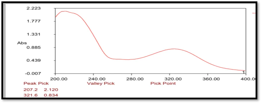
Figure 1:UV spectra Lepdium Satvium.
FTIR Spectroscopy Anaysis:
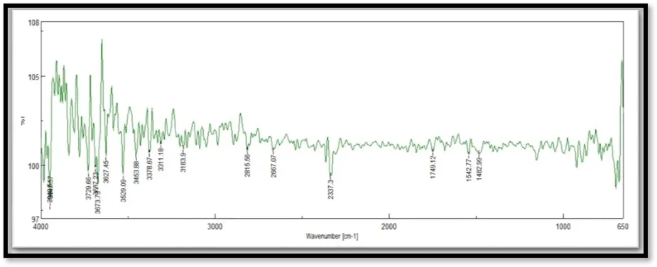
Figure 2: IR spectra of Lepidium Satvium.
This IR spectra is of methanolic extract of Lepdium Satvium which consists of different plants.
Table 6: Interpretation of IR spectra of Lepidium Satvium
|
Peak Position(cm-1) |
Functional Group |
|
3453 |
O-H |
|
2815 |
C-H |
In Vitro Anti-inflammatory Activity
Table 7: In Vitro Anti-inflammatory Activity of Lepidium sativum Extract (Protein Denaturation Method)
|
Sr. No. |
Concentration (µg/mL) |
% Inhibition (Extract) |
% Inhibition (Diclofenac Sodium - Standard) |
|
1 |
100 |
62.45±0.52 |
94.12±0.31 |
|
2 |
200 |
68.87±0.48 |
95.36±0.28 |
|
3 |
300 |
74.53±0.44 |
96.24±0.26 |
|
Sample |
IC₅₀ (µg/mL) |
|
Diclofenac Sodium (Standard) |
182.64 |
|
Lepidium sativum Extract |
228.91 |
The in vitro anti-inflammatory activity of Lepidium sativum extract was evaluated using the protein denaturation method. The extract showed a concentration-dependent inhibition of protein denaturation, with 62.45%, 68.87%, and 74.53% inhibition at 100, 200, and 300 µg/mL, respectively. Although the activity was lower than that of the standard drug, diclofenac sodium, the extract demonstrated significant anti-inflammatory potential. The observed activity may be attributed to the presence of flavonoids, phenolic compounds, alkaloids, and other phytochemicals known to inhibit inflammatory mediators. These findings suggest that Lepidium sativum possesses promising natural anti-inflammatory properties and may serve as a potential herbal source for the development of anti-inflammatory formulations.
CONCLUSION
The present study successfully characterized the methanolic extract of Lepidium sativum through organoleptic evaluation, solubility studies, phytochemical screening, UV-visible spectroscopy, and FTIR analysis. The extract contained important phytochemicals such as flavonoids, alkaloids, and triterpenoids, which are known for their therapeutic properties. UV and FTIR analyses confirmed the characteristic chemical profile of the extract. The protein denaturation assay demonstrated that the extract exhibited concentration-dependent anti-inflammatory activity, indicating its ability to reduce protein denaturation associated with inflammation. Although the activity was lower than that of diclofenac sodium, the extract showed significant natural anti-inflammatory potential. These findings support the traditional medicinal use of Lepidium sativum and suggest that it may serve as a valuable herbal source for the development of anti-inflammatory formulations. Further studies involving isolation of active compounds, in vivo pharmacological evaluation, toxicity assessment, and clinical investigations are recommended.
REFERENCES

Dr. Samrat Khedkar, Dr. Nitin Mali, Amit Chaugule, Phytochemical Characterization and In Vitro Anti-inflammatory Evaluation of Lepidium sativum Seed Extract, Int. J. of Pharm. Sci., 2026, Vol 4, Issue 7, 2874-2878. https://doi.org/10.5281/zenodo.21363153
 10.5281/zenodo.21363153