
We use cookies to ensure our website works properly and to personalise your experience. Cookies policy
Department of Pharmaceutics, Devaki Amma Memorial College of Pharmacy, Kerala 673634, India..
Phytosomes are sophisticated phyto-phospholipid complexes that were designed to overcome constraints faced by standard herbal formulations, including the phytoconstituents' weak solubility, limited permeability, and insufficient bioavailability. Due to limited absorption across biological membranes, many bioactive plant compounds, such as flavonoids, tannins, and glycosides, exhibit considerable therapeutic potential but fall short of achieving optimal clinical efficacy. In order to improve membrane permeability, stability, and systemic availability, phytosome technology combines these phytoconstituents with phospholipids, especially phosphatidylcholine. The basic properties of phytosomes, such as their composition, physicochemical and biological characteristics, preparation methods, characterisation techniques, advantages, and therapeutic applications, are discussed in this paper. Here is also a discussion of many preparation procedures, comprising lyophilization, supercritical fluid extraction, antisolvent precipitation, and solvent evaporation
Over the past century, phytochemical and phytopharmacological sciences have established the compositions, biological activities and health-promoting benefits of numerous botanical products. Most of the biologically active constituents of plants are polar or water-soluble molecules. However, water-soluble phytoconstituents (like flavonoids, tannins, glycosidic aglycones, etc) are poorly absorbed either due to their large molecular size, which cannot be absorbed by passive diffusion, or due to their poor lipid solubility; severely limiting their ability to pass across the lipid-rich biological membranes, resulting in poor bioavailability. [1]
Active chemicals extracted from natural plants have been shown to possess strong in vitro pharmacological effects; however, in vivo absorption has been proven to be insufficient. Several ways have been proposed to address the issue of poor absorption, including the production of emulsions, liposomes, and nanoparticles, as well as chemical structural modification. Phytosomes (also known as 'Phyto-phospholipid complexes,' 'Supra-molecular complexes,' and 'Herbosomes') have emerged as a potential option for enhancing the bioavailability of active ingredients among the many drug delivery systems. [2]
A phytosome is a complex between a natural product and natural phospholipids, like soy phospholipids. Such a complex is obtained by reaction of stoichiometric amounts of phospholipid and the substrate in an appropriate solvent. They are miscible both in water and in oil/ lipid environments, and are well absorbed orally. Phospholipids are small lipid molecules in which the glycerol is bonded only to two fatty acids, instead of three as in triglycerides, with the remaining site occupied by a phosphate group. The phospholipids mainly employed to make phytosomes are phosphatidylcholine, derived from soybean (Glycine max). The phytosome process has been applied successfully to many popular herbal extracts, including Ginkgo biloba, grape seed, hawthorn, milk thistle (Silybum marianum), green tea (Thea sinensis) and ginseng (Panax ginseng). The flavonoids and terpenoid components of these herbal extracts can directly bind to phosphatidylcholine. [3]
COMMONLY USED DRUG DELIVERY SYSTEMS
Phytosomes
Phytosomes are lipid-based nanocarriers that incorporate phospholipids to encapsulate plant-based nutraceuticals and medications. Also referred to as phyto-phospholipid complexes, phytosomes effectively address challenges related to the solubility and bioavailability of these compounds. [4]
Ethosomes
Ethosomes are lipid-based elastic vehicles composed of phospholipids, ethanol, isopropyl alcohol, and water. These nanocarriers enhance the entrapment efficiency, topical administration, and transdermal transfer of hydrophilic, lipophilic, and amphiphilic drugs, facilitating deeper tissue penetration and systemic circulation. Ethosomes signify hopeful progress in transdermal drug delivery systems, proposing enhanced permeation and deeper tissue penetration for numerous therapeutic substances. Their unique composition allows for improved delivery of both small and large molecules, making them suitable for a range of applications, including antifungal treatments and cosmetic agents. [5][6]
Liposomes
Liposomes are among the earliest and most widely utilised drug delivery technologies, consisting of bilayers of phospholipids. These structures encapsulate a variety of substances, including water-soluble, lipid-soluble, and amphiphilic compounds, thereby enhancing the effectiveness of pharmaceuticals, phytopharmaceuticals, nutraceuticals, and other bioactive agents. [7]
Transferosomes
Transferosomes are advanced lipid-based drug delivery systems designed to enhance the transdermal delivery of therapeutic agents. These vesicles possess unique characteristics, including deformability, amphiphilicity, and enhanced permeability, which enable them to effectively penetrate physiological barriers such as the stratum corneum and even the blood-brain barrier (BBB). Despite their advantages, transferosomes face challenges such as chemical instability, susceptibility to oxidative degradation, and high production costs. [8][9]
Dendrimer
Dendrimers consist of several arms originating from a core, which are prepared using sugar, nucleotides and amino acids. Dendrimers are an effective method for treating cancer as it depletes bioavailable copper from tumours. The conventional method for synthesis of dendrimers is a tedious process; an effective method was designed to produce poly(acyl thiourea) and polythiourea dendrimers. This method involves isothiocyanate–amine coupling and thiol?methacrylateMichael addition reaction to produce one dendrimer generation within four hours. [10]
Exosomes
Exosomes are extracellular vesicles composed of proteins, mRNAs, lipids and miRNAs. Exosomes, which are lipid molecules (30–100 nm wide) used for transportation of smaller molecules . Earlier, exosomes were considered as cellular waste, but research has proved their application in intercellular communication, which has an effect on functioning and activity of the recipient cell. Formation of exosomes includes four basic steps: budding, invagination, formation of multivesicular bodies and secretion of multiple vesicle structures. [10]
Aquasomes
Aquasomes are spherical 300nm particles used for drug and antigen delivery. The particle core is composed of non-crystalline calcium phosphate or ceramic diamond, and is covered by a polyhydroxyl oligomeric film. Aquasomes were prepared by self-assembly of hydroxyapatite by the co-precipitation method and thereafter preliminary coated with polyhydroxyl oligomers (cellobiose and trehalose) and subsequently adsorbed with bovine serum albumin (BSA) as a model antigen. BSA-immobilized aquasomes were around 200 nm in diameter and spherical in shape and had approximately 20-30% BSA-loading efficiency. [3]
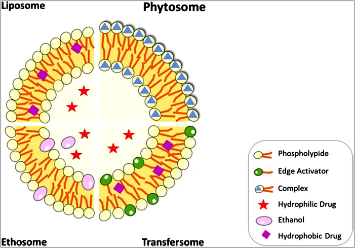
Figure 1: Structure of latest nanocarriers and drug loading. [11]
ADVANTAGES OF PHYTOSOMES
PROPERTIES OF PHYTOSOME
Physico-chemical properties
Phytosomes are prepared by reaction of a stoichiometric amount of phospholipid with the standardised plant extract as substrate. The spectroscopic data reveal that the phospholipid- substrate interaction is due to the formation of hydrogen bonds between the polar head (i.e., phosphate and ammonium group) and the polar functionalities of the substrate. The size of phytosomes varies from 50 nm to a few 100 μm.
Phytosomes, when treated with water, assume a micellar shape resembling a liposome, and Photon Correlation Spectroscopy (PCS) reveals these liposomal structures acquired by Phytosomes. From the 1H NMR and 13C NMR data, it can be deduced that the fatty chain gives unchanged signals both in free phospholipid and in the complex, which indicates that long aliphatic chains are wrapped around the active principle, producing a lipophilic envelope. In liposomes the active principle is dissolved in the internal pocket or it is floating in the layered membrane, while in phytosomes the active principle is anchored to the polar head of phospholipids, becoming an integral part of the membrane for example in the case of the catechindistearoyl phosphatidylcholine complex, in this there is the formation of H-bonds between the phenolic hydroxyls of the flavone moiety and the phosphate ion on the phosphatidylcholine side. [13][14]
Biological properties
Phytosomes are novel complexes which are better absorbed and utilised, hence they produce more bioavailability and better results than the conventional herbal extract or non-complexed extracts, which has been demonstrated by pharmacokinetic studies or by pharmacodynamic tests in experimental animals and in human subjects.
Phytosomes express their behaviour in physical or biological systems because of their physical size, membrane permeability, percentage entrapment, chemical composition, quantity and purity of the materials used. Instead, the phytosomes involve interaction of 1- 4 phospholipid molecules with the phytoconstituents, which are chemically anchored to each other. Several studies have shown phytosomes to be a better alternative for liposomes in terms of membrane permeability and stability. [14][15]
METHOD OF PREPARATION
Antisolvent precipitation method:
In many studies, the drug-phospholipid complex has been precipitated from an organic solvent using the conventional anti-solvent precipitation procedure, which includes the addition of n-hexane as an anti-solvent. A certain amount of soy lecithin and herbal extract was added to a 100 mL round-bottom flask and refluxed with 20 mL of dichloromethane for two hours at a temperature not exceeding 60°C. The mixture was concentrated down to 5 to 10 mL. The precipitate was filtered, collected and kept in vacuum desiccators overnight after the addition of 20 mL of hexane with constant stirring. [16][17]
Solvent Evaporation Method:
The most common method of solvent evaporation is to dissolve the medication and phospholipids in the same flask with a suitable solvent system such as ethanol or tetrahydrofuran. Conduct the reaction at an appropriate constant temperature for a specified period of time to obtain the maximum yield and drug trapping. The liquid was condensed to 5–10 mL, and the precipitate was filtered. The dried precipitate phytosome complex was stored in amber-colored glass jar at room temperature. [18]
The method of rotary evaporation
The complex of plant extracts or specific active principles with dietary phospholipids is typically obtained by solvent evaporation, with alcoholic or organic solvents serving as reaction media. The more popular solvent evaporation method involves placing the medication and the phospholipids into the same flask, with a suitable solvent system such as ethanol or tetrahydrofuran. In a rotary RBF, the exact amount of plant extract and soy lecithin was dissolved in THF and stirred for three hours at a temperature not exceeding forty degrees Celsius. A thin film of the sample was obtained, and n-hexane was added and constantly swirled by a magnetic stirrer. The precipitate was collected and stored in a glass bottle of amber colour in the refrigerator. The reaction was permitted to run for a predetermined time at a suitable fixed temperature to get the maximum possible yield and drug entrapment. [16][19]
Supercritical Fluid Extraction
Supercritical fluids are efficient for the production of large particles (5-2000 nm). The puerarin and phospholipid complexes were prepared by three conventional methods: lyophilisation, solvent evaporation and micronisation. Supercritical fluids (SEDS) can also be used to increase the solubility of poorly soluble drugs. The GAS method involves exposing drug and lipid solutions to supercritical anti-solvent separately and then applying pressure. The yield of this procedure was 93%. [20]
Lyophilisation
Phytoconstituents as well as natural and synthetic phospholipids are dissolved in various solvents. The mixture of phytoconstituents is then added to the phospholipid mixture and agitated until a stable combination forms. This complex is then separated by lyophilisation. Phospholipids used to prepare phytosomes generally have acyl groups derived from fatty acids such as stearic, oleic, palmitic and linoleic acid. These groups may be phosphatidylserine, phosphatidylethanolamine or phosphorylcholine. [20][21]
SELECTION OF DOSAGE FORM FOR DELIVERY OF PHYTOSOMES:
Phytosomes can be formulated in various topical and oral dosage forms, including,
1) Soft gelatin capsules: Phytosomes are prepared using vegetable or semi-synthetic oils as a dispersion medium and phytoconstituents in the form of suspension as a dispersed phase. Then the suspension is filled in soft gelatin capsules. For example, curcumin phytosome
2) Hard gelatin capsules: Phytosomes in powder form can be filled directly into hard gelatin capsules. For low-density phytosomes, such as Ginkgoselect phytosomes, capsule size should not be increased by 300 mg.
3) Tablets: Phytosomes as tablets are obtained by diluting the phytosome complex with 60%–70% excipients. Unit dose forms can be prepared by the direct compression method. For example, Leucoselect phytosome.
4) Topical dosage form: Firstly, an emulsion is prepared using a lipid solvent. Phytosome complexes are added to emulsions, as phyto-phospholipid complexes are dispersible in lipid solvents. [22]
CHARACTERISATION AND EVALUATION OF PHYTOSOMES
The zeta potential and particle size distribution of the phytosome formulae were measured by a computerised system and a dynamic light scattering (DLS) particle size analyser. [23]
Phytosome morphology was observed by transmission electron microscope (TEM). 400 nm wide carbon-coated copper grid was used, and one drop of sample was deposited and dried at room temperature. Then it was stained with a solution of phosphotungstic acid. The sample was dried well and then examined by a microscope at a magnification of 80000 x and an accelerating voltage of 100 kV. [23]
canopy slip of about 5 μL of the phytosomal suspension was mounted on a specimen tab. The formulation was examined and photographed for particle size using a scanning microscope (Sigma scan, Carl Zeiss scan) after drying the samples at room temperature. The particles were coated with platinum by vacuum pressure, and the coated samples were studied and photographed. [24]
for analysis of phospholipid and drug structure and chemical stability. Treatment of the phytosome with potassium bromide produces pellets. FTIR analyses were carried out in the scanning range of 4000- 400 cm-1. [22]
These interactions can be classified based on the transition temperature, melting points, appearance of new bands, disappearance of old bands and change in the related advanced region. [20]
Today, X-ray diffraction is a useful method to study the microstructure of crystalline and some non-crystalline materials. Phyto-phosphatide complexes, PCs, active substances or physical combinations of these are usually subjected to X-ray diffraction. X-ray diffraction indicates hard crystalline peaks of active chemical which is a physical mixtures, indicating a high crystal form. [20]
Entrapment efficiency of nanocarriers can be calculated by direct and indirect methods. The percentage of entrapment efficiency was calculated by the indirect technique as the difference between the initial amount of the drug and the free or unbound amount in the supernatant after centrifugation. The ratio of free or unentrapped drug in the supernatant to the total drug added in the nanocarrier mixture. On the other hand, the direct method involves solubilization of nanocarriers in a suitable solvent and analysis after filtration and dilution for the purpose of estimation of the trapping effectiveness using an appropriate technology. The incorporation efficiency was calculated in terms of percentage of drug content. [16]
The drug content of phytosomes is determined by high-performance liquid chromatography technology. Other spectroscopic methods are possible. [22]
The stability of the phytosome was evaluated by the change in particle size of the phytosome in DD water at room temperature. Then the size of the phytosome was measured with DLS on days 7, 14, 21 and 28. [25]
The models for in vitro and in vivo evaluations are chosen according to the expected therapeutic efficacy of the physiologically active phytoconstituents present in the phytosomes. For instance, the antihepatotoxic activity in vitro can be measured by the phytosomes’ ability to scavenge free radicals and to act as antioxidants. To assess the antihepatotoxic activity in vivo, the effect of produced phytosomes on the animals against thioacetamide-, paracetamol- or alcohol-induced hepatotoxicity can be studied. [12]
CLINICAL USES
Evodiamine is a quinoline alkaloid obtained from Evodia rutaecarpa with several pharmacological activities such as anti-tumour, anti-inflammatory, anti-nociceptive, anti-obesity and thermoregulatory actions. Evodiamine has been shown to have anti-tumour potential in many types of tumour cells through inhibition of proliferation, induction of apoptosis, and reduction of invasion and metastasis. Phytosomes of Evodiamine exhibited greater in vitro dissolution rate, improved absorption, extended duration of action and higher bioavailability. [26]
The increasing usage of prodrugs is in some part attributed to the hydrolysis of N-acetyl carnosine by esterase. L-carnosine and lipoid 75 were exposed to methanol and Milli-Q water and subsequently refluxed for 1hour at 40°C with the aim of producing a phospholipid complex [17]. The penetration rates of L-carnosine phytosomes were 2.4–5.6 times quicker than that of the L-carnosine solution. Phytosome technology increases selective & targeted penetration of phospholipids in the posterior segment of the eye. Furthermore, phytosomes improve the efficiency of drug delivery; however, problems with ocular administration include poor corneal penetration, which may limit the penetration of drugs encapsulated in phytosomes. The delivery of drugs to the eye is hindered by anatomical and physiological barriers, resulting in poor absorption and low ocular bioavailability. Lacrimal secretions may reduce retention time and permeability across the corneal epithelium. [27]
One dependable approach to cross the blood-brain barrier is through the nasal pathway. The olfactory or trigeminal nerves leave the brain at the respiratory epithelium or olfactory neuroepithelium and enter nose cavity. This allows for fast, non-invasive access to the cerebrospinal fluid and interaction with the mucosal tissue. The effective targeted distribution through the nasal route is dependent on formulation procedure, size, zeta potential and therapeutic efficacy of encapsulated medicine. The traditional intranasal delivery strategy involves passive diffusion, carrier-mediated transport and the paracellular pathway to the brain. [28][29]
The Ocimum Basilicum phytosomal gel for topical use was prepared using lecithin, cholesterol and Carbopol 934. Phytosomes were prepared using extract of Ocimum Basilicum and were evaluated for form, yield, stability and effectiveness for suppressing microbial growth [30]. Phospholipid in the phytosomes interacts with the encapsulated phytoconstituent by formation of a hydrogen bond between the polar head of the phospholipid and the polar features of the phytoconstituent. They greatly improve the cutaneous absorption of the included phytoconstituents. Phytosomes possess special nanoscale features that facilitate the quick delivery of the entrapped phytoconstituents across the cell membrane and into the circulation, thus improving their absorption. [31]
The efficacy of 60 mg of Ginkgo biloba terpenes phytosome, 11 mg of coenzyme Q10, and 8.7 mg of vitamin B2 twice daily was investigated in fifty patients with migraine with aura in two studies. [32]
Hepatoprotective action of phyto-phospholipids. When the patient consumes phosphatidylcholine, a synergistic action will be displayed to protect the liver. Under some conditions, phospholipids can be of nutritional benefit. Most of the phytosome research is focused on Silybum marianum Gaertner and its hepatoprotective flavolignans. The fruit of milk thistle (Silybum marianum) contains flavonoids with hepatoprotective activity. Silymarin has been shown to be an effective treatment for many liver diseases including hepatitis, cirrhosis, fatty liver caused by chemicals and alcohol, and bile duct inflammation. Choline has been reported to be essential for normal liver function since 1994. These phospholipids have been demonstrated to enhance hepatic collagenase activity in vitro and may therefore be useful in the prevention of fibrosis and cirrhosis by stimulating collagen degradation. Lecithin has been shown to be protective against non-alcoholic fatty liver disease and many toxins including hepatitis A and B.
Two clinical trials investigated the biological effect of phytosomes on the urinary system. In the first trial, urine from 13 healthy subjects were analysed for its ability to inhibit the growth of Candida albicans after administration of the cranberry extract phytosome or the equivalent standardized extract. In a one-week study, participants received cranberry phytosome or cranberry extract (two capsules daily) and their stool output was measured at several intervals. [33] [34]
The curcumin phytosome significantly decreased lung metastases and production of MMP-9, a protein associated with tumour invasion and metastasis, including breast cancer, without altering tumour size. This was consistent with the findings of the same study on the effect of grape seed phytosomes on in vitro activities of lung cancer cells. Therapy resulted in significant decrease in Ki-67 and bronchial histology grading in bronchial specimens. [35]
Commercial Products of Phytosomes
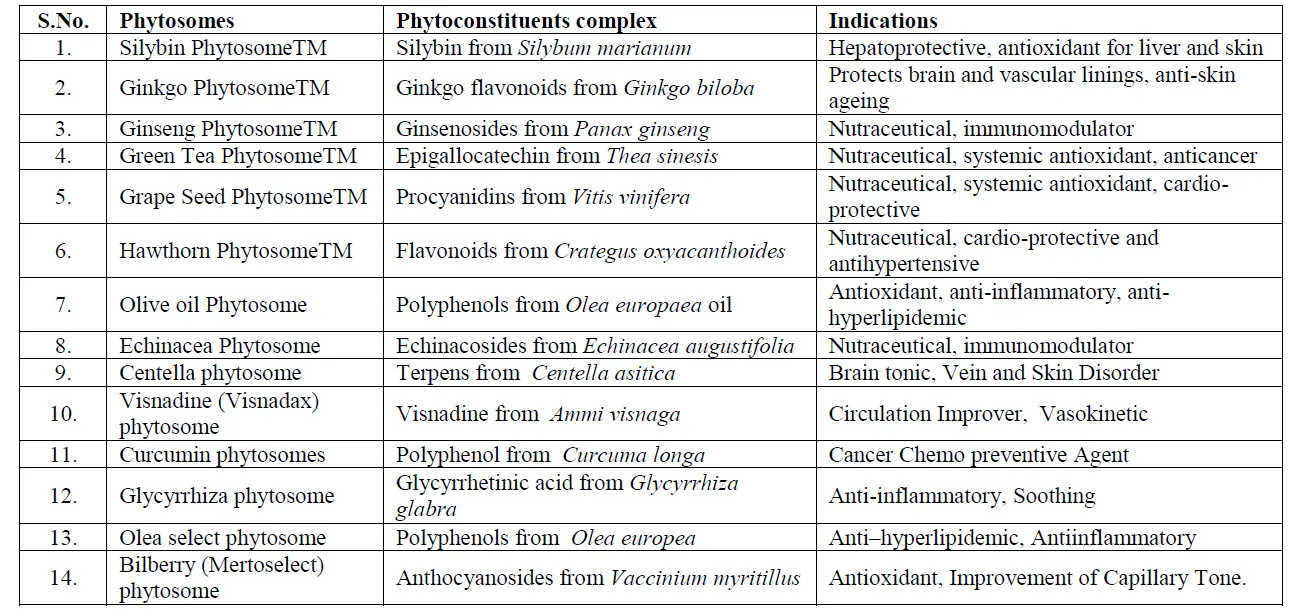
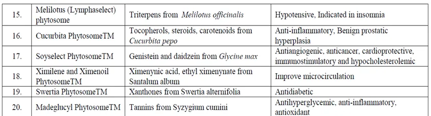
Figure 2: marketed phytosomes [12]
CONCLUSION
Phytosome technology has developed as a unique and effective technique for increasing the delivery and therapeutic efficacy of phytoconstituents. By creating complexes between plant-derived bioactive chemicals and phospholipids, phytosomes successfully overcome major hurdles such as poor solubility, limited membrane permeability and bioavailability. Compared with standard herbal extracts and alternative delivery modalities, phytosomes enable higher absorption, greater stability, enhanced pharmacological action, and better patient compliance. The adaptability of phytosomes enables their inclusion into numerous dosage forms including tablets, capsules, gels, and topical preparations. Numerous studies have proved their promise in the management of liver problems, neurological diseases, respiratory ailments, ophthalmic diseases, and other therapeutic uses. Furthermore, the availability of various commercial phytosomal products demonstrates the expanding adoption of this technique in pharmaceutical and nutraceutical businesses. Despite these developments, further clinical investigations, large-scale manufacturing optimization, and long-term safety evaluations are required to fully utilize their therapeutic potential. In conclusion, phytosomes represent a promising next-generation drug delivery method capable of increasing the effectiveness of herbal medicines and expanding their therapeutic relevance in modern healthcare.
REFERENCES


Abhirami P. P*, Mishahal T. M, Anagha U. V, Haritha K. K, Priyanka P, Nethaji Ramalingam, Phytosomes: A Promising Phospholipid-Based Nanocarrier for Enhanced Bioavailability and Therapeutic Efficacy of Herbal Bioactives -A Comprehensive Review, Int. J. of Pharm. Sci., 2026, Vol 4, Issue 6, 5222-5233, https://doi.org/10.5281/zenodo.20772637
 10.5281/zenodo.20772637