View Article

  • Plant-Derived Bioactive Compounds in Diabetes Mellitus: Mechanisms of Action and Therapeutic Potential

  • 1,2,3 Department of Biochemistry, Presentation College of Applied Sciences, Puthenvelikara, Affiliated to M G University, Kerala, India

    4 Department of Biochemistry, The Cochin College Kochi.

Abstract

Diabetes mellitus is a chronic metabolic disorder characterized by persistent hyperglycemia resulting from impaired insulin secretion, insulin resistance, or both. The increasing global prevalence of diabetes and the limitations associated with conventional antidiabetic therapies have intensified the search for safer and more effective alternatives. Plant-derived bioactive compounds have emerged as promising therapeutic agents due to their diverse pharmacological activities and relatively low toxicity. Various phytochemicals, including polyphenols, flavonoids, alkaloids, terpenoids, saponins, and glycosides, exert antidiabetic effects through multiple mechanisms such as enhancement of insulin secretion, improvement of insulin sensitivity, inhibition of carbohydrate-digesting enzymes, reduction of oxidative stress, stimulation of glucose uptake, and regeneration of pancreatic ?-cells. Medicinal plants including Momordica charantia, Panax ginseng, Gymnemasylvestre, Tinospora cordifolia, and Zingiber officinale have demonstrated significant antidiabetic potential in experimental studies. This review summarizes the major classes of plant-derived bioactive compounds involved in diabetes management, their mechanisms of action, and the therapeutic significance of selected medicinal plants. Challenges associated with phytochemical variability, bioavailability, and clinical standardization are also discussed. Overall, plant-derived bioactive compounds represent promising candidates for the development of alternative and complementary therapies for diabetes mellitus

Keywords

Diabetes mellitus; Medicinal plants; Phytochemicals; Antidiabetic activity; Bioactive compounds; Insulin secretion; ?-glucosidase inhibition; Oxidative stress

Introduction

× Popup Image

Diabetes mellitus (DM) is a multifactorial metabolic disorder characterized by persistent hyperglycemia and disturbances in carbohydrate, protein, and lipid metabolism resulting from defects in insulin secretion, insulin action, or both. Prolonged hyperglycemia is associated with chronic complications, including organ dysfunction and failure [1]. There are three major types of diabetes mellitus [1–5]. Type 1 diabetes mellitus, also known as insulin-dependent diabetes mellitus, is an autoimmune disorder caused by the destruction of pancreatic β-cells, necessitating lifelong insulin administration. It commonly occurs in children and young individuals. Type 2 diabetes mellitus, often referred to as non–insulin-dependent diabetes, accounts for more than 90% of adult cases and is characterized by insulin resistance and/or impaired insulin secretion. Gestational diabetes mellitus (GDM) is a form of glucose intolerance that develops during pregnancy, typically in the second or third trimester, and is associated with hormonal changes and insulin resistance.

Globally, the prevalence of diabetes mellitus is increasing at an alarming rate, with projections estimating that approximately 693 million individuals will be affected by 2045 [6]. This rise is largely attributed to the growing incidence of type 2 diabetes, driven by sedentary lifestyles, unhealthy dietary habits, and increasing obesity rates.

Common symptoms of diabetes mellitus include polyuria, polydipsia, unexplained weight loss, and blurred vision [7]. Chronic hyperglycemia can lead to severe long-term complications affecting multiple organ systems, including diabetic retinopathy, neuropathy, nephropathy, and cardiovascular diseases. In advanced cases, complications such as renal failure, foot ulcers, and amputations may occur [8]. Additionally, diabetes significantly increases the risk of hypertension and associated cardiovascular disorders.

The primary goal of diabetes management is to maintain blood glucose levels as close to normal as possible [9]. Conventional therapeutic approaches act through various mechanisms, including stimulation of insulin secretion from pancreatic β-cells, enhancement of insulin sensitivity, inhibition of hepatic glucose production, and reduction of intestinal glucose absorption [10–12].

A wide range of oral and injectable antidiabetic agents are currently available for the management of type 2 diabetes mellitus [13]. These include biguanides, sulfonylureas, meglitinides, thiazolidinediones, and dipeptidyl peptidase-4 (DPP-4) inhibitors. Metformin, a biguanide, is the most commonly prescribed first-line drug due to its efficacy, safety profile, and cost-effectiveness. However, it may cause gastrointestinal side effects such as nausea and diarrhea, and in rare cases, lactic acidosis. Other classes of antidiabetic drugs are also associated with adverse effects, including hypoglycemia, weight gain, and pancreatitis [13].

Due to the limitations and side effects associated with conventional therapies, there is an increasing demand for safer, cost-effective, and more efficient alternatives. Herbal medicine has been traditionally used in the management of diabetes and continues to gain scientific attention [15].

Numerous medicinal plants have been investigated for their antidiabetic and antihyperlipidemic properties, with over 400 plant species reported to exhibit hypoglycemic activity [18]. Bioactive compounds present in these plants, including flavonoids, terpenoids, alkaloids, phenolics, glycosides, and carotenoids, have demonstrated significant potential in regulating blood glucose levels through diverse mechanisms [19–22]. These phytochemicals offer promising prospects for the development of alternative therapeutic strategies for diabetes management.

2. Bioactive Compounds from Plants with Type 2 Antidiabetic Activity

Several medicinal plants used in traditional systems of medicine have demonstrated significant hypoglycemic and antidiabetic properties. Their mechanisms of action have been extensively investigated, highlighting their potential in diabetes management [23–25]. This review focuses on the mechanisms by which bioactive compounds derived from medicinal plants exert antidiabetic effects.

2.1. Bioactive Compounds Acting as Insulin Mimetics

2.1.1. Momordica charantia (Bitter Melon / Bitter Gourd)           

Figure 2.1(a):Momordica charantia

Momordica charantia (MC), a member of the Cucurbitaceae family, is widely cultivated and consumed in tropical and subtropical regions, including India, China, Southeast Asia, East Africa, and parts of Central and South America [26,27]. In addition to its nutritional value, it is well known for its medicinal properties, including anti-inflammatory, antioxidant, antiviral, antibacterial, anticancer, and notably, antidiabetic activities [28].

MC contains a wide range of bioactive phytochemicals, including steroids, triterpenoids (momordicosides A–L), saponins, alkaloids, phenolic compounds, glycosides, fatty acids, amino acids, vitamins, and minerals [28–36]. Among these, key antidiabetic constituents such as charantin, vicine, and polypeptide-p have been extensively studied.

Polypeptide-p, an insulin-like peptide isolated from the fruits and seeds of MC, exhibits structural and functional similarity to human insulin and has been shown to significantly reduce blood glucose levels in both animal models and humans [37,38]. These compounds act through multiple mechanisms, including stimulation of insulin secretion, enhancement of peripheral glucose uptake, inhibition of hepatic gluconeogenesis, and reduction of intestinal glucose absorption [40].

Experimental studies have demonstrated that MC extracts improve glucose tolerance in both normal and diabetic models [35–39]. In alloxan-induced diabetic rabbits, ethanolic extracts of MC fruit administered at a dose of 200 mg/kg body weight significantly reduced blood glucose levels through insulin-mimetic activity [40]. Additionally, Momordicoside U has been reported to enhance glucose uptake in in vitro insulin secretion assays [41].


Figure 2.1(b): The mechanism in decreasing blood glucose levels of M. charantia


Table1: Bioactive compounds and their effects in Momordica charantia

Bioactive Compounds

Antidiabetic Effects

Polypeptide-p

Act as Insulin-like protein, decrease blood glucose level

Momordicosides

Enhance the uptake of glucose

Saponins

Stimulate insulin secretion, a lower blood glucose level

Conjugated linolenic acid

Release intestinal GLP-1

Momordin

PPAR δ activation

9c, 11t, 13t conjugated linolenic acid

PPAR α activation


In vivo studies further indicate that MC extracts may promote regeneration and repair of pancreatic β-cells. Oral administration of alcoholic fruit extracts (25–75 mg) in alloxan-induced diabetic rats resulted in partial restoration of pancreatic islets of Langerhans [42,43]. Similarly, in streptozotocin-induced diabetic rats, administration of MC fruit extract (10 mL/kg/day for 14 days) improved insulin secretion, enhanced β-cell function, reduced insulin resistance, and increased glucose utilization [44].

Furthermore, aqueous ethanolic extracts of MC seeds have been shown to support long-term preservation of pancreatic β-cells [45]. These findings suggest that MC not only mimics insulin action but may also contribute to pancreatic β-cell regeneration. In addition, MC extracts have demonstrated inhibitory activity against key carbohydrate-digesting enzymes such as α-amylase and α-glucosidase, thereby reducing postprandial hyperglycemia [47].

Overall, Momordica charantia exhibits significant antidiabetic potential through insulin-mimetic activity, enhancement of glucose metabolism, and protective effects on pancreatic β-cells.

 2.1.2. Panax ginseng C.A. Meyer

Figure 2.1.2(a): Panax ginseng C.A. Meyer

Panax ginseng, a member of the Araliaceae family, is one of the most widely used traditional medicinal plants, particularly in Korea and other parts of East Asia [48]. It grows predominantly in colder regions such as Korea, Eastern Siberia, Northeast China, and parts of North America.

The roots of P. ginseng are rich in bioactive compounds, including triterpene glycosides (ginsenosides), panaxans, phenolic compounds, vanillic acid, and salicylates. In addition, the plant contains amino acids, alkaloids, proteins, polypeptides, and vitamins such as B? and B? [49–51]. Among these, ginsenosides are the most pharmacologically active constituents.

Ginsenosides are broadly classified into protopanaxadiol and protopanaxatriol groups Figure 2.1.2(b). These compounds play a crucial role in regulating glucose metabolism. Experimental studies have demonstrated that P. ginseng exhibits significant antidiabetic activity through multiple mechanisms, including enhancement of insulin sensitivity, stimulation of glucose uptake, and protection of pancreatic β-cells [50–52].


An external file that holds a picture, illustration, etc.
Object name is biology-09-00252-i001.jpg         An external file that holds a picture, illustration, etc.
Object name is biology-09-00252-i002.jpg

A. Protopanaxadiol                                                          B. Protopanaxatriol


R2 = Ra1, Ra2, Ra3, Rb1, Rb2, Rb3, Rc, Rd,                         R2 = Re, Rf, Rg1, Rg2, Rh1, etc

                    Rg2, Rg3, Rs1, Rs2, etc.


Figure 2.1.2(b): Structure of ginsenosides (ginseng-specific saponins)


Among the various ginsenosides, ginsenoside Rb? has shown potent hypoglycemic activity in streptozotocin-induced diabetic rats by significantly reducing blood glucose levels [53]. Furthermore, oral administration of fermented red ginseng extract (100–200 mg/kg/day for three weeks) resulted in a significant decrease in blood glucose levels and an increase in plasma insulin levels. This effect has been attributed to higher concentrations of active ginsenosides such as Rg?, Rg?, and Rh? in fermented preparations [54].

Overall, ginseng-derived saponins exert antidiabetic effects by modulating enzyme activity, improving insulin secretion, enhancing glucose utilization, and regulating carbohydrate metabolism Figure 2.1.2(c) [55–61].


Figure 2.1.2(c):Mechanism of Panax ginseng saponins on different organs related to diabetes


2.2. Bioactive Compounds that Increase Insulin Secretion from Pancreatic β-Cells

2.2.1. Allium cepa (Onion)


Allium cepa (garden onion): Go Botany

Figure 2.2.1(a): Allium cepa (Onion)


Allium cepa Linn., commonly known as onion, belongs to the family Liliaceae and is widely consumed across the world, including in countries such as Egypt, China, and India [62]. It is valued not only as a dietary component but also for its medicinal properties.

Onion contains a wide range of bioactive phytochemicals, including flavonoids, phenolic compounds, thiosulfinates, essential oils, amino acids, and vitamins. Key active compounds include quercetin, alliin, allicin, diallyl disulfide, and S-methyl L-cysteine sulfoxide [63,64].

Extracts of A. cepa have demonstrated significant hypoglycemic activity [65]. S-methylcysteine sulfoxide, one of the major active constituents Figure 2.2.1(a), exerts antidiabetic effects through multiple mechanisms:
(1) stimulation of insulin secretion from pancreatic β-cells,
(2) reduction of intestinal glucose absorption, and
(3) enhancement of peripheral glucose utilization [66,67].


Figure 2.2.1(b): Structure of S-methyl cysteine sulfoxide.

2.2.2. Allium sativum (Garlic)


    Free Ornamental Onion Allium photo and picture

Figure 2.2.2(a): Allium sativum (Garlic)


Allium sativum Linn., commonly known as garlic, is a widely used medicinal herb belonging to the family Amaryllidaceae. It is cultivated globally and has been traditionally used for both culinary and therapeutic purposes [68].

Garlic contains numerous bioactive compounds, including alkaloids, flavonoids, glycosides, terpenoids, and sulfur-containing compounds such as alliin, allicin, ajoene, and diallyl sulfides. It also contains proteins, enzymes, vitamins, and minerals [69–72].

Several studies have demonstrated the antidiabetic potential of garlic. Its bioactive compounds help regulate blood glucose levels by enhancing insulin secretion, improving glucose tolerance, and promoting glycogen synthesis [73]. Compounds such as allyl propyl disulfide and S-methylcysteine sulfoxide have been shown to significantly reduce blood glucose levels.

Additionally, ethanol extracts of garlic have been reported to improve delayed insulin response and enhance overall glycemic control [74,75]. These findings highlight the therapeutic potential of garlic in the management of diabetes.

2.2.3. Aloe vera (L.) Burm.f. (Asphodelaceae)

Free Aloe Vera Succulent photo and picture

Figure 2.2.3(a): Aloe vera (L.) Burm.f. (Asphodelaceae)

Aloe vera is one of the most widely used medicinal plants, particularly in the cosmetic and pharmaceutical industries. It belongs to the family Asphodelaceae and has been traditionally used for various therapeutic purposes. The plant is believed to have originated in Africa and the Mediterranean region and is now widely distributed in countries such as India, Cape Verde, Sicily, Malta, and Cyprus [76].

Aloe vera contains a wide range of phytoconstituents, including alkaloids, flavonoids, tannins, phenolic compounds, saponins, carbohydrates, vitamins, minerals, and aromatic compounds [77]. These constituents exhibit diverse pharmacological activities, such as antioxidant, antimicrobial, anticancer, and antidiabetic effects.

Experimental studies have demonstrated that oral administration of Aloe vera aqueous extracts significantly reduces blood glucose levels in diabetic animal models. Additionally, these extracts have been reported to exhibit minimal adverse effects, highlighting their potential as safe antidiabetic agents [78,79]. The relatively low cost and wide availability of Aloe vera further support its potential in the development of antidiabetic therapeutics.

2.3. Bioactive Compounds that Regenerate Pancreatic β-Cells

2.3.1. Pterocarpus marsupium (Fabaceae)

Pterocarpus marsupium, also known as Malabar kino, located at Islamabad near Fasial mosque

Figure 2.3.1(a): Pterocarpus marsupium (Fabaceae)

Pterocarpus marsupium is a large deciduous tree belonging to the family Fabaceae. It is rich in phenolic and flavonoid compounds, along with tannins, terpenoids, alkaloids, proteins, and amino acids. Among its bioactive constituents, epicatechin has been identified as a key antidiabetic compound.

The plant is also abundant in polyphenolic compounds such as pteroside, pterostilbene, marsupsin, pterosupin, and related derivatives, which exhibit significant biological activities, including antioxidant, anti-inflammatory, antibacterial, and antidiabetic effects [81–84].

Studies have demonstrated that oral administration of P. marsupium extracts produces significant antihyperglycemic effects in diabetic models [80,82,83]. Notably, epicatechin isolated from the plant has shown the ability to regenerate pancreatic β-cells in experimental models [85]. Additionally, aqueous extracts have been reported to enhance glucose uptake and stimulate insulin secretion, further supporting its antidiabetic potential.

2.3.2. Tinospora cordifolia (Menispermaceae)


Bratawali (Tinospora cordifolia) Bratawali (Tinospora cordifolia) ali-ali root or brotowali is an Indonesian traditional medicinal plant that is usually planted in the yard or grows wild in the forest. Decoction of the stem is commonly used as an herbal medicine Backgrounds Stock Photo     green leaf tinospora cordifolia bora phet on  medical herb tree  planting nature  background Agriculture Stock Photo    Tinospora cordifolia or Heart-leaved moonseed or Guduchi or Giloy. Alternative Medicine Stock Photo

Figure 2.3.2(a): Tinospora cordifolia (Menispermaceae)


Tinospora cordifolia, commonly known as “Guduchi” or “Amrita,” is a well-known medicinal plant in traditional Indian medicine. It is widely distributed in tropical regions, including India, Sri Lanka, and Myanmar [86].

The plant contains a variety of secondary metabolites, including phenolics, glycosides, terpenoids, alkaloids, essential oils, and polysaccharides. These phytochemicals contribute to its diverse pharmacological properties, including anti-inflammatory, antimicrobial, anticancer, and antidiabetic activities [87–89].

Studies have shown that polysaccharides isolated from T. cordifolia promote regeneration of pancreatic β-cells, suggesting its potential role in diabetes management [90]. In vivo experiments have demonstrated that oral administration of root extracts improves insulin secretion and inhibits glycogenolysis, thereby reducing blood glucose levels [91]. Additionally, alkaloidal fractions of the extract have been reported to enhance insulin sensitivity and inhibit gluconeogenesis [92].

2.3.3. Tinospora crispa (Menispermaceae)

https://thumbs.dreamstime.com/z/leaves-vines-tinospora-crispa-tree-garden-45952423.jpg?ct=jpeg

Figure 2.3.3(a): Tinospora crispa (Menispermaceae)

Tinospora crispa is a climbing shrub known for its rich phytochemical composition. It contains various bioactive compounds, including alkaloids, flavonoids, lignans, sterols, and terpenoids [93].

Several alkaloids and flavonoids, such as magnoflorine, luteolin derivatives, genkwanin, and diosmetin, have been isolated from the plant [93–97]. These compounds contribute to its pharmacological activities, including antidiabetic effects.

Experimental studies have shown that T. crispa extracts exert hypoglycemic effects by enhancing insulin secretion. The mechanism involves modulation of intracellular Ca²? levels in pancreatic β-cells, leading to increased insulin release [94]. Additionally, the extract has been reported to upregulate glucose transporter expression (GLUT1), thereby enhancing glucose uptake in muscle cells [98,99].


2.3.4. Gymnemasylvestre (Apocynaceae)


https://4.bp.blogspot.com/-QsQkASxAfrE/UMWzWI6Cg2I/AAAAAAAAF_E/6u4XqQ8pVZk/s1600/Gymnema-sylvestre.jpg    https://melhorcomsaude.com.br/wp-content/uploads/2021/06/gymnemna-silvestre.jpg

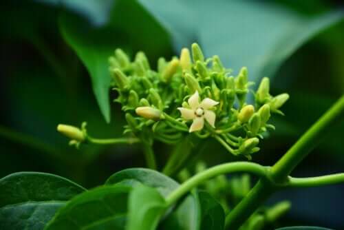
Figure 2.3.4(a): Gymnemasylvestre (Apocynaceae)


Gymnemasylvestre is a climbing plant native to the tropical forests of India and Sri Lanka. It has been widely used in traditional medicine for the treatment of diabetes [100].

The primary bioactive constituents of G. sylvestre are gymnemic acids, a group of triterpenoid saponins responsible for its antidiabetic activity. Other constituents include gymnemasaponins, gymnemasins, flavonoids, and sterols

Studies have shown that aqueous leaf extracts of G. sylvestre exhibit significant hypoglycemic effects in both normal and diabetic models [102]. The plant exerts its antidiabetic effects through multiple mechanisms, including stimulation of insulin secretion, regeneration of pancreatic β-cells, enhancement of insulin sensitivity, and inhibition of glucose absorption in the intestine [101,103–106].


Figure 2.3.4(b): Mechanism of Gymnemasylvestre in the Antidiabetic Activity


Gymnemic acids have also been reported to reduce blood glucose levels by inhibiting intestinal glucose absorption and suppressing hepatic glucose production. These properties make G. sylvestre a promising candidate for diabetes management.


Table 2.3.4: Phytoconstituents of Gymnemasylvestre

Phytoconstituents

Classification

Triterpene saponins

Gymnemic acids-acylated (tiglolyl, methylbutyroyl) derivatives of deacylgymnemic acid (DAGA) which is a 3-O-β-glucouronide of gymnemagenin (3β, 16β, 21β, 22α, 23, 28-hexahydroxy-olean-12-ene)

Oleanane saponins

Gymnemic acids and gymnemasaponins

Dammarene saponins

Gymnemosides A, B, C, D, E, and F

Gurmarin

A novel 35-amino-acid peptide with a 4209 molecular weight

Gymnemasins A

3-O [β-d-glucopyranosyl (1-3)-β-d-glucopyranosyl]-22-O-tiglyol gymnemanol

Gymnemasins B

3-O-[β-d-glucopyranosyl-(1-3)-β-d-glucuro-nopyranosyl]-gymnemanol

Gymnemasins C

glucuronopyranosyl-22-O-tigloyl-gymnemanol

Gymnemasins D

3-O-β-d-glucopyranosyl-gymnemanol

Gymnemanol

3, β-16,β-22, α-23-28-pentahydroxyolean-12-ene

Gymmestrogenin

Pentahydroxytriterpene

Flavonol glycoside

Kaempferol 3-O-β-d-glucopyranosyl-(1-4)-α-l-rhamnopyranosyl-(1-6)- β-d-galactopyranoside

Sterols

Stigmasterol


2.4. Bioactive Compounds that Reduce Glucose Absorption from the Gastrointestinal Tract

2.4.1. Cyamopsis tetragonoloba (Fabaceae)


https://c7.alamy.com/comp/ET09CN/cyamopsis-tetragonoloba-linn-taub-cyamopsis-psoralioides-fabaceae-ET09CN.jpg    https://c7.alamy.com/comp/2GXJK34/guar-or-cluster-bean-field-strong-sunlight-falls-on-guar-or-cluster-bean-plants-2GXJK34.jpg

Figure 2.4.1(a):Cyamopsis tetragonoloba (Fabaceae)


Cyamopsis tetragonoloba, commonly known as guar or cluster bean, is a drought-resistant plant widely cultivated in India. It is rich in dietary fiber and carbohydrates [107].

The plant contains several phytochemicals, including phenolic acids, flavonoids, galactomannans, tannins, and ascorbic acid. These compounds contribute to its biological activities, including antihyperglycemic effects.

Studies have demonstrated that C. tetragonoloba exhibits significant glucose-lowering effects in diabetic animal models. Its bioactive compounds help improve insulin secretion, reduce blood glucose levels, and lower HbA1c levels [108,109]. Polyphenols present in the plant have also been shown to protect pancreatic β-cells and contribute to its hypoglycemic activity [110].

2.4.2. Ocimum sanctum L. (Lamiaceae)

https://as1.ftcdn.net/v2/jpg/03/72/69/06/1000_F_372690608_y1gokjzB8YkKeCO4TYsJYy0Cxn28Y5ON.jpg

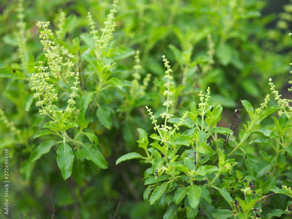
Figure 2.4.2(a): Ocimum sanctum L. (Lamiaceae)

Ocimum sanctum (holy basil) is a widely used medicinal plant cultivated in many parts of the world, particularly in India and Southeast Asia [111,112].

The plant contains a diverse range of phytochemicals, including eugenol, methyl eugenol, linalool, caryophyllene, flavonoids, phenolics, terpenoids, and fatty acids [113,114]. These compounds exhibit various pharmacological activities, including antioxidant, anti-inflammatory, antimicrobial, and antidiabetic effects.

Experimental studies have shown that different extracts of O. sanctum (ethanol, aqueous, butanol, and ethyl acetate) enhance insulin secretion and exhibit antihyperglycemic activity [115]. In vivo studies further demonstrate that the plant extract improves glycogen storage in the liver, reduces blood glucose levels, and enhances glucose tolerance [116].

Additionally, specific bioactive compounds such as eugenol and β-sitosterol have been associated with hypoglycemic effects. A triterpenoid compound isolated from O. sanctum has also been reported to enhance insulin secretion and improve glucose utilization, indicating its potential as a therapeutic agent for diabetes [117,118].

2.5. Bioactive Compounds with Oxygen Radical Scavenging Activity

The antioxidant activity of plant-derived bioactive compounds plays a crucial role in mitigating oxidative stress associated with diabetes mellitus. The butanol fraction of Momordica charantia extract has demonstrated strong free radical scavenging activity, indicating its antioxidant potential [119].

Further studies by Tsai T.H. et al. (2014) reported that M. charantia exhibits significant antioxidant and cytoprotective effects, which contribute to its antidiabetic activity [120].

Several Ayurvedic plants, including Momordica charantia, Eugenia jambolana, Tinospora cordifolia, and Mucuna pruriens, have been evaluated for their protective effects against diabetic cataract in vivo. Among these, M. charantia and E. jambolana demonstrated superior protective activity compared to the other plants studied [121].

2.6. Bioactive Compounds that Inhibit α-Glucosidase and α-Amylase Activities

α-Amylase and α-glucosidase are key digestive enzymes involved in carbohydrate metabolism. α-Amylase catalyzes the hydrolysis of starch into oligosaccharides, while α-glucosidase further breaks down these products into glucose in the small intestine.

Inhibition of these enzymes delays carbohydrate digestion and glucose absorption, thereby reducing postprandial hyperglycemia. This mechanism is considered an important therapeutic strategy in the management of diabetes mellitus [122–126].

Numerous plant-derived phytochemicals have demonstrated inhibitory activity against these enzymes, increasing interest in their use as natural antidiabetic agents.

2.6.1. Costus pictus D. Don (Zingiberaceae)


Figure 2.6.1(a): Costus pictus D. Don (Zingiberaceae)


Costus pictus, commonly known as the “insulin plant,” is widely used in Southern India as a traditional remedy for diabetes [127].

Phytochemical analysis has revealed the presence of various bioactive compounds, including alkaloids, flavonoids, glycosides, terpenoids, steroids, tannins, and phenolic compounds [128–131]. Trace elements such as potassium, calcium, chromium, manganese, copper, and zinc are also present and may contribute to its biological activity [132].

Experimental studies have demonstrated that C. pictus exhibits antidiabetic effects primarily through inhibition of α-amylase and α-glucosidase enzymes. In vivo studies using streptozotocin- and alloxan-induced diabetic models showed significant reductions in blood glucose levels following administration of plant extracts [132–134].

Additionally, studies have reported enhanced insulin secretion and improved glucose utilization following treatment with C. pictus extracts [135,136].

2.6.2. Phaseolus vulgaris L. (Fabaceae)

Figure 2.6.2(a): Phaseolus vulgaris L. (Fabaceae)

Phaseolus vulgaris (common bean) is an important dietary legume widely consumed across the world [137].

The plant is rich in bioactive compounds, including phenolic acids, flavonoids, anthocyanins, tannins, and proteins such as α-amylase inhibitors [137–139]. These inhibitors, including isoforms α-AI1 (phaseolamin), α-AI2, and α-AIL, have been extensively studied for their ability to inhibit α-amylase activity [140,143].

Phytohaemagglutinin, a lectin present in the plant, has also been reported to influence glucagon-like peptide-1 (GLP-1) activity, contributing to improved glycemic control [140–142].

By inhibiting carbohydrate-digesting enzymes, P. vulgaris reduces glucose absorption and lowers postprandial blood glucose levels. This effect may also help reduce insulin resistance and prevent diabetes-related complications [143–145].


Table 2.6.2: Major phytoconstituents of Phaseolus vulgaris

Group

Phytochemical Compound

Phenolic acids

Hydroxybezoic acid and derivatives flavonoids, anthocyanins, flavonols, flavanols, isoflavones, flavanones, proanthocyanidins, and tannins

Hydroxycinnamic acid and derivatives

Flavonoids

Orientin, isoerientin, rutin, myricetin, luteolin, quercetin, kaempferol, myricetin-3-rhamnoside, hyperoside, isorhamnetin-3-glucoside, isoquercitirn

Proteins

Vicilin, phytohenmagglutinin, alpha-amylase inhibitor (α-AI1, α-AI2, and α-AIL)


2.6.3. Euphorbia hirta L. (Euphorbiaceae)

Figure 2.6.3(a):Euphorbia hirta L. (Euphorbiaceae)

Euphorbia hirta is a widely distributed medicinal herb found in tropical and subtropical regions, including India, Southeast Asia, and Africa [146].

The plant contains a wide range of phytochemicals, including flavonoids (quercetin, quercitrin, rutin), terpenoids, phenolic compounds, tannins, and sterols [147,148].

Studies have demonstrated that ethanolic and ethyl acetate extracts of E. hirta exhibit significant hypoglycemic activity in alloxan-induced diabetic models [146–148]. These extracts also show strong inhibitory effects on α-glucosidase and α-amylase enzymes.

Flavonoids such as quercetin and rutin have been identified as potent enzyme inhibitors contributing to this activity. Although their inhibitory effect is weaker than that of the standard drug Acarbose, they offer the advantage of fewer side effects.

Further studies, including in vitro, in vivo, and in silico analyses, have confirmed the potential of E. hirta bioactive compounds in targeting enzymes involved in carbohydrate metabolism [149–152].


Table 2.6.3: some major isolated phytoconstituents on Euphorbia hirta L.


Group

Compound Name

Structure of Compound


Flavonols

Quercetin


Kaempferol


Leucoanthocyanidins

Leucocyanidin


Flavonoid glycosides

Rutin


Myricitrin


Afzelin


Luteolin-7-O-glucoside


Isoquercitrin


Acids

Syringic acid


Ellagic acid


Gallic acid


Shikimic acid


2β,16α-dihydroxy-ent-kaurane


β-sitosterol


Terpenoids


Sterols


Stigmasterol


2.7. Bioactive Compounds that Increase Glucose Utilization

Certain medicinal plants enhance peripheral glucose utilization, thereby contributing to improved glycemic control. Examples include Zingiber officinale, Grewia asiatica, and Cyamopsis tetragonoloba [153–155].

Among these, Zingiber officinale is one of the most extensively studied and widely used medicinal plants.

2.7.1. Zingiber officinale Roscoe (Zingiberaceae)

Figure 2.7.1(a): Zingiber officinale Roscoe (Zingiberaceae)

Zingiber officinale (ginger) is a widely used medicinal and culinary plant cultivated in tropical and subtropical regions [156,157].

Ginger contains a variety of bioactive compounds, including phenolics (gingerols, shogaols), terpenoids, volatile oils, amino acids, and organic acids [156–158]. These compounds exhibit strong antioxidant and antidiabetic properties.

Experimental studies have shown that ginger reduces blood glucose levels in both normal and diabetic animal models [159]. The hypoglycemic effect is attributed to delayed gastric emptying, enhanced glucose uptake, and improved insulin sensitivity.

Additionally, ginger has been shown to increase glycogen storage in tissues and enhance peripheral glucose utilization [160]. In vitro studies have demonstrated increased glucose uptake in muscle cells, primarily due to gingerol compounds [161].

Furthermore, ginger extracts have been reported to inhibit α-amylase and α-glucosidase activities, contributing to reduced postprandial hyperglycemia [162,163].


CONCLUSION

Plant-derived bioactive compounds possess significant therapeutic potential in the management of diabetes mellitus through multiple mechanisms, including stimulation of insulin secretion, enhancement of glucose uptake, inhibition of carbohydrate-digesting enzymes, antioxidant activity, and regeneration of pancreatic β-cells. Medicinal plants such as Momordica charantia, Panax ginseng, Gymnema sylvestre, Tinospora cordifolia, Aloe vera, and Zingiber officinale have demonstrated promising antidiabetic effects in experimental and preclinical studies. The diverse phytoconstituents present in these plants, including flavonoids, alkaloids, terpenoids, glycosides, and saponins, contribute to improved glycemic control and reduction of diabetes-related complications.

Despite encouraging findings, challenges related to phytochemical variability, standardization, bioavailability, dosage optimization, and long-term clinical safety remain important concerns. Further clinical investigations and pharmaceutical studies are necessary to validate their efficacy and support the development of safe, effective, and affordable plant-based antidiabetic therapies. Overall, medicinal plant-derived bioactive compounds represent promising candidates for future antidiabetic drug development and complementary therapeutic strategies.

FUTURE PERSPECTIVES

Plant-derived bioactive compounds have emerged as promising therapeutic agents for the management of diabetes mellitus due to their multitarget mechanisms, low toxicity, and wide availability. Future research should focus on the isolation and characterization of novel phytochemicals with potent antidiabetic activity and improved pharmacological efficacy. Advanced analytical techniques such as metabolomics, proteomics, and molecular docking studies may help identify specific molecular targets involved in glucose metabolism and insulin signaling pathways.

Further investigations are required to improve the bioavailability, stability, and targeted delivery of phytoconstituents. In this context, nanoformulation-based approaches such as nanoparticles, liposomes, phytosomes, nanoemulsions, and polymeric drug delivery systems offer significant potential for enhancing therapeutic effectiveness and reducing dosage-related limitations. Integration of nanotechnology with herbal medicine may provide innovative strategies for controlled and sustained drug release in diabetes therapy.

Although numerous experimental studies have demonstrated encouraging antidiabetic effects, large-scale clinical trials are still limited. Therefore, future studies should emphasize clinical validation, long-term safety evaluation, toxicity assessment, and standardization of herbal formulations to ensure reproducibility and regulatory acceptance. In addition, the development of standardized herbal formulations with defined active constituents may improve consistency in therapeutic outcomes.

The combination of plant-derived bioactive compounds with conventional antidiabetic drugs also represents an important area for future exploration, as such combinations may enhance efficacy while minimizing adverse effects. Moreover, personalized medicine approaches based on genetic and metabolic profiling could help optimize phytochemical-based therapies for individual patients.

Overall, continued interdisciplinary research involving pharmacology, biotechnology, nanotechnology, and clinical sciences may facilitate the development of safe, effective, and affordable plant-based therapeutics for diabetes mellitus in the future.

Declarations

Conflict of Interest

The authors declare no conflict of interest.

Funding

Not applicable.

Author Contributions

All authors contributed equally to literature collection, manuscript preparation, data analysis, and final manuscript revision.

Ethical Approval

This review article does not involve experiments on human participants or animals.

REFERENCES

  1. World Health Organization. Definition, diagnosis and classification of diabetes mellitus and its complications. Geneva: WHO; 1999.
  2. American Diabetes Association. Classification and diagnosis of diabetes: Standards of medical care in diabetes—2019. Diabetes Care. 2019;42(Suppl 1):S13-S28.
  3. Heise T, Nosek L, Rønn BB, et al. Lower within-subject variability of insulin detemir compared to NPH insulin and insulin glargine in type 1 diabetes. Diabetes. 2004;53:1614-20.
  4. McIntyre HD, Catalano P, Zhang C, et al. Gestational diabetes mellitus. Nat Rev Dis Primers. 2019;5:47.
  5. Faselis C, Katsimardou A, Imprialos K, et al. Microvascular complications of type 2 diabetes mellitus. Curr Vasc Pharmacol. 2020;18:117-24.
  6. Roden M, Shulman GI. The integrative biology of type 2 diabetes. Nature. 2019;576:51-60.
  7. Chatterjee S, Khunti K, Davies MJ. Type 2 diabetes. Lancet. 2017;389:2239-51.
  8. Lotfy M, Adeghate J, Kalasz H, et al. Chronic complications of diabetes mellitus: A mini review. Curr Diabetes Rev. 2017;13:3-10.
  9. Knight K. A systematic review of diabetes disease management programs. Am J Manag Care. 2005;11:242-50.
  10. Bhoyar PK, Tripathi AK, Baheti JR, et al. Herbal antidiabetics: A review. Int J Res Pharm Sci. 2011;2:30-7.
  11. Wang Z, Wang J, Chan P. Treating type 2 diabetes mellitus with traditional Chinese and Indian medicinal herbs. Evid Based Complement Alternat Med. 2013;2013:343594.
  12. Thulé PM. Mechanisms of current therapies for type 2 diabetes mellitus. Adv Physiol Educ. 2012;36:275-83.
  13. Feingold KR. Approach to the patient with dyslipidemia. In: Endotext. South Dartmouth (MA): MDText.com; 2020.
  14. Chaudhury A, Duvoor C, Dendi VSR, et al. Clinical review of antidiabetic drugs. Front Endocrinol (Lausanne). 2017;8:6.
  15. Choudhury H, Pandey M, Hua CK, et al. Natural compounds in diabetes management: A systematic review. J Tradit Complement Med. 2018;8:361-76.
  16. Lai WF. Development of hydrogels with self-healing properties. Mol Pharm. 2021;18:1833-41.
  17. Obireddy SR, Lai WF. Multi-component hydrogel beads for controlled delivery. Pharmaceutics. 2021;13:313.
  18. Patel DK, Prasad SK, Kumar R, et al. An overview on antidiabetic medicinal plants having insulin mimetic property. Asian Pac J Trop Biomed. 2012;2:320-30.
  19. Malviya N. Antidiabetic potential of medicinal plants. Acta Pol Pharm. 2010;67:113-8.
  20. Grover JK, Yadav S, Vats V. Medicinal plants of India with anti-diabetic potential. J Ethnopharmacol. 2002;81:81-100.
  21. Aggarwal N, Aggarwal S. Medicinal herbs with antidiabetic properties. J Nutr Disord Ther. 2011;1:102.
  22. Kayarohanam S, Kavimani S. Current trends of plants having antidiabetic activity: A review. J Bioanal Biomed. 2015;7:55-65.
  23. Sidhu MC, Sharma T. Medicinal plants from North-West India with antidiabetic activity. Am J PharmTech Res. 2013;3:36-52.
  24. Li WL, Zheng HC, Bukuru J, et al. Natural medicines used in the traditional Chinese medical system for therapy of diabetes mellitus. J Ethnopharmacol. 2004;92:1-21.
  25. Rathi SS, Grover JK, Vats V. The effect of Momordica charantia and Mucuna pruriens in experimental diabetes and their effect on key metabolic enzymes involved in carbohydrate metabolism. Phytother Res. 2002;16:236-43.
  26. Saeed F, Afzaal M, Niaz B, et al. Bitter melon (Momordica charantia): A natural healthy vegetable. Int J Food Prop. 2018;21:1270-90.
  27. Dandawate PR, Subramaniam D, Padhye SB, et al. Bitter melon: A panacea for inflammation and cancer. Chin J Nat Med. 2016;14:81-100.
  28. Ibrahim TA, El-Hefnawy HM, El-Hela AA. Antioxidant potential of cucurbitaceous plants. Nat Prod Res. 2010;24:1537-45.
  29. Tan SP, Stathopoulos C, Parks S, et al. Extraction of flavonoids from bitter melon. Antioxidants (Basel). 2014;3:814-29.
  30. Popovich DG, Li L, Zhang W. Bitter melon triterpenoid extract reduces blood glucose. Food Chem Toxicol. 2010;48:1619-26.
  31. Chuang CY, Hsu C, Chao CY, et al. Fractionation and identification of conjugated linolenic acid as an activator of PPARα in Momordica charantia. J Biomed Sci. 2006;13:763-72.
  32. Ahmad Z, Zamhuri KF, Yaacob A, et al. In vitro antidiabetic activities and chemical analysis of polypeptide-K from Momordica charantia. Molecules. 2012;17:9631-40.
  33. Keller AC, Ma J, Kavalier A, et al. Saponins from Momordica charantia stimulate insulin secretion in vitro. Phytomedicine. 2011;19:32-7.
  34. Sasa M, Inoue I, Shinoda Y, et al. Activation of PPARδ by bitter melon extract. J Atheroscler Thromb. 2009;16:888-92.
  35. Khanna P, Jain SC, Panagariya A, et al. Hypoglycemic activity of polypeptide-p from Momordica charantia. J Nat Prod. 1981;44:648-55.
  36. Dans AM, Villarruz MV, Jimeno CA, et al. The effect of Momordica charantia capsule preparation on glycemic control in type 2 diabetes mellitus. J Clin Epidemiol. 2007;60:554-9.
  37. Jiang B, Ji M, Liu W, et al. Antidiabetic activity of triterpenoids from Momordica charantia. Mol Med Rep. 2016;14:4865-72.
  38. Akhtar N, Khan BA, Majid A, et al. Hypoglycemic responses of plant extracts in normal and diabetic rats. Acta Pol Pharm. 2011;68:919-25.
  39. Ma J, Whittaker P, Keller AC, et al. Cucurbitane-type triterpenoids from Momordica charantia. Planta Med. 2010;76:1758-61.
  40. Singh N, Gupta M, Sirohi P. Effects of Momordica charantia fruit extract on pancreatic islets in diabetic rats. J Environ Biol. 2008;29:101-6.
  41. Singh N, Gupta M. Regeneration of β-cells by Momordica charantia in diabetic rats. Indian J Exp Biol. 2007;45:1055-62.
  42. Mahmoud MF, El Ashry FE, El Maraghy NN, et al. Antidiabetic activity of Momordica charantia fruit juice in streptozotocin-induced diabetic rats. Pharm Biol. 2017;55:758-65.
  43. Kim K, Kim HY. Bitter melon extract suppresses MAPK activation. Food Sci Biotechnol. 2011;20:531-5.
  44. Paul A, Raychaudhuri SS. Medicinal uses and molecular identification of Momordica charantia. Electron J Biol. 2010;6:43-51.
  45. Poovitha S, Parani M. In vitro and in vivo α-amylase and α-glucosidase inhibitory activities of bitter gourd extracts. BMC Complement Altern Med. 2016;16:185.
  46. Attele AS, Zhou YP, Xie JT, et al. Antidiabetic effects of Panax ginseng berry extract and the identification of an effective component. Diabetes. 2002;51:1851-8.
  47. Park JD, Rhee DK, Lee YH. Biological activities and chemistry of saponins from Panax ginseng. Phytochem Rev. 2005;4:159-67.
  48. Ratan ZA, Haidere MF, Hong YH, et al. Pharmacological potential of ginseng and its major component ginsenosides. J Ginseng Res. 2021;45:199-210.
  49. Konno C, Murakami M, Oshima Y, et al. Isolation and hypoglycemic activity of Panaxans from Panax ginseng. J Ethnopharmacol. 1985;14:69-74.
  50. Kimura M, Waki I, Tanaka O, et al. Pharmacological studies on ginseng saponins. J Pharmacobiodyn. 1981;4:402-9.
  51. Yokozawa T, Kobayashi T, Oura H, et al. Studies on the mechanism of the hypoglycemic activity of ginsenoside-Rb2 in streptozotocin-diabetic rats. Chem Pharm Bull (Tokyo). 1985;33:869-72.
  52. Kim HJ, Chae IG, Lee SG, et al. Effects of red ginseng extract on blood glucose and lipid profiles in diabetic mice. J Ginseng Res. 2010;34:104-12.
  53. Shao JW, Jiang JL, Zou JJ, et al. Therapeutic potential of ginsenosides in diabetes mellitus. J Funct Foods. 2020;64:103630.
  54. Bai L, Gao J, Wei F, et al. Therapeutic potential of ginsenosides as an adjuvant treatment for diabetes. Front Pharmacol. 2018;9:423.
  55. Chakrabarti S, Sen S, Lui E. Ginseng in diabetes and related complications: Evidence from experimental studies. J Altern Complement Med. 2017;23:215-23.
  56. Bao TT. Effect of ginseng saponins on blood glucose levels. Acta Pharm Sin. 1981;16:618-20.
  57. Chen W, Balan P, Popovich DG. Review of ginseng anti-diabetic studies. Molecules. 2019;24:4501.
  58. Hyun SH, Kim SW, Won H, et al. Physiological and pharmacological features of non-saponin components in Korean ginseng. J Ginseng Res. 2020;44:527-37.
  59. Zhou P, Xie W, He S, et al. Ginsenoside Rb1 as an anti-diabetic agent and its underlying mechanism. Cells. 2019;8:204.
  60. World Health Organization. WHO monographs on selected medicinal plants. Vol. 2. Geneva: WHO; 1999.
  61. Kumari K, Mathew BC, Augusti KT. Antidiabetic and hypolipidemic effects of S-methyl cysteine sulfoxide isolated from Allium cepa Linn. Indian J Biochem Biophys. 1995;32:49-54.
  62. Lakshmi MS, Rani KSS, Reddy UTK. Diabetes mellitus and herbal medicines. Asian J Pharm Clin Res. 2012;5:15-21.
  63. Galal EE, Gawad MAA. Antidiabetic activity of Allium cepa extract. J Egypt Med Assoc. 1965;48:14-45.
  64. Kumari K, Augusti KT. Antidiabetic and antioxidant effects of onion compounds. Indian J Exp Biol. 2002;40:1005-9.
  65. Das S. Garlic as a natural source of anticancer agents. Asian Pac J Cancer Prev. 2002;3:305-11.
  66. Singh V, Kumar R. Phytochemical and antioxidant activity of Allium sativum. Int J Life Sci Sci Res. 2017;3:1451-8.
  67. El-Saber Batiha G, Beshbishy AM, Wasef LG, et al. Chemical constituents and pharmacological activities of garlic. Nutrients. 2020;12:872.
  68. Nasir A, Fatma G, Neshat N, et al. Therapeutic attributes of garlic. J Med Plants Stud. 2020;8:6-9.
  69. Chauhan A, Sharma PK, Srivastava P, et al. Plants having potential antidiabetic activity: A review. Der Pharm Lett. 2010;2:369-87.
  70. Bnouham M, Ziyyat A, Mekhfi H, et al. Medicinal plants with potential antidiabetic activity. Int J Diabetes Metab. 2006;14:1-25.
  71. Eidi A, Eidi M, Esmaeili E. Antidiabetic effect of garlic in normal and streptozotocin-induced diabetic rats. Phytomedicine. 2006;13:624-9.
  72. Martha T, Zainab AA, Khaled AQ, et al. Antidiabetic properties of garlic. Int J Diabetes Metab. 2007;15:108-15.
  73. Adinortey MB, Agbeko R, Boison D, et al. Phytomedicines used for diabetes mellitus in Ghana. Evid Based Complement Alternat Med. 2019;2019:6021209.
  74. Ali A. Effect of Aloe vera on blood glucose levels in diabetic patients. J Ethnopharmacol. 1990;28:215-20.
  75. Patel DK, Patel K, Dhanabal SP. Standardization of Aloe vera extract. J Acute Dis. 2012;1:47-50.
  76. Yimam M, Zhao J, Corneliusen B, et al. Aloe vera-based composition for management of diabetes mellitus. Diabetol Metab Syndr. 2014;6:61.
  77. Kim K, Kim H, Kwon J, et al. Hypoglycemic and hypolipidemic effects of processed Aloe vera gel in diabetic mice. Phytomedicine. 2009;16:856-63.
  78. Gupta R, Gupta RS. Effect of Pterocarpus marsupium in experimental diabetes mellitus. Diabetol Croat. 2009;38:39-45.
  79. Rahman MS, Mujahid MD, Siddiqui MA, et al. Pterocarpus marsupium Roxb.: A review on phytochemistry and pharmacology. Pharmacogn J. 2018;10:1077-83.
  80. Chakravarthy BK, Gupta S, Gambhir SS, et al. Pancreatic β-cell regeneration by Pterocarpus marsupium. Indian J Pharmacol. 1980;12:123-7.
  81. Adinarayana D, Syamasundar KV. Sesquiterpene alcohol from Pterocarpus marsupium. Phytochemistry. 1982;21:1083-5.
  82. Halagappa K, Girish HN, Srinivasan BP. The effect of Pterocarpus marsupium on TNF-α levels in diabetic rats. Indian J Pharmacol. 2010;42:392-6.
  83. Ahmad F, Khalid P, Khan MM, et al. Insulin-like activity of epicatechin isolated from Pterocarpus marsupium. Acta Diabetol Lat. 1989;26:291-300.
  84. Tiwari P, Nayak P, Prusty SK, et al. Tinospora cordifolia: Phytochemistry and pharmacology. Syst Rev Pharm. 2018;9:70-82.
  85. Sharma P, Dwivedee BP, Bisht D, et al. The chemical constituents and diverse pharmacological importance of Tinospora cordifolia. Heliyon. 2019;5:e02437.
  86. Kumar DV, Geethanjali B, Avinash KO, et al. Antimicrobial activity of Tinospora cordifolia. J Bacteriol Mycol. 2017;5:363-71.
  87. Rajalakshmi M, Anita R. β-cell regeneration by polysaccharides of Tinospora cordifolia. Chem Biol Interact. 2016;243:45-53.
  88. Sangeetha MK, Raghavendran HRB, Gayathri V, et al. Tinospora cordifolia and oxidative stress in experimental diabetes. J Nat Med. 2011;65:544-50.
  89. Patel MB, Mishra S. Hypoglycemic activity of alkaloidal fraction of Tinospora cordifolia. Phytomedicine. 2011;18:1045-52.
  90. Ahmad W, Jantan I, Bukhari SNA. Tinospora crispa (L.) Hook. f. & Thomson: A review of its ethnobotanical, phytochemical, and pharmacological aspects. Front Pharmacol. 2016;7:59.
  91. Pathak AK, Jain DC, Sharma RP. Chemistry and biological activities of the genus Tinospora. Pharm Biol. 1995;33:277-87.
  92. Noor H, Ashcroft SJH. Antihyperglycemic effects of Tinospora crispa in experimental diabetes. J Ethnopharmacol. 1998;62:7-13.
  93. Choudhary MI, Ismail M, Ali Z, et al. Alkaloidal constituents of Tinospora crispa. Nat Prod Commun. 2010;5:1747-50.
  94. Umi Kalsom Y, Noor H. Flavone O-glycosides from Tinospora crispa. Fitoterapia. 1995;66:280-1.
  95. Koay YC, Amir F. A review of the secondary metabolites and biological activities of Tinospora crispa. Trop J Pharm Res. 2013;12:641-9.
  96. Noipha K, Ratanachaiyavong S, Purintrapiban J, et al. Effect of Tinospora crispa on glucose uptake activity. Asian Biomed. 2011;5:361-9.
  97. Klangjareonchai T, Roongpisuthipong C. The effect of Tinospora crispa on serum glucose and insulin levels. J Biomed Biotechnol. 2012;2012:808762.
  98. Tiwari P, Mishra BN, Sangwan NS. Gymnema sylvestre: An alternative therapeutic agent for management of diabetes. Biomed Res Int. 2014;2014:830285.
  99. Khan F, Sarker MMR, Ming LC, et al. Comprehensive review on Gymnema sylvestre. Front Pharmacol. 2019;10:1223.
  100. Sathya S, Kokilavani R, Gurusamy K. Hypoglycemic effect of Gymnema sylvestre in experimental diabetes. Anc Sci Life. 2008;28:12-4.
  101. Persaud SJ, Al-Majed H, Raman A, et al. Gymnema sylvestre stimulates insulin release in vitro by increased membrane permeability. J Endocrinol. 1999;163:207-12.
  102. Ananda U, Nandy S, Mundhra A, et al. Antimicrobial botanicals from Apocynaceae family. Drug Resist Updat. 2020;51:100695.
  103. Spasov AA, Samokhina MP, Bulanov AE. Antidiabetic properties of Gymnema sylvestre. Pharm Chem J. 2008;42:626-9.
  104. Laha S, Paul S. Gymnema sylvestre: Antidiabetic and antioxidant potential. Pharmacogn J. 2019;11:201-6.
  105. Sharma P, Dubey G, Kaushik S. Cyamopsis tetragonoloba: A comprehensive review. J Appl Pharm Sci. 2011;1:32-7.
  106. Rahman AU, Zaman K. Medicinal plants with hypoglycemic activity. J Ethnopharmacol. 1989;26:1-55.
  107. Hayat M, Mukhtar SH, Ansari ZAB, et al. Antihyperglycemic activity of Cyamopsis tetragonoloba seeds. Pharm Biol. 2006;44:10-3.
  108. Gandhi GR, Vanlalhruaia P, Stalin A, et al. Polyphenols from Cyamopsis tetragonoloba and their antioxidant activity. Food Chem Toxicol. 2014;66:358-65.
  109. Das SK, Vasudevan DM. Tulsi: The Indian holy plant. Nat Prod Radiance. 2006;5:279-83.
  110. Verma S. Pharmacological activities of Ocimum sanctum. J Phytopharmacol. 2016;5:205-7.
  111. Rahman S, Islam R, Kamruzzaman M, et al. Ocimum sanctum L.: A phytochemical and pharmacological review. Am J Drug Discov Dev. 2011;1:1-15.
  112. Singh D, Chaudhuri PK. A review on phytochemical and pharmacological properties of Ocimum sanctum. Ind Crops Prod. 2018;118:367-82.
  113. Hannan JMA, Marenah L, Ali L, et al. Ocimum sanctum leaf extract stimulates insulin secretion from perfused pancreas. J Endocrinol. 2006;189:127-36.
  114. Hannan JMA, Ojo OO, Ali L, et al. Antidiabetic effects of Ocimum sanctum in experimental diabetes. Eur J Med Plants. 2014;5:1-12.
  115. Patil R, Patil R, Ahirwar B, et al. Antidiabetic activity of Ocimum sanctum. Asian Pac J Trop Med. 2011;4:278-82.
  116. Kapoor S. Ocimum sanctum and diabetes mellitus. Horm Metab Res. 2008;40:296.
  117. Kim HY, Sin SM, Lee S, et al. Antioxidant activity of Momordica charantia extracts. Prev Nutr Food Sci. 2013;18:18-22.
  118. Tsai TH, Huang CJ, Wu WH, et al. Antioxidant and protective effects of bitter melon. Bot Stud. 2014;55:78.
  119. Rathi SS, Grover JK, Vikrant V, et al. Prevention of diabetic cataract by Momordica charantia extracts. Phytother Res. 2002;16:774-7.
  120. Kwon YI, Apostolidis E, Kim YC, et al. Health benefits of traditional foods in diabetes management. J Med Food. 2007;10:266-75.
  121. Kazeem MI, Ashafa AOT. In vitro antioxidant and antidiabetic activities of Dianthus basuticus extract. J Herb Med. 2015;5:158-64.
  122. Bhutkar M, Bhise SB. In vitro assay of alpha-amylase inhibitory activity of selected indigenous plants. Int J Chem Sci. 2012;10:457-62.
  123. Mahmood N. α-Amylase inhibitors and their role in glycemic control and obesity management. Comp Clin Path. 2016;25:1253-64.
  124. de Souza PM, de Oliveira e Magalhães P. Application of microbial α-amylase in industry: A review. Braz J Microbiol. 2010;41:850-60.
  125. Radha Devi GM. A comprehensive review on Costus pictus D Don. Int J Pharm Sci Res. 2019;10:3187-95.
  126. Prakash K, Harini H, Rao A, et al. A review on insulin plant (Costus igneus Nak). Pharmacogn Rev. 2014;8:67-72.
  127. Jothivel N, Ponnusamy SP, Appachi M, et al. Anti-diabetic activity of methanol leaf extract of Costus pictus D Don in alloxan-induced diabetic rats. J Health Sci. 2007;53:655-63.
  128. Peasari JR, Motamarry SS, Varma KS, et al. Chromatographic analysis of phytochemicals in Costus igneus and computational studies of flavonoids. Inform Med Unlocked. 2018;13:34-40.
  129. Sidhu AK, Wani S, Tamboli PS, et al. In vitro evaluation of antidiabetic activity of leaf and callus extracts of Costus pictus. Int J Recent Sci Res. 2014;3:1622-5.
  130. Jayasri MA, Gunasekaran S, Radha A, et al. Antidiabetic effect of Costus pictus leaves in normal and streptozotocin-induced diabetic rats. Int J Diabetes Metab. 2008;16:117-22.
  131. Radha JMA, Mathew TL. α-Amylase and α-glucosidase inhibitory activity of Costus pictus D Don in diabetes management. J Herb Med Toxicol. 2009;3:91-4.
  132. Ramu FA, Kumar SR. Scientific evaluation of traditionally known insulin plant Costus species for treatment of diabetes. Int J Curr Res Biosci Plant Biol. 2016;3:87-91.
  133. Gireesh G, Thomas SK, Joseph B, et al. Antihyperglycemic and insulin secretory activity of Costus pictus leaf extract in streptozotocin-induced diabetic rats. J Ethnopharmacol. 2009;123:470-4.
  134. Al-Romaiyan A, Jayasri MA, Mathew TL, et al. Costus pictus extracts stimulate insulin secretion from mouse and human islets of Langerhans. Cell Physiol Biochem. 2010;26:1051-8.
  135. Abu-Reidah IM, Arráez-Román D, Lozano-Sánchez J, et al. Phytochemical characterization of green beans (Phaseolus vulgaris L.) using HPLC-TOF-MS. Phytochem Anal. 2013;24:105-16.
  136. Bollini R, Chrispeels MJ. Characterization and localization of vicilin and phytohemagglutinin in Phaseolus vulgaris L. Planta. 1978;142:291-8.
  137. Los FGB, Zielinski AAF, Wojeicchowski JP, et al. Beans (Phaseolus vulgaris L.): Whole seeds with complex chemical composition. Curr Opin Food Sci. 2018;19:63-71.
  138. Ishimoto M, Kitamura K. Growth inhibitory effects of alpha-amylase inhibitor from Phaseolus vulgaris on Bruchid species. Appl Entomol Zool. 1989;24:281-6.
  139. Ngoh Y, Gan CY. Identification of Pinto bean peptides with inhibitory effects on alpha-amylase and angiotensin-converting enzyme activities. Food Chem. 2018;267:124-31.
  140. Nolan R, Shannon OM, Robinson N, et al. A review of white kidney bean extract on body composition and metabolic health. Nutrients. 2020;12:1398.
  141. Pueyo JJ, Hunt DC, Chrispeels MJ. Activation of bean α-amylase inhibitor requires proteolytic processing. Plant Physiol. 1993;101:1341-8.
  142. Barrett ML, Udani JK. A proprietary alpha-amylase inhibitor from white bean (Phaseolus vulgaris): Review of clinical studies on weight loss and glycemic control. Nutr J. 2011;10:24.
  143. Micheli L, Lucarini E, Trallori E, et al. Phaseolus vulgaris extract and alpha-amylase inhibition against metabolic syndrome in mice. Nutrients. 2019;11:1778.
  144. Sivaji A. Euphorbia hirta Linn: A review on traditional uses, phytochemistry and pharmacology. World J Pharm Res. 2014;3:180-205.
  145. Widharna RM, Soemardji AA, Wirasutisna KR, et al. Antidiabetic activity of ethanolic extract and ethyl acetate fraction of Euphorbia hirta herb. Int J Pharmacol. 2010;6:231-40.
  146. Kumar S, Malhotra R, Kumar D. Euphorbia hirta: Chemistry, traditional uses and pharmacological activities. Pharmacogn Rev. 2010;4:58-61.
  147. Tran N, Tran M, Truong H, et al. Spray-drying microencapsulation of bioactive compounds from Euphorbia hirta extract. Foods. 2020;9:881.
  148. Sheliya MA, Rayhana B, Ali A, et al. Inhibition of α-glucosidase by prenylated flavonoids from Euphorbia hirta. J Ethnopharmacol. 2015;176:1-8.
  149. Sheliya MA, Begum R, Pillai KK, et al. In vitro α-glucosidase and α-amylase inhibitory activity of Euphorbia hirta extracts. Drug Dev Ther. 2016;7:26-30.
  150. Trinh Q, Le L. Antidiabetic activities of bioactive compounds in Euphorbia hirta using molecular docking and pharmacophore analysis. Med Chem Res. 2014;23:2033-45.
  151. Nasry MR, Abo-Youssef AM, Abd El-Latif HA. Antidiabetic activity of petroleum ether extract of guar gum in streptozotocin-induced diabetic rats. Beni Suef Univ J Basic Appl Sci. 2013;2:51-9.
  152. Khattab HA, El-Shitany NA, Abdallah IZ, et al. Antihyperglycemic potential of Grewia asiatica fruit extract against streptozotocin-induced hyperglycemia in rats. Oxid Med Cell Longev. 2015;2015:549743.
  153. Wei CK, Tsai YH, Korinek M, et al. 6-Paradol and 6-shogaol from ginger promote glucose utilization and reduce blood glucose in diabetic mice. Int J Mol Sci. 2017;18:168.
  154. Ali BH, Blunden G, Tanira MO, et al. Phytochemical, pharmacological and toxicological properties of ginger (Zingiber officinale Roscoe): A review. Food Chem Toxicol. 2008;46:409-20.
  155. Imtiyaz S, Rahman K, Sultana A, et al. Zingiber officinale Roscoe: A traditional herb with medicinal properties. Tang Humanit Med. 2013;3:27.1-27.6.
  156. Liu Y, Liu J, Zhang Y. Research progress on chemical constituents of Zingiber officinale Roscoe. Biomed Res Int. 2019;2019:5370823.
  157. Sharma M, Shukla S. Hypoglycemic effect of ginger in experimental diabetes. J Res Indian Yoga Homoeop. 1977;12:127-30.
  158. Abdulrazaq NB, Cho MM, Win NN, et al. Beneficial effects of ginger on carbohydrate metabolism in streptozotocin-induced diabetic rats. Br J Nutr. 2012;108:1194-201.
  159. Li Y, Tran VH, Duke CC, et al. Gingerols of Zingiber officinale enhance glucose uptake through GLUT4 translocation in cultured L6 myotubes. Planta Med. 2012;78:1549-55.
  160. Oboh G, Akinyemi AJ, Ademiluyi AO, et al. Inhibitory effects of aqueous extracts of ginger on enzymes linked to type 2 diabetes. J Food Nutr Res. 2010;49:14-20.
  161. Akinyemi AJ, Ademiluyi AO, Oboh G. Inhibition of angiotensin-converting enzyme activity by ginger in rats fed a high cholesterol diet. J Med Food. 2014;17:317-23.
  162. Sahardi NFNM, Makpol S. Ginger (Zingiber officinale Roscoe) in prevention of ageing and degenerative diseases. Evid Based Complement Alternat Med. 2019;2019:5054395.
  163. Nguyen-Vo TH, Le T, Pham D, et al. VIETHERB: A database for Vietnamese herbal species. J Chem Inf Model. 2019;59:1-9.

Reference

  1. World Health Organization. Definition, diagnosis and classification of diabetes mellitus and its complications. Geneva: WHO; 1999.
  2. American Diabetes Association. Classification and diagnosis of diabetes: Standards of medical care in diabetes—2019. Diabetes Care. 2019;42(Suppl 1):S13-S28.
  3. Heise T, Nosek L, Rønn BB, et al. Lower within-subject variability of insulin detemir compared to NPH insulin and insulin glargine in type 1 diabetes. Diabetes. 2004;53:1614-20.
  4. McIntyre HD, Catalano P, Zhang C, et al. Gestational diabetes mellitus. Nat Rev Dis Primers. 2019;5:47.
  5. Faselis C, Katsimardou A, Imprialos K, et al. Microvascular complications of type 2 diabetes mellitus. Curr Vasc Pharmacol. 2020;18:117-24.
  6. Roden M, Shulman GI. The integrative biology of type 2 diabetes. Nature. 2019;576:51-60.
  7. Chatterjee S, Khunti K, Davies MJ. Type 2 diabetes. Lancet. 2017;389:2239-51.
  8. Lotfy M, Adeghate J, Kalasz H, et al. Chronic complications of diabetes mellitus: A mini review. Curr Diabetes Rev. 2017;13:3-10.
  9. Knight K. A systematic review of diabetes disease management programs. Am J Manag Care. 2005;11:242-50.
  10. Bhoyar PK, Tripathi AK, Baheti JR, et al. Herbal antidiabetics: A review. Int J Res Pharm Sci. 2011;2:30-7.
  11. Wang Z, Wang J, Chan P. Treating type 2 diabetes mellitus with traditional Chinese and Indian medicinal herbs. Evid Based Complement Alternat Med. 2013;2013:343594.
  12. Thulé PM. Mechanisms of current therapies for type 2 diabetes mellitus. Adv Physiol Educ. 2012;36:275-83.
  13. Feingold KR. Approach to the patient with dyslipidemia. In: Endotext. South Dartmouth (MA): MDText.com; 2020.
  14. Chaudhury A, Duvoor C, Dendi VSR, et al. Clinical review of antidiabetic drugs. Front Endocrinol (Lausanne). 2017;8:6.
  15. Choudhury H, Pandey M, Hua CK, et al. Natural compounds in diabetes management: A systematic review. J Tradit Complement Med. 2018;8:361-76.
  16. Lai WF. Development of hydrogels with self-healing properties. Mol Pharm. 2021;18:1833-41.
  17. Obireddy SR, Lai WF. Multi-component hydrogel beads for controlled delivery. Pharmaceutics. 2021;13:313.
  18. Patel DK, Prasad SK, Kumar R, et al. An overview on antidiabetic medicinal plants having insulin mimetic property. Asian Pac J Trop Biomed. 2012;2:320-30.
  19. Malviya N. Antidiabetic potential of medicinal plants. Acta Pol Pharm. 2010;67:113-8.
  20. Grover JK, Yadav S, Vats V. Medicinal plants of India with anti-diabetic potential. J Ethnopharmacol. 2002;81:81-100.
  21. Aggarwal N, Aggarwal S. Medicinal herbs with antidiabetic properties. J Nutr Disord Ther. 2011;1:102.
  22. Kayarohanam S, Kavimani S. Current trends of plants having antidiabetic activity: A review. J Bioanal Biomed. 2015;7:55-65.
  23. Sidhu MC, Sharma T. Medicinal plants from North-West India with antidiabetic activity. Am J PharmTech Res. 2013;3:36-52.
  24. Li WL, Zheng HC, Bukuru J, et al. Natural medicines used in the traditional Chinese medical system for therapy of diabetes mellitus. J Ethnopharmacol. 2004;92:1-21.
  25. Rathi SS, Grover JK, Vats V. The effect of Momordica charantia and Mucuna pruriens in experimental diabetes and their effect on key metabolic enzymes involved in carbohydrate metabolism. Phytother Res. 2002;16:236-43.
  26. Saeed F, Afzaal M, Niaz B, et al. Bitter melon (Momordica charantia): A natural healthy vegetable. Int J Food Prop. 2018;21:1270-90.
  27. Dandawate PR, Subramaniam D, Padhye SB, et al. Bitter melon: A panacea for inflammation and cancer. Chin J Nat Med. 2016;14:81-100.
  28. Ibrahim TA, El-Hefnawy HM, El-Hela AA. Antioxidant potential of cucurbitaceous plants. Nat Prod Res. 2010;24:1537-45.
  29. Tan SP, Stathopoulos C, Parks S, et al. Extraction of flavonoids from bitter melon. Antioxidants (Basel). 2014;3:814-29.
  30. Popovich DG, Li L, Zhang W. Bitter melon triterpenoid extract reduces blood glucose. Food Chem Toxicol. 2010;48:1619-26.
  31. Chuang CY, Hsu C, Chao CY, et al. Fractionation and identification of conjugated linolenic acid as an activator of PPARα in Momordica charantia. J Biomed Sci. 2006;13:763-72.
  32. Ahmad Z, Zamhuri KF, Yaacob A, et al. In vitro antidiabetic activities and chemical analysis of polypeptide-K from Momordica charantia. Molecules. 2012;17:9631-40.
  33. Keller AC, Ma J, Kavalier A, et al. Saponins from Momordica charantia stimulate insulin secretion in vitro. Phytomedicine. 2011;19:32-7.
  34. Sasa M, Inoue I, Shinoda Y, et al. Activation of PPARδ by bitter melon extract. J Atheroscler Thromb. 2009;16:888-92.
  35. Khanna P, Jain SC, Panagariya A, et al. Hypoglycemic activity of polypeptide-p from Momordica charantia. J Nat Prod. 1981;44:648-55.
  36. Dans AM, Villarruz MV, Jimeno CA, et al. The effect of Momordica charantia capsule preparation on glycemic control in type 2 diabetes mellitus. J Clin Epidemiol. 2007;60:554-9.
  37. Jiang B, Ji M, Liu W, et al. Antidiabetic activity of triterpenoids from Momordica charantia. Mol Med Rep. 2016;14:4865-72.
  38. Akhtar N, Khan BA, Majid A, et al. Hypoglycemic responses of plant extracts in normal and diabetic rats. Acta Pol Pharm. 2011;68:919-25.
  39. Ma J, Whittaker P, Keller AC, et al. Cucurbitane-type triterpenoids from Momordica charantia. Planta Med. 2010;76:1758-61.
  40. Singh N, Gupta M, Sirohi P. Effects of Momordica charantia fruit extract on pancreatic islets in diabetic rats. J Environ Biol. 2008;29:101-6.
  41. Singh N, Gupta M. Regeneration of β-cells by Momordica charantia in diabetic rats. Indian J Exp Biol. 2007;45:1055-62.
  42. Mahmoud MF, El Ashry FE, El Maraghy NN, et al. Antidiabetic activity of Momordica charantia fruit juice in streptozotocin-induced diabetic rats. Pharm Biol. 2017;55:758-65.
  43. Kim K, Kim HY. Bitter melon extract suppresses MAPK activation. Food Sci Biotechnol. 2011;20:531-5.
  44. Paul A, Raychaudhuri SS. Medicinal uses and molecular identification of Momordica charantia. Electron J Biol. 2010;6:43-51.
  45. Poovitha S, Parani M. In vitro and in vivo α-amylase and α-glucosidase inhibitory activities of bitter gourd extracts. BMC Complement Altern Med. 2016;16:185.
  46. Attele AS, Zhou YP, Xie JT, et al. Antidiabetic effects of Panax ginseng berry extract and the identification of an effective component. Diabetes. 2002;51:1851-8.
  47. Park JD, Rhee DK, Lee YH. Biological activities and chemistry of saponins from Panax ginseng. Phytochem Rev. 2005;4:159-67.
  48. Ratan ZA, Haidere MF, Hong YH, et al. Pharmacological potential of ginseng and its major component ginsenosides. J Ginseng Res. 2021;45:199-210.
  49. Konno C, Murakami M, Oshima Y, et al. Isolation and hypoglycemic activity of Panaxans from Panax ginseng. J Ethnopharmacol. 1985;14:69-74.
  50. Kimura M, Waki I, Tanaka O, et al. Pharmacological studies on ginseng saponins. J Pharmacobiodyn. 1981;4:402-9.
  51. Yokozawa T, Kobayashi T, Oura H, et al. Studies on the mechanism of the hypoglycemic activity of ginsenoside-Rb2 in streptozotocin-diabetic rats. Chem Pharm Bull (Tokyo). 1985;33:869-72.
  52. Kim HJ, Chae IG, Lee SG, et al. Effects of red ginseng extract on blood glucose and lipid profiles in diabetic mice. J Ginseng Res. 2010;34:104-12.
  53. Shao JW, Jiang JL, Zou JJ, et al. Therapeutic potential of ginsenosides in diabetes mellitus. J Funct Foods. 2020;64:103630.
  54. Bai L, Gao J, Wei F, et al. Therapeutic potential of ginsenosides as an adjuvant treatment for diabetes. Front Pharmacol. 2018;9:423.
  55. Chakrabarti S, Sen S, Lui E. Ginseng in diabetes and related complications: Evidence from experimental studies. J Altern Complement Med. 2017;23:215-23.
  56. Bao TT. Effect of ginseng saponins on blood glucose levels. Acta Pharm Sin. 1981;16:618-20.
  57. Chen W, Balan P, Popovich DG. Review of ginseng anti-diabetic studies. Molecules. 2019;24:4501.
  58. Hyun SH, Kim SW, Won H, et al. Physiological and pharmacological features of non-saponin components in Korean ginseng. J Ginseng Res. 2020;44:527-37.
  59. Zhou P, Xie W, He S, et al. Ginsenoside Rb1 as an anti-diabetic agent and its underlying mechanism. Cells. 2019;8:204.
  60. World Health Organization. WHO monographs on selected medicinal plants. Vol. 2. Geneva: WHO; 1999.
  61. Kumari K, Mathew BC, Augusti KT. Antidiabetic and hypolipidemic effects of S-methyl cysteine sulfoxide isolated from Allium cepa Linn. Indian J Biochem Biophys. 1995;32:49-54.
  62. Lakshmi MS, Rani KSS, Reddy UTK. Diabetes mellitus and herbal medicines. Asian J Pharm Clin Res. 2012;5:15-21.
  63. Galal EE, Gawad MAA. Antidiabetic activity of Allium cepa extract. J Egypt Med Assoc. 1965;48:14-45.
  64. Kumari K, Augusti KT. Antidiabetic and antioxidant effects of onion compounds. Indian J Exp Biol. 2002;40:1005-9.
  65. Das S. Garlic as a natural source of anticancer agents. Asian Pac J Cancer Prev. 2002;3:305-11.
  66. Singh V, Kumar R. Phytochemical and antioxidant activity of Allium sativum. Int J Life Sci Sci Res. 2017;3:1451-8.
  67. El-Saber Batiha G, Beshbishy AM, Wasef LG, et al. Chemical constituents and pharmacological activities of garlic. Nutrients. 2020;12:872.
  68. Nasir A, Fatma G, Neshat N, et al. Therapeutic attributes of garlic. J Med Plants Stud. 2020;8:6-9.
  69. Chauhan A, Sharma PK, Srivastava P, et al. Plants having potential antidiabetic activity: A review. Der Pharm Lett. 2010;2:369-87.
  70. Bnouham M, Ziyyat A, Mekhfi H, et al. Medicinal plants with potential antidiabetic activity. Int J Diabetes Metab. 2006;14:1-25.
  71. Eidi A, Eidi M, Esmaeili E. Antidiabetic effect of garlic in normal and streptozotocin-induced diabetic rats. Phytomedicine. 2006;13:624-9.
  72. Martha T, Zainab AA, Khaled AQ, et al. Antidiabetic properties of garlic. Int J Diabetes Metab. 2007;15:108-15.
  73. Adinortey MB, Agbeko R, Boison D, et al. Phytomedicines used for diabetes mellitus in Ghana. Evid Based Complement Alternat Med. 2019;2019:6021209.
  74. Ali A. Effect of Aloe vera on blood glucose levels in diabetic patients. J Ethnopharmacol. 1990;28:215-20.
  75. Patel DK, Patel K, Dhanabal SP. Standardization of Aloe vera extract. J Acute Dis. 2012;1:47-50.
  76. Yimam M, Zhao J, Corneliusen B, et al. Aloe vera-based composition for management of diabetes mellitus. Diabetol Metab Syndr. 2014;6:61.
  77. Kim K, Kim H, Kwon J, et al. Hypoglycemic and hypolipidemic effects of processed Aloe vera gel in diabetic mice. Phytomedicine. 2009;16:856-63.
  78. Gupta R, Gupta RS. Effect of Pterocarpus marsupium in experimental diabetes mellitus. Diabetol Croat. 2009;38:39-45.
  79. Rahman MS, Mujahid MD, Siddiqui MA, et al. Pterocarpus marsupium Roxb.: A review on phytochemistry and pharmacology. Pharmacogn J. 2018;10:1077-83.
  80. Chakravarthy BK, Gupta S, Gambhir SS, et al. Pancreatic β-cell regeneration by Pterocarpus marsupium. Indian J Pharmacol. 1980;12:123-7.
  81. Adinarayana D, Syamasundar KV. Sesquiterpene alcohol from Pterocarpus marsupium. Phytochemistry. 1982;21:1083-5.
  82. Halagappa K, Girish HN, Srinivasan BP. The effect of Pterocarpus marsupium on TNF-α levels in diabetic rats. Indian J Pharmacol. 2010;42:392-6.
  83. Ahmad F, Khalid P, Khan MM, et al. Insulin-like activity of epicatechin isolated from Pterocarpus marsupium. Acta Diabetol Lat. 1989;26:291-300.
  84. Tiwari P, Nayak P, Prusty SK, et al. Tinospora cordifolia: Phytochemistry and pharmacology. Syst Rev Pharm. 2018;9:70-82.
  85. Sharma P, Dwivedee BP, Bisht D, et al. The chemical constituents and diverse pharmacological importance of Tinospora cordifolia. Heliyon. 2019;5:e02437.
  86. Kumar DV, Geethanjali B, Avinash KO, et al. Antimicrobial activity of Tinospora cordifolia. J Bacteriol Mycol. 2017;5:363-71.
  87. Rajalakshmi M, Anita R. β-cell regeneration by polysaccharides of Tinospora cordifolia. Chem Biol Interact. 2016;243:45-53.
  88. Sangeetha MK, Raghavendran HRB, Gayathri V, et al. Tinospora cordifolia and oxidative stress in experimental diabetes. J Nat Med. 2011;65:544-50.
  89. Patel MB, Mishra S. Hypoglycemic activity of alkaloidal fraction of Tinospora cordifolia. Phytomedicine. 2011;18:1045-52.
  90. Ahmad W, Jantan I, Bukhari SNA. Tinospora crispa (L.) Hook. f. & Thomson: A review of its ethnobotanical, phytochemical, and pharmacological aspects. Front Pharmacol. 2016;7:59.
  91. Pathak AK, Jain DC, Sharma RP. Chemistry and biological activities of the genus Tinospora. Pharm Biol. 1995;33:277-87.
  92. Noor H, Ashcroft SJH. Antihyperglycemic effects of Tinospora crispa in experimental diabetes. J Ethnopharmacol. 1998;62:7-13.
  93. Choudhary MI, Ismail M, Ali Z, et al. Alkaloidal constituents of Tinospora crispa. Nat Prod Commun. 2010;5:1747-50.
  94. Umi Kalsom Y, Noor H. Flavone O-glycosides from Tinospora crispa. Fitoterapia. 1995;66:280-1.
  95. Koay YC, Amir F. A review of the secondary metabolites and biological activities of Tinospora crispa. Trop J Pharm Res. 2013;12:641-9.
  96. Noipha K, Ratanachaiyavong S, Purintrapiban J, et al. Effect of Tinospora crispa on glucose uptake activity. Asian Biomed. 2011;5:361-9.
  97. Klangjareonchai T, Roongpisuthipong C. The effect of Tinospora crispa on serum glucose and insulin levels. J Biomed Biotechnol. 2012;2012:808762.
  98. Tiwari P, Mishra BN, Sangwan NS. Gymnema sylvestre: An alternative therapeutic agent for management of diabetes. Biomed Res Int. 2014;2014:830285.
  99. Khan F, Sarker MMR, Ming LC, et al. Comprehensive review on Gymnema sylvestre. Front Pharmacol. 2019;10:1223.
  100. Sathya S, Kokilavani R, Gurusamy K. Hypoglycemic effect of Gymnema sylvestre in experimental diabetes. Anc Sci Life. 2008;28:12-4.
  101. Persaud SJ, Al-Majed H, Raman A, et al. Gymnema sylvestre stimulates insulin release in vitro by increased membrane permeability. J Endocrinol. 1999;163:207-12.
  102. Ananda U, Nandy S, Mundhra A, et al. Antimicrobial botanicals from Apocynaceae family. Drug Resist Updat. 2020;51:100695.
  103. Spasov AA, Samokhina MP, Bulanov AE. Antidiabetic properties of Gymnema sylvestre. Pharm Chem J. 2008;42:626-9.
  104. Laha S, Paul S. Gymnema sylvestre: Antidiabetic and antioxidant potential. Pharmacogn J. 2019;11:201-6.
  105. Sharma P, Dubey G, Kaushik S. Cyamopsis tetragonoloba: A comprehensive review. J Appl Pharm Sci. 2011;1:32-7.
  106. Rahman AU, Zaman K. Medicinal plants with hypoglycemic activity. J Ethnopharmacol. 1989;26:1-55.
  107. Hayat M, Mukhtar SH, Ansari ZAB, et al. Antihyperglycemic activity of Cyamopsis tetragonoloba seeds. Pharm Biol. 2006;44:10-3.
  108. Gandhi GR, Vanlalhruaia P, Stalin A, et al. Polyphenols from Cyamopsis tetragonoloba and their antioxidant activity. Food Chem Toxicol. 2014;66:358-65.
  109. Das SK, Vasudevan DM. Tulsi: The Indian holy plant. Nat Prod Radiance. 2006;5:279-83.
  110. Verma S. Pharmacological activities of Ocimum sanctum. J Phytopharmacol. 2016;5:205-7.
  111. Rahman S, Islam R, Kamruzzaman M, et al. Ocimum sanctum L.: A phytochemical and pharmacological review. Am J Drug Discov Dev. 2011;1:1-15.
  112. Singh D, Chaudhuri PK. A review on phytochemical and pharmacological properties of Ocimum sanctum. Ind Crops Prod. 2018;118:367-82.
  113. Hannan JMA, Marenah L, Ali L, et al. Ocimum sanctum leaf extract stimulates insulin secretion from perfused pancreas. J Endocrinol. 2006;189:127-36.
  114. Hannan JMA, Ojo OO, Ali L, et al. Antidiabetic effects of Ocimum sanctum in experimental diabetes. Eur J Med Plants. 2014;5:1-12.
  115. Patil R, Patil R, Ahirwar B, et al. Antidiabetic activity of Ocimum sanctum. Asian Pac J Trop Med. 2011;4:278-82.
  116. Kapoor S. Ocimum sanctum and diabetes mellitus. Horm Metab Res. 2008;40:296.
  117. Kim HY, Sin SM, Lee S, et al. Antioxidant activity of Momordica charantia extracts. Prev Nutr Food Sci. 2013;18:18-22.
  118. Tsai TH, Huang CJ, Wu WH, et al. Antioxidant and protective effects of bitter melon. Bot Stud. 2014;55:78.
  119. Rathi SS, Grover JK, Vikrant V, et al. Prevention of diabetic cataract by Momordica charantia extracts. Phytother Res. 2002;16:774-7.
  120. Kwon YI, Apostolidis E, Kim YC, et al. Health benefits of traditional foods in diabetes management. J Med Food. 2007;10:266-75.
  121. Kazeem MI, Ashafa AOT. In vitro antioxidant and antidiabetic activities of Dianthus basuticus extract. J Herb Med. 2015;5:158-64.
  122. Bhutkar M, Bhise SB. In vitro assay of alpha-amylase inhibitory activity of selected indigenous plants. Int J Chem Sci. 2012;10:457-62.
  123. Mahmood N. α-Amylase inhibitors and their role in glycemic control and obesity management. Comp Clin Path. 2016;25:1253-64.
  124. de Souza PM, de Oliveira e Magalhães P. Application of microbial α-amylase in industry: A review. Braz J Microbiol. 2010;41:850-60.
  125. Radha Devi GM. A comprehensive review on Costus pictus D Don. Int J Pharm Sci Res. 2019;10:3187-95.
  126. Prakash K, Harini H, Rao A, et al. A review on insulin plant (Costus igneus Nak). Pharmacogn Rev. 2014;8:67-72.
  127. Jothivel N, Ponnusamy SP, Appachi M, et al. Anti-diabetic activity of methanol leaf extract of Costus pictus D Don in alloxan-induced diabetic rats. J Health Sci. 2007;53:655-63.
  128. Peasari JR, Motamarry SS, Varma KS, et al. Chromatographic analysis of phytochemicals in Costus igneus and computational studies of flavonoids. Inform Med Unlocked. 2018;13:34-40.
  129. Sidhu AK, Wani S, Tamboli PS, et al. In vitro evaluation of antidiabetic activity of leaf and callus extracts of Costus pictus. Int J Recent Sci Res. 2014;3:1622-5.
  130. Jayasri MA, Gunasekaran S, Radha A, et al. Antidiabetic effect of Costus pictus leaves in normal and streptozotocin-induced diabetic rats. Int J Diabetes Metab. 2008;16:117-22.
  131. Radha JMA, Mathew TL. α-Amylase and α-glucosidase inhibitory activity of Costus pictus D Don in diabetes management. J Herb Med Toxicol. 2009;3:91-4.
  132. Ramu FA, Kumar SR. Scientific evaluation of traditionally known insulin plant Costus species for treatment of diabetes. Int J Curr Res Biosci Plant Biol. 2016;3:87-91.
  133. Gireesh G, Thomas SK, Joseph B, et al. Antihyperglycemic and insulin secretory activity of Costus pictus leaf extract in streptozotocin-induced diabetic rats. J Ethnopharmacol. 2009;123:470-4.
  134. Al-Romaiyan A, Jayasri MA, Mathew TL, et al. Costus pictus extracts stimulate insulin secretion from mouse and human islets of Langerhans. Cell Physiol Biochem. 2010;26:1051-8.
  135. Abu-Reidah IM, Arráez-Román D, Lozano-Sánchez J, et al. Phytochemical characterization of green beans (Phaseolus vulgaris L.) using HPLC-TOF-MS. Phytochem Anal. 2013;24:105-16.
  136. Bollini R, Chrispeels MJ. Characterization and localization of vicilin and phytohemagglutinin in Phaseolus vulgaris L. Planta. 1978;142:291-8.
  137. Los FGB, Zielinski AAF, Wojeicchowski JP, et al. Beans (Phaseolus vulgaris L.): Whole seeds with complex chemical composition. Curr Opin Food Sci. 2018;19:63-71.
  138. Ishimoto M, Kitamura K. Growth inhibitory effects of alpha-amylase inhibitor from Phaseolus vulgaris on Bruchid species. Appl Entomol Zool. 1989;24:281-6.
  139. Ngoh Y, Gan CY. Identification of Pinto bean peptides with inhibitory effects on alpha-amylase and angiotensin-converting enzyme activities. Food Chem. 2018;267:124-31.
  140. Nolan R, Shannon OM, Robinson N, et al. A review of white kidney bean extract on body composition and metabolic health. Nutrients. 2020;12:1398.
  141. Pueyo JJ, Hunt DC, Chrispeels MJ. Activation of bean α-amylase inhibitor requires proteolytic processing. Plant Physiol. 1993;101:1341-8.
  142. Barrett ML, Udani JK. A proprietary alpha-amylase inhibitor from white bean (Phaseolus vulgaris): Review of clinical studies on weight loss and glycemic control. Nutr J. 2011;10:24.
  143. Micheli L, Lucarini E, Trallori E, et al. Phaseolus vulgaris extract and alpha-amylase inhibition against metabolic syndrome in mice. Nutrients. 2019;11:1778.
  144. Sivaji A. Euphorbia hirta Linn: A review on traditional uses, phytochemistry and pharmacology. World J Pharm Res. 2014;3:180-205.
  145. Widharna RM, Soemardji AA, Wirasutisna KR, et al. Antidiabetic activity of ethanolic extract and ethyl acetate fraction of Euphorbia hirta herb. Int J Pharmacol. 2010;6:231-40.
  146. Kumar S, Malhotra R, Kumar D. Euphorbia hirta: Chemistry, traditional uses and pharmacological activities. Pharmacogn Rev. 2010;4:58-61.
  147. Tran N, Tran M, Truong H, et al. Spray-drying microencapsulation of bioactive compounds from Euphorbia hirta extract. Foods. 2020;9:881.
  148. Sheliya MA, Rayhana B, Ali A, et al. Inhibition of α-glucosidase by prenylated flavonoids from Euphorbia hirta. J Ethnopharmacol. 2015;176:1-8.
  149. Sheliya MA, Begum R, Pillai KK, et al. In vitro α-glucosidase and α-amylase inhibitory activity of Euphorbia hirta extracts. Drug Dev Ther. 2016;7:26-30.
  150. Trinh Q, Le L. Antidiabetic activities of bioactive compounds in Euphorbia hirta using molecular docking and pharmacophore analysis. Med Chem Res. 2014;23:2033-45.
  151. Nasry MR, Abo-Youssef AM, Abd El-Latif HA. Antidiabetic activity of petroleum ether extract of guar gum in streptozotocin-induced diabetic rats. Beni Suef Univ J Basic Appl Sci. 2013;2:51-9.
  152. Khattab HA, El-Shitany NA, Abdallah IZ, et al. Antihyperglycemic potential of Grewia asiatica fruit extract against streptozotocin-induced hyperglycemia in rats. Oxid Med Cell Longev. 2015;2015:549743.
  153. Wei CK, Tsai YH, Korinek M, et al. 6-Paradol and 6-shogaol from ginger promote glucose utilization and reduce blood glucose in diabetic mice. Int J Mol Sci. 2017;18:168.
  154. Ali BH, Blunden G, Tanira MO, et al. Phytochemical, pharmacological and toxicological properties of ginger (Zingiber officinale Roscoe): A review. Food Chem Toxicol. 2008;46:409-20.
  155. Imtiyaz S, Rahman K, Sultana A, et al. Zingiber officinale Roscoe: A traditional herb with medicinal properties. Tang Humanit Med. 2013;3:27.1-27.6.
  156. Liu Y, Liu J, Zhang Y. Research progress on chemical constituents of Zingiber officinale Roscoe. Biomed Res Int. 2019;2019:5370823.
  157. Sharma M, Shukla S. Hypoglycemic effect of ginger in experimental diabetes. J Res Indian Yoga Homoeop. 1977;12:127-30.
  158. Abdulrazaq NB, Cho MM, Win NN, et al. Beneficial effects of ginger on carbohydrate metabolism in streptozotocin-induced diabetic rats. Br J Nutr. 2012;108:1194-201.
  159. Li Y, Tran VH, Duke CC, et al. Gingerols of Zingiber officinale enhance glucose uptake through GLUT4 translocation in cultured L6 myotubes. Planta Med. 2012;78:1549-55.
  160. Oboh G, Akinyemi AJ, Ademiluyi AO, et al. Inhibitory effects of aqueous extracts of ginger on enzymes linked to type 2 diabetes. J Food Nutr Res. 2010;49:14-20.
  161. Akinyemi AJ, Ademiluyi AO, Oboh G. Inhibition of angiotensin-converting enzyme activity by ginger in rats fed a high cholesterol diet. J Med Food. 2014;17:317-23.
  162. Sahardi NFNM, Makpol S. Ginger (Zingiber officinale Roscoe) in prevention of ageing and degenerative diseases. Evid Based Complement Alternat Med. 2019;2019:5054395.
  163. Nguyen-Vo TH, Le T, Pham D, et al. VIETHERB: A database for Vietnamese herbal species. J Chem Inf Model. 2019;59:1-9.

Photo
Geena Jose
Corresponding author

Department of Biochemistry, Presentation College of Applied Sciences, Puthenvelikara, Affiliated to M G University, Kerala, India

Photo
Sivapriya K N
Co-author

Department of Biochemistry, Presentation College of Applied Sciences, Puthenvelikara, Affiliated to M G University, Kerala, India

Photo
Rosmi Joy
Co-author

Department of Biochemistry, Presentation College of Applied Sciences, Puthenvelikara, Affiliated to M G University, Kerala, India

Photo
Nayana Jose C
Co-author

Department of Biochemistry, The Cochin College Kochi

Geena Jose, Sivapriya K N, Rosmi Joy, Nayana Jose C, Plant-Derived Bioactive Compounds in Diabetes Mellitus: Mechanisms of Action and Therapeutic Potential, Int. J. of Pharm. Sci., 2026, Vol 4, Issue 6, 1023-1048, https://doi.org/10.5281/zenodo.20539105

More related articles
Evaluation of Antioxidant Activity of Punica grana...
Suhani Saste, Rajendra Patil, Mayur Sonar, Saurabh shendage, Adit...
To Review “RP-HPLC Method Development and Valida...
Gayatri V. Maneri , Nagaraju Potnuri, Kodalkar S. V., Davkare S. ...
A Rare Case of Multifocal Demyelination Involving ...
Grace N Raju, Shaiju S Dharan, Ardra S A...
Anticoronary Activity of Lens culinaris (Lentils) Natural Extract...
Shubhangi Deshpande, Nikhil Kunjir, Prajakta Sangale, Neha Mangnale, Vedanti Sonone, Sanika Chartha...
Related Articles
Herbal Face Mask Formulation Using Chia Seeds for Skin Nourishment...
Payal Bisai, Dr. Gyanesh Kumar Sahu, Akansha Sahu, Ujjawal Dubey, Rupesh Nirmalkar, Vishal Shrivasta...
Role of Artificial Intelligence in Modern Pharmaceutical Formulation Development...
Shubhangi Kandalkar , Pramod Ingale , Poonam Purkar, Shruti Jorwar, Sandhya Desai ...
Evaluation of Antioxidant Activity of Punica granatum Leaves using DPPH Assay...
Suhani Saste, Rajendra Patil, Mayur Sonar, Saurabh shendage, Aditi Salve...
More related articles
Evaluation of Antioxidant Activity of Punica granatum Leaves using DPPH Assay...
Suhani Saste, Rajendra Patil, Mayur Sonar, Saurabh shendage, Aditi Salve...
To Review “RP-HPLC Method Development and Validation of Upadacitinib (UDB) And...
Gayatri V. Maneri , Nagaraju Potnuri, Kodalkar S. V., Davkare S. A....