

We use cookies to ensure our website works properly and to personalise your experience. Cookies policy
1Assistant Professor, Department of Biotechnology, Sri Shakthi Institute of Engineering & Technology, Coimbatore-641062, Tamil Nadu
2Department of Biotechnology, Sri Shakthi Institute of Engineering & Technology, India
Probiotics have gained attention for their role in maintaining oral health by inhibiting pathogenic microorganisms and supporting a balanced oral microbiome. The present study aimed to formulate and evaluate chitosan-coated probiotic tablets containing Saccharomyces boulardii and Lactobacillus rhamnosus. The tablets were prepared using suitable excipients and coated with chitosan to enhance stability and control release in the oral environment. FTIR analysis was performed to evaluate compatibility between formulation components. UV–Visible spectroscopy was used for analytical assessment of the formulation. In vitro drug release studies were carried out to determine the release behavior of the tablets. The results showed a controlled and sustained release pattern due to the chitosan coating. CFU analysis indicated that probiotic viability was maintained in the formulation. Overall, the developed system demonstrated good stability and release characteristics. The findings suggest that chitosan-coated probiotic tablets may be an effective oral delivery system for probiotics.
The oral cavity contains a diverse microbial community that plays a crucial role in maintaining oral health. Disruption of this microbial balance can lead to various oral diseases such as dental caries, gingivitis, and periodontal infections. Conventional treatments commonly involve the use of antimicrobial agents and antiseptic mouthwashes. Although effective, prolonged use of these agents may lead to undesirable effects including microbial resistance, disturbance of normal oral flora, and mucosal irritation. Therefore, alternative strategies that help maintain the natural balance of oral microbiota are being actively explored (Lu Gao, 2018).
Probiotics are defined as live microorganisms that provide health benefits to the host when administered in adequate amounts. In oral health applications, probiotics can inhibit pathogenic microorganisms, compete for adhesion sites, and modulate immune responses within the oral cavity. These beneficial effects contribute to the maintenance of a balanced oral microbiome and help prevent the colonization of harmful bacteria (F Inchingolo, 2023).
Among the probiotic microorganisms, Lactobacillus rhamnosus and Saccharomyces boulardii have demonstrated significant therapeutic potential. Lactobacillus rhamnosus is known for its ability to inhibit oral pathogens through the production of organic acids and antimicrobial substances, while Saccharomyces boulardii has been widely studied for its antimicrobial and immunomodulatory properties. These microorganisms have shown promising results in supporting microbial balance and improving oral health.
However, maintaining the viability and stability of probiotic organisms during formulation and storage remains a major challenge. Probiotics are sensitive to environmental conditions such as temperature, moisture, and oxygen exposure. Therefore, the development of protective delivery systems is essential to enhance probiotic survival and effectiveness.
Natural polymers such as Chitosan have gained considerable attention in pharmaceutical formulations due to their biocompatibility, biodegradability, and mucoadhesive properties. Chitosan also possesses antimicrobial activity and excellent film-forming ability, making it a suitable coating material for probiotic formulations. Chitosan coating can provide protection to probiotic cells, improve their stability, and enhance their retention in the oral cavity (A Paradowska-Stolarz, 2023).
Among various probiotic delivery systems, tablets offer advantages such as accurate dosing, improved stability, and ease of administration. However, limited studies have focused on the development of chitosan-coated probiotic tablets specifically intended for oral health applications. Therefore, the present study aims to prepare and evaluate chitosan-coated probiotic tablets containing Lactobacillus rhamnosus and Saccharomyces boulardii for potential oral health benefits.
Fresh shrimp shells were used as the raw material for the preparation of Chitosan. The probiotic microorganisms Saccharomyces boulardii and Lactobacillus rhamnosus were employed for the formulation of probiotic tablets. Starch was used as a binder and filler, while Vitamin D was incorporated as an excipient.
The chemicals used in the study included sodium hydroxide (NaOH) in concentrations of 4% and 50% for deproteinization and deacetylation processes, hydrochloric acid (HCl, 1 M) for demineralization, ethanol (95%) for decolorization, and acetic acid (1%) for the preparation of chitosan solution. Distilled water was used throughout the study. All chemicals and reagents used were of analytical grade.
2.2.1 Preparation of Shrimp shell powder:
Fresh shrimp shells were collected and thoroughly washed with distilled water to remove adhering impurities such as dirt, tissues, and soluble proteins. The cleaned shells were then dried under hot air oven at 50–60°C until a constant weight was obtained. The dried shells were crushed and ground into a fine powder using a mechanical grinder. The resulting powder was sieved to obtain uniform particle size and stored in airtight containers for further processing in the extraction of Chitosan (Arafat A,2015).
2.2.2 Deproteinization:
The prepared shrimp shell powder was subjected to deproteinization to remove protein content. The powder was treated with 4% sodium hydroxide (NaOH) solution and heated at 60–70°C for 1–2 hours with continuous stirring. This process facilitated the breakdown and removal of proteins present in the shell matrix. The resulting mixture was filtered, and the residue was washed repeatedly with distilled water until a neutral pH was achieved. The deproteinized material was then dried for further processing in the extraction of Chitosan (Arafat A,2015).
2.2.3 Demineralization:
The deproteinized shrimp shell powder was subjected to demineralization to remove inorganic components, primarily calcium carbonate. The material was treated with 1 M hydrochloric acid (HCl) at room temperature for 24 hours. During this process, effervescence was observed due to the release of carbon dioxide, indicating the removal of minerals. The resulting product was filtered and washed thoroughly with distilled water until a neutral pH was obtained. The demineralized material was then dried and used for further processing.
2.2.4 Decolorization:
The demineralized material was subjected to decolorization to remove pigments and obtain a lighter-colored product. This was achieved by treating the sample with 95% ethanol for a suitable period under mild stirring. The material was then filtered and washed with distilled water to remove residual solvent. The obtained product (chitin) was dried for subsequent conversion into Chitosan.
2.2.5 Deacetylation:
The dried chitin obtained from the previous step was converted into Chitosan by the process of deacetylation. The material was treated with 50% sodium hydroxide (NaOH) solution and heated at 90–100°C for 2–3 hours. This process removed acetyl groups from chitin, resulting in the formation of chitosan. The final product was washed thoroughly with distilled water until neutral pH was reached, followed by drying. The dried chitosan was stored in airtight containers for further use in tablet formulation (Arafat A,2015).
Probiotic tablets were formulated by the direct compression method using Saccharomyces boulardii and Lactobacillus rhamnosus. The required quantities of probiotic powder and excipients were accurately weighed. Starch was used as a binder and filler, while Vitamin D was incorporated as an excipient.
The final blend was evaluated for pre-compression parameters and compressed into tablets using a tablet compression machine. The prepared tablets were stored in airtight containers under controlled conditions to maintain probiotic viability (A.Hoffmann, 2020).
Table 1: Composition of Probiotic Tablets
|
Ingredients |
Formulations (mg) |
Roles |
|
Lactobacillus rhamnosus |
100mg |
Probiotic |
|
Saccharomyces boulardii |
50mg |
Probiotic |
|
Starch |
300mg |
Binder/Filler |
|
Vitamin (400IU) |
0.01mg |
Excipient |
|
Total weight |
450mg |
|
A chitosan solution was prepared using the previously extracted Chitosan. Accurately weighed chitosan was gradually added to 1% acetic acid solution under continuous stirring. The mixture was stirred using a magnetic stirrer for several hours until a clear and homogeneous solution was obtained.
The solution was then filtered to remove any undissolved particles and allowed to stand to eliminate entrapped air bubbles. The prepared chitosan solution was used for coating of probiotic tablets (PJ do Amaral Sobral, 2022).
The prepared probiotic tablets containing Lactobacillus rhamnosus and Saccharomyces boulardii were coated using the previously prepared Chitosan solution. The coating was carried out by the dip-coating method.The tablets were individually immersed in the chitosan solution for a few seconds to ensure uniform coating, followed by removal and draining of excess solution. The coated tablets were then dried in a hot air oven at 40°C for a specified period until a uniform film was formed on the tablet surface.
The final coated tablets were allowed to cool and stored in airtight containers under controlled conditions to maintain probiotic viability and stability (MT Cook, 2011).
2.6.1 Fourier Transform Infrared (FTIR) Analysis:
Fourier Transform Infrared (FTIR) spectroscopy was performed to evaluate the compatibility between the formulation components and the polymer. Samples of the chitosan-coated probiotic tablets were prepared and analyzed using an FTIR spectrophotometer. The spectra were recorded in the range of 4000–600 cm⁻¹. The obtained spectra were analyzed for characteristic peaks corresponding to functional groups present in the formulation. Any significant shift or disappearance of peaks was monitored to assess possible interactions between components.
2.6.2 UV–Visible Spectroscopy:
UV–Visible spectroscopic analysis was performed to determine the absorbance of the probiotic formulation and to assist in the in vitro drug release study. The samples were prepared by dissolving the coated tablets in a suitable solvent and filtering to obtain a clear solution. The absorbance was measured using a UV–Visible spectrophotometer over an appropriate wavelength range (200–270 nm). The measurements were carried out at different concentrations, and the corresponding absorbance values were recorded to evaluate the analytical behavior of the formulation.
2.6.3 In Vitro Drug Release Study:
The in vitro drug release study of the chitosan- coated probiotic tablets was carried out using a suitable dissolution outfit. The study was performed in phosphate buffer( pH 6.8) as the dissolution medium to pretend oral conditions. The temperature was maintained at 37 ± 0.5 °C throughout the trial. At destined time intervals, samples were withdrawn and replaced with fresh dissolution medium to maintain sink conditions. The collected samples were filtered and analyzed using UV – Visible spectrophotometry at a wavelength of 270 nm. The accretive chance drug release was calculated and plotted against time.
2.6.4 Probiotic Viability (CFU Count):
The viability of probiotic microorganisms in the chitosan-coated tablets was determined by the colony forming unit (CFU) method. The tablets containing Lactobacillus rhamnosus and Saccharomyces boulardii were aseptically dispersed in sterile phosphate buffer (pH 6.8). The resulting solution was subjected to serial dilution using sterile diluent. Appropriate dilutions were plated onto selective growth media using the spread plate technique. The plates were incubated at suitable temperature conditions for 24–48 hours. After incubation, the number of colonies formed was counted, and the results were expressed as colony forming units (CFU) per tablet.
3.1 Effect of pH on Deproteinization Efficiency:
The pH variation observed during the deproteinization process across 12 experimental trials is presented in Figure 1. The initial pH was recorded at 12.16, which progressively decreased to 7.11 by the final trial, indicating a systematic reduction in alkalinity throughout the reaction.
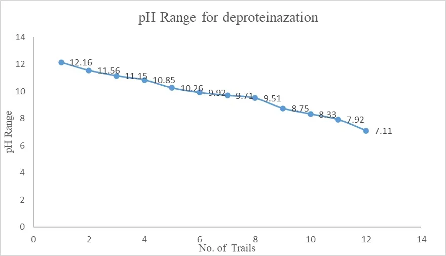
Fig 1 : Variation of pH during the Deproteinization Process
3.2 Effect of pH on Demineralization Efficiency:
The pH variation during the demineralization process was systematically analyzed across four experimental trials in Figure 2. The observed pH values decreased progressively from 8.32 (Trial 1) to 7.01 (Trial 4), indicating a gradual shift from mildly alkaline to near-neutral conditions.
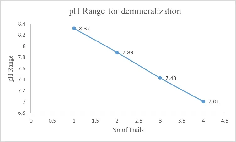
Fig 2 : Variation of pH during the Demineralization Process
3.3 Effect of pH on Deacetylation Efficiency:
The pH variation across different trials (Trail 1 – Trail 16) is presented in Figure 3. A gradual decrease in pH was observed from 13.86 (Trail 1) to 7.12 (Trail 16), indicating a controlled progression of the deacetylation reaction.
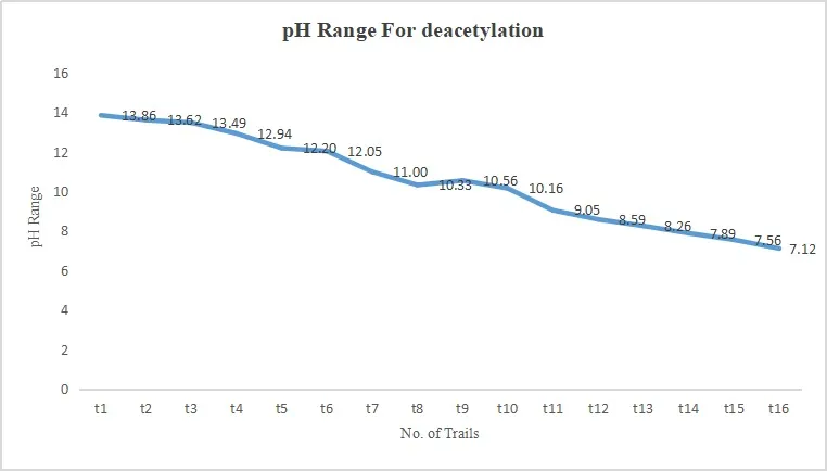
Fig 3 : Variation of pH during the Deacetylation Process
3.4 FTIR Analysis:
The Fourier Transform Infrared (FTIR) spectrum of the chitosan-coated probiotic tablets was analyzed to evaluate the compatibility between formulation components. The FTIR spectrum is presented in Figure 4, and the corresponding peak assignments are summarized in Table 2.
The spectrum exhibited a broad absorption peak in the region of 3270–3300 cm⁻¹, which is attributed to O–H and N–H stretching vibrations, characteristic of Chitosan. A distinct peak observed near 2920 cm⁻¹ corresponds to C–H stretching vibrations. The absorption band around 1640–1650 cm⁻¹ is assigned to amide I (C=O stretching), while the peak near 1540 cm⁻¹ corresponds to amide II (N–H bending vibrations).
Peaks observed in the region of 1400–1300 cm⁻¹ indicate C–N stretching and CH bending vibrations. Strong peaks in the range of 1150–1000 cm⁻¹ are associated with C–O–C and C–O stretching, confirming the polysaccharide backbone structure.
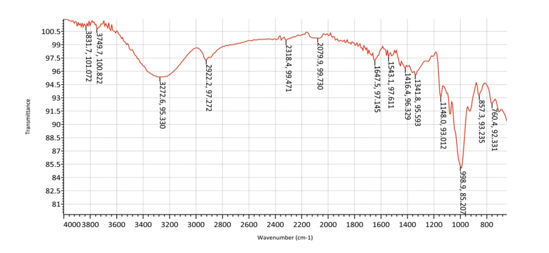
Fig 4 : FTIR Spectrum
Table 2: FTIR Peak Assignment
|
Wavenumber (cm⁻¹) |
Functional Group |
Assignment |
|
~3270–3300 |
O–H / N–H |
Stretching vibrations |
|
~2920 |
C–H |
Aliphatic stretching |
|
~1640–1650 |
C=O |
Amide I |
|
~1540 |
N–H |
Amide II |
|
~1400–1300 |
C–N / CH |
Stretching and bending |
|
~1150–1000 |
C–O–C / C–O |
Polysaccharide structure |
The retention of all characteristic peaks without significant shift or disappearance indicates the absence of chemical interaction between the probiotic components and the polymer. This confirms the compatibility of the formulation components and suggests that the formulation process did not alter the chemical structure of Chitosan.
3.5 UV–Visible Analysis:
The UV–Visible spectroscopic analysis of the chitosan-coated probiotic tablets was carried out to evaluate the analytical behavior of the formulation. The obtained spectrum/graph is presented in Figure 5.
The analysis showed measurable absorbance values across the trials, indicating that the formulation exhibits a consistent analytical response. The absorbance values ranged from 0.197nm to 0.124nm, demonstrating a gradual variation in absorbance.
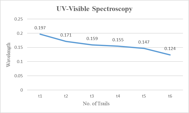
Fig 5 : UV-Visible Spectrum
The observed variation in absorbance may be attributed to differences in concentration or dilution during analysis. The results indicate that the formulation can be effectively analyzed using UV–Visible spectroscopy at the selected wavelength. The UV–Visible method was further utilized in the in vitro drug release study for quantification of the released components, confirming its suitability as an analytical technique for the evaluation of the formulation.
3.6 In Vitro Drug Release Study:
The in vitro drug release profile of the chitosan-coated probiotic tablets was evaluated to study the release behavior of the formulation. The release profile is presented in Figure 6.
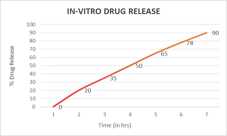
Fig 6 : In vitro drug release profile
The release study demonstrated a gradual and controlled release pattern over a period of 7 hours. An initial release of approximately 20% within 2 hours was observed, followed by a steady increase in drug release. At 4 hours, about 50% of the drug was released, indicating a moderate release rate. The release further increased to 65% at 5 hours and 78% at 6 hours, reaching approximately 90% drug release at 7 hours.
The sustained release behavior can be attributed to the presence of Chitosan coating, which acts as a polymeric barrier and controls the diffusion of probiotic components. The release profile suggests a controlled drug delivery system, which is advantageous for maintaining prolonged therapeutic activity in oral health applications.
3.7 Probiotic Viability (CFU Analysis):
The probiotic viability of the chitosan-coated tablets was evaluated using the colony forming unit (CFU) method to determine the number of viable microorganisms present in the formulation. The results of the CFU analysis are presented in Table 3, Table 4, and Table 5.
Table 3: Probiotic Composition (CFU per Tablet):
|
Ingredient |
Specification |
|
Saccharomyces boulardii |
5 Billion CFU |
|
Lactobacillus rhamnosus |
10 Billion CFU |
|
Total Probiotic Count |
15 Billion CFU |
|
Vitamin D3 |
400 IU |
|
Starch |
Binder/Filler |
The formulation exhibited a total probiotic count of approximately 15 billion CFU per tablet, indicating the presence of a sufficient number of viable microorganisms required for therapeutic activity.
Table 4: CFU Standardization Parameters:
|
Parameter |
Specification |
|
Label Claim (End of Shelf Life) |
≥ 15 Billion CFU |
|
Manufacturing Overfill |
18–20 Billion CFU |
|
CFU Loss Allowance |
20–30% |
The CFU standardization parameters indicate that an overfill was maintained during formulation to compensate for potential loss in viability during processing and storage.
Table 5: Stability and Quality Control Parameters:
|
Test |
Specification |
|
Total Viable Count |
≥ 15 Billion CFU |
|
Microbial Purity |
As per pharmacopeial limits |
|
Moisture Content |
≤ 5% |
|
Disintegration Time |
≤ 20 minutes |
|
Hardness |
3–5 kg/cm² |
|
Friability |
≤ 1% |
The stability and quality control parameters confirm that the formulation meets acceptable pharmacopeial limits, ensuring product stability and safety.
Overall, the results demonstrate that the formulation successfully maintained probiotic viability. The presence of Chitosan coating likely contributed to the protection of probiotic cells against environmental stress, thereby enhancing their stability.
The CFU values obtained are within the acceptable range, indicating that the developed formulation is capable of delivering viable probiotics effectively for oral health applications.
CONCLUSION:
Chitosan-coated probiotic tablets were successfully formulated using Lactobacillus rhamnosus and Saccharomyces boulardii. Chitosan provided an effective protective coating that improved stability and performance of the formulation. FTIR analysis confirmed compatibility of the components, while UV–Visible spectroscopy supported evaluation of the system. The in vitro drug release study showed a controlled and sustained release profile, demonstrating the effectiveness of the coating. CFU results confirmed good probiotic viability, ensuring therapeutic potential. Overall, the formulation showed promising characteristics for oral delivery with improved stability, controlled release, and maintained viability. In conclusion, chitosan-coated probiotic tablets represent an effective drug delivery system for probiotic administration.
DECLARATIONS:
Conflict of interest: The authors report no conflicts of interest.
Acknowledgement: This research is a part of B. Tech project work of the second to Sixth authors. Authors gratefully acknowledge Department of Biotechnology, Sri Shakthi Institute of Engineering and Technology.
Funding: The Source of funding is nil.
Ethical Clearance: Nil.
Permission To Reproduce: Nil.
REFERENCES





Yuvarani S., Ashwin Raj P., Devi S., Kamudesh M., Preparation And Evaluation Of Chitosan-Coated Probiotic Tablets For Oral Health Applications, Int. J. of Pharm. Sci., 2026, Vol 4, Issue 8, 3340-3351. https://doi.org/10.5281/zenodo.22045741
 10.5281/zenodo.22045741