

We use cookies to ensure our website works properly and to personalise your experience. Cookies policy
SS Institute of Medical Science and Research Center (SSIMS & RC), Janashankara, NH-4 Bypass Road, Davangere, Karnataka, India 577005
Primary ciliary dyskinesia (PCD) is a rare, often underdiagnosed genetic disorder that impairs the normal clearance of mucus from the airways, leading to recurrent infections and progressive lung damage. Because its symptoms frequently overlap with other chronic respiratory conditions, and many patients lack the classical hallmark of situs inversus, the diagnosis is often missed or delayed, particularly in adolescents. We describe the case of a 16-year-old boy, previously diagnosed with PCD and bronchiectasis in early childhood, who was admitted with a short history of fever, worsening cough, and vomiting, later complicated by blood in his urine and abdominal pain. On examination, he was found to be underweight for his age, with reduced air entry and crackles over the left side of his chest. Imaging told a clear story: his chest X-ray showed a classic "honeycomb" pattern, while a CT scan confirmed widespread bronchiectasis, and an abdominal ultrasound revealed a distended bladder with debris, pointing toward a urinary tract infection alongside his respiratory illness. He responded well to intravenous antibiotics, nebulization, and supportive care, improving steadily before discharge. This case is a reminder that PCD can present quite variably, and the absence of textbook features like organ mirror-imaging should never rule it out, especially in a young patient with a history of recurring chest infections. Recognizing this early, backed by consistent clinical and radiological clues even without genetic testing, allows for timely, coordinated care that can meaningfully slow disease progression and protect long-term lung health.
Primary ciliary dyskinesia (PCD) is an rare genetic condition that leads to abnormalities in the structure and function of cilia, tiny hair-like attachment that help move out mucus and debris from the respiratory passages. Any defects in the functioning of the cilia result in reduced mucus clearance, leading to frequent respiratory infections in individuals with primary ciliary dyskinesia (1).
Primary ciliary dyskinesia usually occurs at birth with the incidence being around 1 in 10,000 to 1 in 20,000 births. It is however an underdiagnosed condition because of its varied presentations and unawareness about the disease. Patients usually suffer from continuous cough, recurrent sinusitis, and frequent ear infections, which might lead to progressive lung damage (1).
The proteins associated with ciliary structure and functions are abnormal in people with primary ciliary dyskinesia due to mutations in specific genes. Some of the gene mutations responsible for primary ciliary dyskinesia include mutations of DNAH5, CCNO, FOXJ1, and ODAD4 genes. Mutations of any of these genes lead to abnormalities such as defective dynein arms, improper development of cilia, or abnormal ciliary movement, resulting in ineffective removal of the mucus from airways (2, 3, 4, 5).
Diagnosis of PCD is challenging because it generally needs to be done through a combination of clinical assessment along with some specific tests. Late diagnosis may lead to deterioration of respiratory function and complications. Early diagnosis and proper treatment are crucial for improving outcomes. (1, 2, 3, 4, 5).
CASE PRESENTATION
A 16-year-old male, born to non-consanguineous parents and developed normally with appropriate immunization for his age, presented with fever, cough and vomiting for 1 day. Fever was insidious in onset, gradually progressive, high-grade, and without associated features of chills or rigors. Similarly, cough was progressive and he had a single episode of vomiting without any indication of blood or other features. During hospitalization the patient had complaints of 3 episodes of vomiting and 1 episode of blood in urine and pain in abdomen.
The child had a history of multiple previous hospitalization for fever, cough, vomiting and reduced appetite for which at the age of 6 the patient was diagnosed with Primary Ciliary dyskinesia (PCD) with Bronchiectasis and treated with antibiotics including Cefixime for 5 days and 2 puffs of MDI Foracort (Budesonide and Formoterol) per day. The patient was also advised to use Hypertonic nebulization and also to continue to weekly chest physiotherapy.
On examination, the child was conscious and oriented with an IV cannula in place. His vitals were pulse rate 130/min, respiratory rate 28cycles/min and oxygen saturation 96% on room. Similarly, respiratory examination, percussion revealed impaired note over the left infra-axillary, infrascapular and interscapular regions. On auscultation, bilateral crepts and wheeze were present in lung fields, vocal resonance was decreased over the left infra-axillary, suprascapular and infrascapular regions. The other systemic examination parameters were within normal limits. +-x
The anthropometric parameters revealed that the weight was 30 kg (<3rd centile), height was 156cm and BMI was 12.33kg/m2 (<3rd centile). The Chest X-ray showed a “HONEY COMB” appearance, suggestive of chronic lung disease. The Computed tomography (CT) revealed bilateral bronchiectasis, confirming chronic suppurative lung disease. The patient showed gradual clinical improvement and was discharged in a satisfactory condition after receiving intravenous fluids and antibiotics, antipyretics, antiviral, hyper saline nebulization and supportive management.
Table no:1 Abnormal laboratory investigations
|
Test parameters |
Result(s) |
Reference range |
|
Neturophils |
82% |
34-75% |
|
C-Reactive protein (CRP) |
11.00mg/L |
<5mgL |
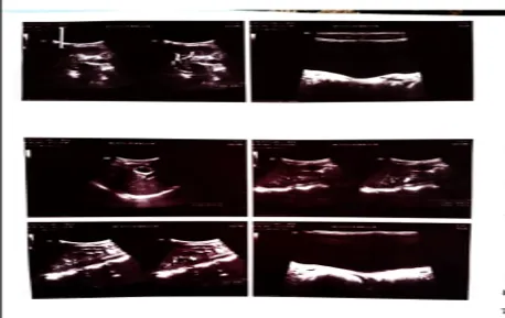
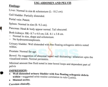
Figure no.1 USG ABDOMEN AND PELVIS
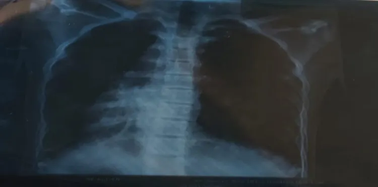
FIGURE NO 2 CHEST X – RAY
DISCUSSION
Primary ciliary dyskinesia (PCD) is a rare and genetically heterogeneous ciliopathy, characterized by impaired mucociliary clearance, resulting in recurrent sino-pulmonary infections, persistent airway inflammation and progressive bronchiectasis . However, its clinical signs sometimes overlap other chronic pulmonary conditions which complicate early detection and result in permanent lung damage (1).
The present case was a 16-year-old male patient with history of fever, chronic productive cough, recurrent respiratory tract infections diagnosed as bronchiectasis due to primary ciliary dyskinesia. Despite the absence of situs abnormalities and other extra-pulmonary signs and symptoms, the patient presented with the characteristic respiratory phenotype of PCD, including recurrent lower respiratory infections and chronic airway disease, as seen in other studies. This confirms previous findings that about half of the individuals with PCD may not have problems with dominance and underlines the importance of a medical facility for diagnosis (1).
Delayed diagnosis is still common, because PCD mimics recurrent pneumonia, asthma or other chronic pus-forming lung diseases, especially in children (2). Our patient also had a history of many respiratory infections before the diagnosis was made. This highlights the importance of considering PCD in teenagers with bronchiectasis and chronic respiratory symptoms.
Although several genetic variations such as DNAH5, CCNO and FOXJ1 have been associated with PCD, we did not find molecular evidence for the PCD in our case. Similar limitations have been reported in resource-poor settings where radiological findings and distinctive clinical features rather than genetic analysis were the main diagnostic tools 2,3,4. The clinical picture of the patient and the radiological data strongly supported the diagnosis even without genetic testing.
Our patient exhibited mild respiratory involvement with progressive bronchiectasis, contrary to previously described cases associated with hydrocephalus, laterality defects or other congenital anomalies (3,4). This highlights the phenotypic variability of PCD and emphasizes that absence of syndromic features should not preclude the diagnosis.
Finding the disease early is important because the right treatments can slow the progress of the illness and preserve lung function. This includes airway clearance therapy, early treatment of respiratory infections, immunization, pulmonary rehabilitation and long-term multidisciplinary follow-up. The severity in patients with this disease is highly variable, and prompt management is required to improve long-term respiratory outcome (5).
This case highlights the importance of suspecting primary ciliary dyskinesia in the differential diagnosis of children and adolescents with idiopathic bronchiectasis and recurrent respiratory infections without situs inversus or other classical features. A better long-term prognosis and an earlier diagnosis through prompt multidisciplinary management may result from increased clinician awareness (1,2,5).
The urinary findings in the present case are clinically relevant as a concurrent urinary tract infection (UTI). During hospitalization, the patient developed hematuria and abdominal pain, and ultrasonography of the abdomen and pelvis revealed a well-distended urinary bladder with free-floating echogenic debris, suggestive of cystitis. UTI in children may present with fever, vomiting, abdominal pain, and hematuria, while cystitis commonly presents with lower urinary tract symptoms and hematuria. In the present case, the clinical and ultrasonographic findings suggest a concurrent UTI; however, in the absence of documented urinalysis and urine culture results, microbiological confirmation cannot be established. Appropriate urine testing and culture are recommended when UTI is suspected in children, particularly in the presence of fever. (6)
CONCLUSION
This case highlights on 16-year-old with known case of Primary Ciliary dyskinesia (PCD) with Bronchiectasis complicated by pneumonia, who presented with presented with fever, cough and vomiting, during hosplization he had complaints of 3 episodes of vomiting and 1 episode of blood in urine and pain in abdomen.
Early radiological findings with chest x ray and CT thorax played crucial role in confirming diagnosis , a “honey comb” appearance and bilateral bronchiectasis suggestive of chronic suppurative lung disease. USG abdomen and pelvis showed well distented urinary bladder with free floating echogenic debris within- suggested that cystitis and minimal ascites.
Multidisciplinary management including intravenous antibiotics , antipyretics, hypertonic saline nebulization, and supportive therapy resulted in gradually clinical improvement.
This case emphasizes the importance of early recognition of acute exacerbation of PCD with UTI with Pneumonia with cholelithiasis, timely imaging and appropriate medical therapy, regular follow-up with chest physiotherapy can prevent disease progression, reduced complications and improves clinical outcomes in paediatric patients with PCD with UTI with Pneumonia with cholelithiasis.
REFERENCES




Pujari Sandhya, Ramkumar B, Pallavi P G, Manoj S Gowda, Primary Ciliary Dyskinesia with Bronchiectasis Presenting as Pneumonia and Urinary Tract Infection in an Adolescent: A Rare Case Report, Int. J. of Pharm. Sci., 2026, Vol 4, Issue 10, 914-918. https://doi.org/10.5281/zenodo.23202616
 10.5281/zenodo.23202616