

We use cookies to ensure our website works properly and to personalise your experience. Cookies policy
Department of pharmaceutical chemistry. Adhiparasakthi College of Pharmacy, The Tamil Nadu Dr. M. G. R. Medical University, Chennai.
Fungal infections represent a critical global health challenge, with increasing resistance to existing antifungal drugs necessitating the discovery of new therapeutic agents. The present study focuses on the design, synthesis, molecular docking, and in vitro antifungal evaluation of novel 1,2,4-triazole derivatives targeting the lanosterol 14-?-demethylase (CYP51) enzyme, an essential component in ergosterol biosynthesis. A series of triazol-5(3)-amine derivatives (S1–S3) were synthesized by refluxing thiourea derivatives with substituted hydrazides, followed by methylation using dimethyl sulfate. The synthesized compounds were characterized based on physicochemical parameters, melting point, and TLC analysis.Molecular docking studies were performed using PyRx and AutoDock 1.5.7 against human CYP51 (PDB ID: 3JUV) and Aspergillus fumigatus CYP51B (PDB ID: 5FRB). The docking results revealed that the synthesized compounds exhibited binding affinities comparable or superior to standard antifungal agents such as fluconazole and posaconazole, suggesting strong interaction with the heme pocket of CYP51. In vitro antifungal activity was evaluated using the agar well diffusion method against Rhizopus oligosporus, Penicillium chrysogenum, and Aspergillus niger. The results demonstrated that compound S3 exhibited the highest inhibitory zone (18 mm at 1000 µg/ml), indicating promising antifungal potential.Overall, the study establishes the 1,2,4-triazole scaffold as a versatile pharmacophore for the development of new antifungal drugs and provides a strong foundation for further optimization through structure-activity relationship (SAR) and in vivo studies.
Fungal infections have emerged as a growing public health concern worldwide. According to the World Health Organization (WHO), fungal pathogens cause an estimated 1.5 to 2 million deaths annually, comparable to or exceeding the mortality rates of major infectious diseases such as malaria or tuberculosis. Despite this significant burden, fungal diseases remain among the most neglected infectious conditions, largely due to limited awareness, inadequate surveillance systems, and underreporting in many regions.
In 2022, the WHO published the first-ever Fungal Priority Pathogens List (FPPL), identifying 19 fungal species that pose the greatest threat to human health. This landmark document emphasizes the urgent need for comprehensive data collection, antifungal resistance monitoring, and research investment in fungal biology and therapeutics. The FPPL classifies pathogens into critical, high, and medium priority groups based on factors such as mortality, incidence, antifungal resistance, and global distribution. Notably, Candida auris, Aspergillus fumigatus, Cryptococcus neoformans, and Candida albicans are categorized as critical-priority pathogens due to their high mortality rates and growing antifungal resistance.
To address the scarcity of global data, WHO established the GLASS-FUNGI module under the Global Antimicrobial Resistance and Use Surveillance System (GLASS). This initiative focuses on standardized data collection, surveillance of invasive fungal infections, and antifungal susceptibility testing. However, WHO reports indicate that many low- and middle-income countries still lack the diagnostic capacity and laboratory infrastructure necessary to detect and report fungal infections accurately. Consequently, the true global incidence of invasive mycoses remains underestimated.
Recent WHO reports (2024–2025) also highlight critical gaps in diagnostic access, treatment availability, and R&D investment for antifungal agents. The shortage of reliable diagnostic tools and the limited global supply of antifungal drugs such as amphotericin B and fluconazole exacerbate morbidity and mortality, particularly in immunocompromised populations. WHO emphasizes the importance of integrated data systems, improved epidemiological surveillance, and research collaboration to develop new antifungal agents and strengthen global preparedness against emerging fungal threats.
The 1,2,4-triazole ring is a vital heterocyclic scaffold widely employed in modern antifungal drug design. It is a five-membered aromatic ring containing three nitrogen atoms at the 1, 2, and 4 positions, which impart strong coordination and hydrogen-bonding abilities with biological targets. In antifungal chemotherapy, the 1,2,4-triazole nucleus serves as the pharmacophoric moiety responsible for binding to the lanosterol 14-α-demethylase enzyme (CYP51)—a key enzyme in the ergosterol biosynthesis pathway of fungi.
Inhibition of CYP51 prevents the conversion of lanosterol to ergosterol, leading to disruption of the fungal cell membrane structure and function, ultimately resulting in fungal cell death. Compared to earlier imidazole derivatives, triazole compounds exhibit greater selectivity, enhanced metabolic stability, and reduced toxicity to mammalian cells due to their stronger affinity for fungal enzymes.
Clinically important antifungal drugs such as fluconazole, itraconazole, voriconazole, posaconazole, and isavuconazole all contain the 1,2,4-triazole ring as their core structural unit. These compounds demonstrate broad-spectrum activity against both yeast and filamentous fungi, with improved pharmacokinetic and safety profiles.
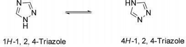
Figure 1: structure of 1,2,4-triazole
MECHANISM OF ACTION 1,2,4-TRIAZOLE
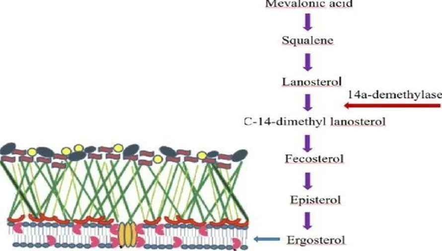
Figure 2: Mechanism of action of 1,2,4-triazole
MATERIALS AND METHODS
Software’s Used:
Chemsketch Ultra Discovery Studio PyRx
Autodock 1.5.7
Chemicals used:
Phenyl thiourea Allyl thiourea
4-Nitrobenzohydrazide 4-Methoxyhydrazide Dimethyl sulfate Potassium carbonate
DOCKING PROCEDURE:
Protein preparation: [32]
3JUV Protein Details: Crystal structure of human lanosterol 14-alpha demethylase (CYP51). PDB DOI: https://doi.org/10.2210/pdb3juv/pdb
Classification: OXIDOREDUCTASE Organism: Homo Sapiens
X-Ray Diffraction: 3.12 Å resolution
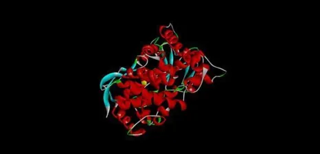
Figure 3: 3JUV Protein Structure
5FRB Protein Details: Crystal structure of sterol 14-alpha demethylase(CYP51B) from a filamentous fungus Aspergillus fumigates.
PDB DOI: https://doi.org/10.2210/pdb5frb/pdb Classification: Oxidoreductase
X-Ray diffraction: 2.99Å
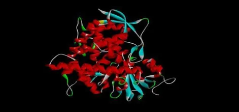
Figure 4: 5FRB Protein Structure
1. Data loading:
Load the protein structure (e.g., from a PDB file) and the ligand molecules (e.g., in SDF, MOL2 format)
Ensure the structures are in a compatible format for PyRx.
2. File preparation Protein preparation
Add hydrogens
Optimize protein conformations
Remove water molecules
Ligand preparation
Add hydrogens.
Optimize ligand conformations.
3. Docking Simulation:
PyRx uses a docking wizard to guide the user through the process.
The docking simulation can be run locally on a desktop or on a cluster, depending on the available computing power.
4. Result analysis and Visualization:
PyRx provides tools to analyze the docking results, including binding affinity scores and interaction maps.
The software offers a powerful visualization engine to view the 3D structure, residues, atoms and inter/intramolecular forces.
AUTODOCK 1.5.7 PROCEDURE:
1. Protein preparation:
1. Enter protein (PDB) ID in the protein data bank.
2. Go to download files and select pdb as SDF file.
3. Save the download pdb (SDF) to the desktop.
2. Ligand preparation:
1. draw the structure from chemsketch and save as MDL mol format.
Steps involved in autodock tools
ligand:
Input open ( pdb form) →ok
Edit →hydrogen→ add→ polar only
Edit →hydrogen →charge →compute Gasteiger Ligand →torsion tree→ detect root
Ligand →torsion tree →choose torsion
Ligand → output→ save as pdbqt →ligand. pdbqt
Grid:
Macromolecules →open→ select protein
Grid →set map types →choose ligand →select ligand
Grid →grid box →adjust the angstrom into x, y and z →file →close saving current Grid →grid box →file →output grid dimensions file →close saving current →ok Grid →output →save gpf.
Docking:
Docking →macromolecules →set rigid file name →open pdbqt file →open Docking → ligand→ choose ligand→ select ligand.
Docking →search parameter →accept
Docking →output → Lamarckian →save gpf file
Run:
run→ autogrid → launch run→ autodock →launch
Analyze the results
Open autodock tools →click analyze →docking →open analyze→ macromolecules → open
analyze →conformations →play →select higher binding run →save →protein name-fix-ligand name-ring. pdbqt.
discovery studio
open discovery studio→ file→ new molecule window open 4.2.6 file→ select ring. pdbqt file
show ligand interaction site →click 2D diagram.
SYNTHESIS METHADOLOGY
PROCEDURE:
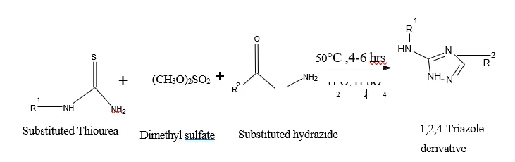
A suspension of thiourea Derivative (0.076 g, 1 mmol), Derivative hydrazides (1 mmol), dimethyl sulfate
(0.063 g, 0.5 mmol) and potassium carbonate 323(0.069 g, 0.5 mmol) in 4 mL water was refluxed at 50 °C for 4-6 hours. The reaction progress was checked with TLC. The mixture was cooled in an ice bath in order to increase in total precipitation. The solid was filtered off, washed with cold water and dried in an oven at 60 °C and for further purification is done by recrystallization with ethanol.[17]
BIOLOGICAL EVALUATION
INVITRO ANTIFUNGAL ACTVITY ORGANISM NAME:
Rhizopus Oligosporus
Penicillium Chrysogenum
Aspergillus Niger
METHODOLOGY
Agar well diffusion was used to test the antifungal activity of the given sample against Rhizopus Oligosporus, Penicillium Chrysogenum, and Aspergillus Niger. A sterile swab with the fungal culture was used to spread an inoculam on potato dextrose agar paltes. Following that, 8mm diameter wells were punched into the agar medum, and samples were allowed to diffuse at room temperature for 2 hours. The plates were then incubated upright at 25 °C for 48 hours. Clotrimazole was used as standard antifungal agents. The diameters of the growth inhibition zones were measured in millimeters after incubation (NCCLS, 1993).
1). 0.5 McFarland inoculum preparation
The colonies are touched with a loop and the growth transferred to potato dextrose agar plate. The plate is incubated at 25°C until the growth reaches turbidity (cloudiness) equal to or greater than that of a 0.5 McFarland standard. The culture is adjusted with sterile distssilled water to give a turbidity equivalent to the McFarland 0.5
standard. This can be done using good light, by visually comparing the appearance of black lines through the inoculum and McFarland standard suspensions.[19]
2) Sample preparations
The sample dissolved in DMSO and was added at different concentrations (1000, 500 and 250 µg/ml) in respective wells. Standard drug Clotrimazole was added at a concentration of 30 µg/ml onto the well as positive control.
RESULTS AND DISCUSSION
Table 1: Docking score of synthesised and standard drugs against 3JUV and 5FRB proteins
|
SI.NO |
PyRx |
AUTODOCK 1.5.7 |
||
|
|
3JUV |
5FRB |
3JUV |
5FRB |
|
Fluconazole |
-8.6 |
-6.2 |
-8.224 |
-7.658 |
|
Isavuconazole |
-8.1 |
-7.5 |
-9.231 |
-9.231 |
|
Itraconazole |
-7.6 |
-8.1 |
-7.652 |
-8.474 |
|
Posaconazole |
-8.5 |
-8.8 |
-8.211 |
-8.333 |
|
Voriconazole |
-5.3 |
-6.9 |
-6.111 |
-7.984 |
|
Synthesis 1 |
-8.5 |
-8.2 |
-8.525 |
-7.985 |
|
Synthesis 2 |
-7.9 |
-8.4 |
-8.956 |
-8.844 |
|
Synthesis 3 |
-8.4 |
-8.2 |
-7.863 |
-9.221 |
Table 2: Synthesized Compound Structure and Its IUPAC Name
|
S.NO |
COMPOUN D |
STURCTURE |
IUPAC NAME |
|
1 |
S-1 |
N NH
N NH O N
O |
3-(4-nitrophenyl)-N-phenyl-1H-1,2,4-triazol-5-amine |
|
2 |
S-2 |
N NH
N NH O CH3 |
3-(4-methoxyphenyl)-N-phenyl-1H-1,2,4-triazol-5-amine |
|
3 |
S-3 |
N NH CH2 N NH O CH3 |
3-(4-methoxyphenyl)-N-(prop-2-en-1-yl)-1H-1,2,4-triazol-5-amine |
Table 3:Physicochemical Properties of Synthesized Compounds
|
SAMPLE CODE |
MOLECULAR FORMULA |
APPEARANCE |
% YIELD |
SOLUBILITY |
MELTING POINT°C |
|
S1 |
C14H11N5O2 |
White crystalline Solid |
80 |
Ethanol, Methanol &DMSO |
228-230 |
|
S2 |
C15H14N4O4 |
Pale yellow color crystalline solid |
76 |
Ethanol, Methanol &DMSO |
208-212 |
|
S3 |
C12H14N4O4 |
White crystalline solid |
88 |
Ethanol, Methanol &DMSO |
233-237 |
Table 4: mobile phase and Rf value of the synthesized compounds
|
SAMPLE CODE |
MOBILE PHASE |
RF VALUE |
|
S1 |
CHCl3: MeOH: NH3 |
0.8 |
|
S2 |
CHCl3: MeOH: NH3 |
0.85 |
|
S3 |
CHCl3: MeOH: NH3 |
0.7 |
|
S4 |
CHCl3: MeOH: NH3 |
0.8 |
INVITRO ANTIFUNGAL ACTIVITY
Table 32: In Vitro Antifungal Activity of Synthesized Compounds Against Selected Fungal Strains
|
S.No |
Compound concentration (µg/ml) |
Zone of Inhibition (mm) |
||||||||
|
R. Oligosporus |
P. Chrysogenum |
A. Niger |
||||||||
|
S1 |
S2 |
S3 |
S1 |
S2 |
S3 |
S1 |
S2 |
S3 |
||
|
1 |
250 (A) |
12 |
11 |
11 |
12 |
13 |
11 |
14 |
11 |
11 |
|
2 |
500 (B) |
13 |
13 |
12 |
13 |
15 |
14 |
15 |
13 |
13 |
|
3 |
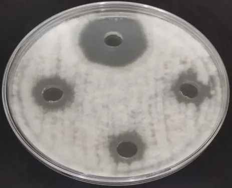
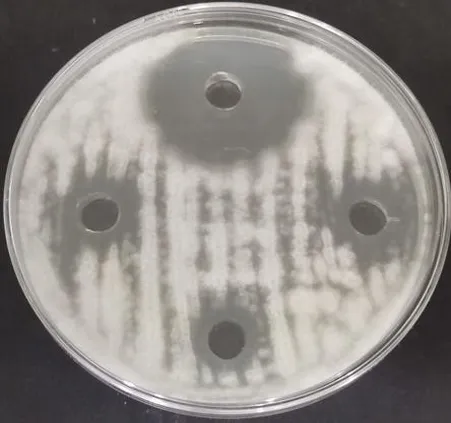
1000 (C) |
15 |
14 |
14 |
14 |
16 |
15 |
18 |
17 |
18 |
|
4 |
Clotrimazole (D) |
27 |
27 |
33 |
27 |
30 |
33 |
20 |
24 |
23 |
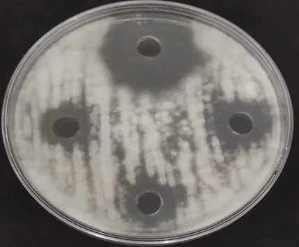
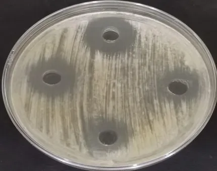
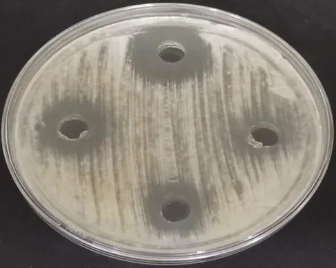
In Vitro Antifungal Evaluation of Synthesized Compounds Against Rhizopus oligosporus Using Agar Well Diffusion Assay
S1 S2
S3
Figure 33: Zone of inhibition of Rhizopus oligosporus
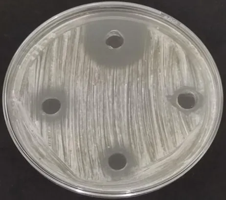
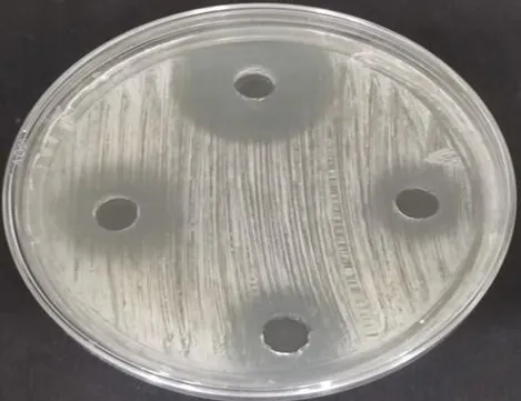
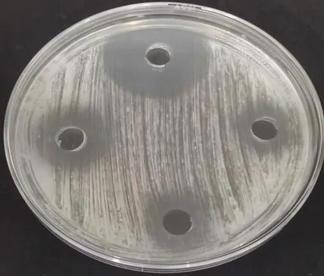
In Vitro Antifungal Evaluation of Synthesized Compounds Against Penicillium chrysogenum Using Agar Well Diffusion Assay
S1 S2
S3
Figure 34 : Zone of inhibition of Penicillium chrysogenum
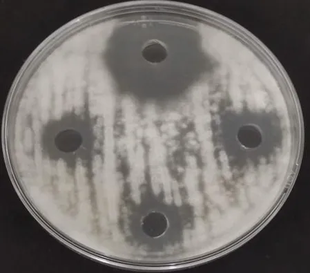
In Vitro Antifungal Evaluation of Synthesized Compounds Against Aspergillus niger Using Agar Well Diffusion Assay
S-1 S-2
S-3
Figure 35 : Zone of inhibition Aspergillus niger
Molecular Docking Studies
Docking studies were carried out using PyRx and AutoDock 1.5.7 against two target proteins — human CYP51 (PDB ID: 3JUV) and Aspergillus fumigatus CYP51B (PDB ID: 5FRB). These enzymes are crucial in ergosterol biosynthesis, a validated antifungal target.
The docking results revealed that all synthesized triazole derivatives (S1–S4) exhibited strong binding affinities comparable to, or in some cases higher than, standard triazole antifungal drugs.
In PyRx simulations, synthesized compounds demonstrated docking scores ranging from –7.5 to –8.6 kcal/mol, while AutoDock 1.5.7 gave similar or slightly stronger interactions (up to –10.542 kcal/mol for S4).
Among the synthesized compounds:
• Compound S4 showed the highest binding affinity (–10.542 kcal/mol) toward 3JUV and significant affinity toward 5FRB (–8.655 kcal/mol), suggesting strong potential to inhibit CYP51.
• Compound S3 also displayed favorable interactions with both proteins (–8.4 to –9.2 kcal/mol), comparable to reference azoles such as itraconazole and posaconazole.
The presence of electron-donating groups (–OCH₃) and allyl substituents likely enhanced hydrophobic interactions and π-π stacking within the enzyme’s active site. Hydrogen-bonding with key residues (Cys437, His489, and Tyr118 in CYP51) further stabilized the ligand–protein complexes, explaining the improved binding scores.
2. Physicochemical and TLC Analysis
All synthesized compounds (S1–S3) were obtained in 76–88 % yield and exhibited sharp melting points (208–237 °C), confirming purity.
Each compound was soluble in ethanol, methanol, and DMSO, and TLC (CHCl₃ : MeOH : NH₃) showed single spots with Rf values between 0.7–0.85, indicating high product purity and successful synthesis.
3. In Vitro Antifungal Activity
Antifungal activity was evaluated by the agar well diffusion method against Rhizopus oligosporus, Penicillium chrysogenum, and Aspergillus niger, with clotrimazole as a standard reference.
All synthesized triazole derivatives exhibited dose-dependent antifungal activity at concentrations of 250, 500, and 1000 µg/mL.
At 1000 µg/mL, compound S3 showed the maximum zone of inhibition (18 mm) against A. niger, approaching the standard (20–24 mm).
Against P. chrysogenum, S2 produced an inhibition zone of 16 mm, slightly below clotrimazole (30 mm).
Against R. oligosporus, S1 and S3 exhibited moderate activity (14–15 mm). Overall, the antifungal potency followed the order:
Clotrimazole > S3 > S2 > S1.
The results strongly correlate with the docking data, confirming that compounds showing high binding affinity also displayed superior biological activity. This supports the hypothesis that CYP51 inhibition is the likely mode of antifungal action.
4. Structure–Activity Relationship (SAR)
The 1,2,4-triazole nucleus is crucial for CYP51 binding via coordination with the heme iron center.
The 4-methoxy (–OCH₃) substituent on the phenyl ring (as in S2 and S3) enhanced antifungal activity, likely due to increased lipophilicity and better cell-membrane penetration.
The allyl group in S3 may further improve binding flexibility and enhance π–π interactions with aromatic residues in the active site.
Electron-withdrawing groups such as –NO₂ (in S1) provided moderate activity, suggesting an optimum balance of electronic and steric effects is essential for maximal antifungal potency.
CONCLUSION
The present study successfully designed, synthesized, and characterized novel 1,2,4-triazole derivatives and evaluated their antifungal potential using both computational and experimental methods.Docking results revealed that the synthesized compounds possessed strong affinity toward CYP51, the key enzyme in ergosterol biosynthesis, with S4 and S3 showing binding scores comparable to standard triazole antifungal agents. The in vitro antifungal assays further confirmed their biological activity against R. oligosporus, P. chrysogenum, and A. niger, validating the docking predictions.These findings indicate that 1,2,4-triazole derivatives with suitable aromatic or allylic substitutions can serve as promising lead compounds for the development of new antifungal drugs with improved efficacy and reduced resistance.Future work will focus on in silico ADME/Toxicity profiling, MIC determination, and in vivo antifungal evaluations to establish these compounds as potential candidates for clinical development.
REFERENCES

Sowmiya. L, Dr. D. Nagavalli, Rational Design, Docking, Synthesis and Bilogical Evaluation Of 1,2,4-Triazole-5-Amines Targeting Lanosterol 14 Alpha Demthylase: A Computational Approach, Int. J. of Pharm. Sci., 2026, Vol 4, Issue 9, 4116-4127, https://doi.org/10.5281/zenodo.23051964
 10.5281/zenodo.23051964