View Article

  • Rats With Epilepsy Caused by Kainic Acid or Quinolinic Acid Were Used to Test the Neuroprotective and Cognitive-Enhancing Effects of A Herbal Extract

  • 1. Late Laxmibai Phadtare College of Pharmacy, DBATU University, Kalamb, Pune 413114, Maharashtra, India.
    2. Guide, Late Laxmibai Phadtare College Of Pharmacy, DBATU University, Kalamb, Pune 413114, Maharashtra, India.
    3. Principal, Late Laxmibai Phadtare College Of Pharmacy, DBATU University, Kalamb, Pune 413114, 

Abstract

Background: Recurrent seizures, neuronal degeneration, oxidative stress, neuroinflammation, and cognitive impairment are the hallmarks of epilepsy, a long-term neurological condition. Because they resemble the neuropathological and behavioural characteristics of temporal lobe epilepsy, excitotoxic substances like quinolinic acid (QA) and kainic acid (KA) are frequently employed to create experimental epilepsy in mice. In experimental models of epilepsy, herbal extracts rich in polyphenols, flavonoids, and alkaloids have shown encouraging neuroprotective, antioxidant, anti-inflammatory, and cognition-enhancing qualities. Objective: The current study sought to assess the neuroprotective and cognitive-enhancing benefits of a standardized herbal extract in rats with epilepsy caused by KA or QA and to explore potential mechanisms of action. Methods: Five groups (n = 6–8) of adult Wistar rats were randomly assigned to receive either standard therapy, epileptic control, normal control, or two treatments with herbal extract. According to the experimental methodology, a single injection of either QA or KA (10 mg/kg, i.p.) caused epilepsy. For 14–21 days, the herbal extract was taken orally. Seizure scoring, locomotor activity, passive avoidance, Y-maze, and Morris water maze tests were among the behavioral evaluations used to assess memory and learning. To identify oxidative stress indicators including malondialdehyde (MDA), reduced glutathione (GSH), superoxide dismutase (SOD), and catalase, biochemical studies were carried out.Additionally assessed were neuroinflammatory indicators such as interleukin-1? (IL-1?), tumor necrosis factor-? (TNF-?), and neuronal damage markers. To evaluate neuronal deterioration and neuroprotection, hippocampus areas were examined histopathologically. Results: When compared to the epileptic control group, treatment with the herbal extract significantly decreased the frequency and severity of seizures (p < 0.05). By improving memory retention and spatial learning in behavioral paradigms, the extract enhanced cognitive function. MDA levels were significantly lowered and endogenous antioxidant defenses, such as GSH, SOD, and catalase levels, were significantly restored. Additionally, the herbal extract shielded hippocampus neurons from excitotoxic injury and reduced the generation of neuroinflammatory cytokines. The treated animals' hippocampus architecture was preserved and there was less neuronal loss, according to histopathological results. These effects were similar to those seen with the common antiepileptic medication.Conclusion: The results indicate that the herbal extract has important neuroprotective and cognitive-enhancing properties against seizures in rats caused by KA or QA. Its anti-inflammatory, anti-apoptotic, and antioxidant qualities may be responsible for the reported advantages. As a result, the herbal extract could be a viable treatment option for the treatment of neuronal damage and cognitive impairment brought on by epilepsy

Keywords

Epilepsy, Kainic Acid, Quinolinic Acid, Herbal Extract, Neuroprotection, Cognitive Enhancement, Oxidative Stress, Neuroinflammation, Rats

Introduction

× Popup Image

One of the most common chronic neurological conditions in the world, epilepsy affects around 50 million individuals and has a significant impact on society, the economy, and healthcare. Recurrent, unprovoked seizures brought on by aberrant, excessive, and synchronized brain neuronal discharges are the hallmark of the illness. Almost one-third of patients still have uncontrollable seizures despite the availability of various antiepileptic medications (AEDs), and many endure side effects such as drowsiness, cognitive impairment, mental disorders, hepatotoxicity, and drug resistance. Therefore, one of the biggest challenges in epilepsy research continues to be the creation of safer and more successful treatment techniques. [1-8]

Epilepsy is becoming more widely acknowledged as a condition linked to oxidative stress, neuroinflammation, progressive neuronal damage, and cognitive impairment in addition to repeated seizures. Deficits in learning, memory, attention, and executive function are common in patients with temporal lobe epilepsy, which greatly lowers their quality of life. Excitotoxic neuronal damage, a pathogenic process driven by excessive activation of glutamate receptors that results in calcium overload, mitochondrial malfunction, oxidative stress, and ultimately neuronal death, is mostly responsible for these neurological problems. As a result, current research focuses on preventing neurodegeneration and maintaining cognitive function in addition to seizure management.[9-16]

To study the processes behind epileptogenesis and to assess possible treatments, experimental models of epilepsy have been widely used. Because they closely mimic numerous clinical and behavioral characteristics of real temporal lobe epilepsy, kainic acid (KA) and quinolinic acid (QA)-induced epilepsy are the most extensively recognized of these models. As a strong agonist of ionotropic glutamate receptors, especially kainate receptors, kainic acid causes oxidative stress, gliosis, neuronal degeneration, extended seizure activity, and hippocampus injury. Learning and memory losses are severe when KA is administered because it causes selective neuronal death in hippocampus areas including CA1, CA3, and the dentate gyrus. Similar to this, quinolinic acid, an endogenous metabolite of the kynurenine pathway, functions as a selective agonist of the N-methyl-D-aspartate (NMDA) receptor and causes excitotoxic neuronal damage by generating reactive oxygen species and excessive calcium influx. QA-induced neurotoxicity is a useful paradigm for researching neuroprotective therapies since it is linked to neuronal degeneration, neuroinflammation, mitochondrial malfunction, and cognitive impairment.[17-34]

A growing body of research indicates that oxidative stress is a key factor in the pathophysiology of epilepsy and neuronal damage brought on by seizures. Lipid peroxidation, protein oxidation, DNA damage, and neuronal death are the outcomes of natural antioxidant defense mechanisms being overloaded by excessive formation of reactive oxygen species (ROS) and reactive nitrogen species (RNS) during epileptic episodes. Both clinical research and experimental epilepsy models have shown elevated levels of malondialdehyde (MDA) and decreased activity of antioxidant enzymes such glutathione (GSH), catalase (CAT), and superoxide dismutase (SOD). Moreover, pro-inflammatory cytokines such as interleukin-1 beta (IL-1β), interleukin-6 (IL-6), and tumor necrosis factor-alpha (TNF-α) are released when seizures activate microglia and astrocytes. These cytokines cause neuronal hyperexcitability and neurodegeneration. Thus, medicinal substances with anti-inflammatory and antioxidant qualities may be quite helpful in reducing epileptic brain damage.[34-40]

Due to their long history of traditional usage, acceptable safety profiles, and multitarget pharmacological activities, medicinal plants and herbal medicines have garnered significant attention as complementary and alternative therapy alternatives for neurological illnesses. Preclinical studies have shown that a variety of phytochemicals, including as flavonoids, polyphenols, alkaloids, terpenoids, and phenolic acids, have neuroprotective, anticonvulsant, antioxidant, anti-inflammatory, and cognition-enhancing properties. These bioactive substances have the ability to control calcium homeostasis, limit oxidative stress, inhibit neuroinflammatory signaling pathways, alter neurotransmitter systems, and stop neuronal death. As a result, plant extracts are intriguing options for treating epilepsy and the neurological issues it causes. [37-45]

Recent studies have shown that a number of herbal extracts provide protection against cognitive impairments and experimentally produced seizures. Enhancement of endogenous antioxidant defenses, decrease of lipid peroxidation, attenuation of inflammatory cytokine production, regulation of glutamatergic and GABAergic neurotransmission, and maintenance of hippocampal neuronal integrity have all been linked to these effects. However, there is still much to learn about the therapeutic potential of many medicinal plants, especially when it comes to neurodegeneration and cognitive impairment linked to seizures.[41-47]

Many bioactive phytoconstituents with antioxidant and neuroprotective qualities are found in the herbal extract used for this study. According to preliminary pharmacological research, the extract may have strong anti-inflammatory, free radical scavenging, and cognitively enhancing properties. Nevertheless, a thorough assessment of its effectiveness against excitotoxin-induced seizures and associated neuronal injury has not yet been conducted. It is thus necessary to conduct a thorough evaluation of its potential for neuroprotection and cognitive enhancement. The goal of this study was to assess the herbal extract's neuroprotective and cognitive-enhancing properties in rats with epilepsy caused by kainic acid or quinolinic acid. Learning, memory, and seizure severity were all assessed behaviorally. Oxidative stress and antioxidant levels were assessed biochemically, while hippocampus architecture and neuronal integrity were examined histopathologically. Treatment with the herbal extract was predicted to improve cognitive function, reduce seizure-induced neuronal damage, enhance antioxidant defences, and reduce neuroinflammatory responses. The results of this study may aid in the creation of innovative plant-based treatment approaches for the treatment of epilepsy and related cognitive deficits.[44-50]

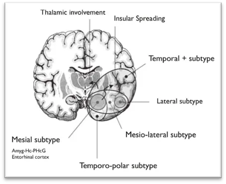
Image No.01: Epilepsy and Hippocampal

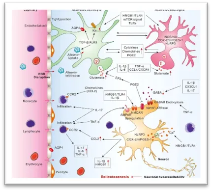
Image No.02: Quinolinic Acid-Induced Excitotoxicity

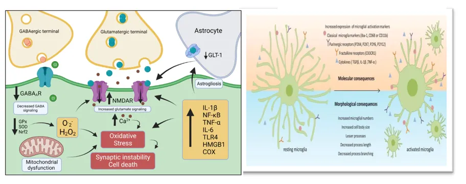
Image No.03: Oxidative Stress and Neuroinflammation in Epilepsy

Types of epilepsy:

A. Based on Seizure Onset

  1. Focal Epilepsy (Partial Epilepsy)
  • Focal aware seizures
  • Focal impaired awareness seizures
  • Focal to bilateral tonic-clonic seizures
  1. Generalized Epilepsy
  • Absence seizures
  • Tonic-clonic seizures
  • Myoclonic seizures
  • Tonic seizures
  • Clonic seizures
  • Atonic seizures
  1. Combined Generalized and Focal Epilepsy
  • Dravet syndrome
  • Lennox-Gastaut syndrome
  1. Unknown Epilepsy
  • Unclassified or undetermined seizure onset

B. Based on Etiology (Cause)

  1. Structural epilepsy
  2. Genetic epilepsy
  3. Infectious epilepsy
  4. Metabolic epilepsy
  5. Immune-mediated epilepsy
  6. Unknown etiology epilepsy

C. Clinically Important Types of Epilepsy

  1. Temporal lobe epilepsy (TLE)
  2. Frontal lobe epilepsy
  3. Occipital lobe epilepsy
  4. Parietal lobe epilepsy
  5. Juvenile myoclonic epilepsy
  6. Childhood absence epilepsy
  7. Benign Rolandic epilepsy
  8. Reflex epilepsy
  9. Status epilepticus
  10. Drug-resistant (refractory) epilepsy

Signs and Symptoms of Epilepsy

  • Recurrent seizures (repeated episodes of abnormal brain activity)
  • Loss of consciousness or awareness
  • Uncontrolled jerking movements of the arms and legs (convulsions)
  • Muscle stiffness or rigidity
  • Staring spells (blank stare for a few seconds)
  • Confusion during or after a seizure
  • Temporary loss of responsiveness
  • Sudden falls due to loss of muscle tone
  • Tongue biting
  • Loss of bladder or bowel control
  • Memory problems
  • Difficulty concentrating
  • Fatigue and drowsiness after a seizure (postictal state)
  • Headache following a seizure
  • Behavioral, emotional, or mood changes such as anxiety or irritability

Models used in the treatment of epilepsy:

1. Maximal Electroshock Seizure (MES) Model

For testing antiepileptic medications against generalized tonic-clonic seizures, the MES model is among the most established and trustworthy experimental models. In this technique, a regulated electrical shock is applied through corneal or ear electrodes to Wistar rats in order to produce seizures. Tonic hind-limb extension, which is the main endpoint for evaluating anticonvulsant activity, is produced by the electric shock. Medications that stop or lessen hind-limb extension are thought to be useful in preventing generalized seizures. Since several therapeutically useful antiepileptic medications, such as carbamazepine and phenytoin, exhibit action in this test, the model is highly repeatable, easy to apply, and has great predictive validity. Compounds that limit the spread of seizures by blocking sodium channels or altering excitatory neurotransmission can be found using MES.

Advantages

  • Simple and reproducible.
  • Suitable for screening anticonvulsant drugs.
  • Mimics generalized tonic-clonic seizures.

Limitation

  • Does not reproduce chronic epilepsy or cognitive impairment.

2. Pentylenetetrazole (PTZ) Model:

One of the most used models for chemically inducing seizures in Wistar rats is the PTZ model. PTZ, a GABA_A receptor antagonist, causes neuronal hyperexcitability and seizures by reducing inhibitory neurotransmission in the brain. Animals have myoclonic jerks, clonic convulsions, tonic seizures, and even status epilepticus after intraperitoneal injection. This model is very helpful for researching myoclonic epilepsy and absence seizures, as well as for assessing medications that increase GABAergic neurotransmission. The paradigm is ideal for anticonvulsant screening because PTZ-induced seizures have a quick onset and reliable behavioral symptoms. A kindling paradigm that simulates epileptogenesis may also be established by repeatedly administering subconvulsive doses of PTZ.

Advantages

  • Easy to establish.
  • Useful for evaluating GABA-mediated anticonvulsant activity.
  • Suitable for acute and kindling studies.

Limitation

  • Limited ability to mimic hippocampal neurodegeneration.

3. Kainic Acid (KA) Model:

One of the finest experimental models for temporal lobe epilepsy (TLE) is the Kainic Acid model. A strong glutamate receptor agonist, kainic acid causes excitotoxicity and excessive neuronal excitement. When KA is administered, it causes status epilepticus, which is followed by a latent phase and the emergence of spontaneous recurring seizures that resemble temporal lobe epilepsy in humans. Selective hippocampal neuronal loss, mossy fiber sprouting, gliosis, oxidative stress, neuroinflammation, and cognitive impairments are the hallmarks of the model. The KA model is often used to assess neuroprotective therapeutics, antioxidant treatments, anti-inflammatory substances, and medications that improve cognition because of these pathological characteristics. It is particularly pertinent to research on hippocampal injury and memory loss.

Advantages

  • Closely mimics human temporal lobe epilepsy.
  • Produces hippocampal neurodegeneration.
  • Useful for studying cognitive impairment and neuroprotection.

Limitation

  • Higher mortality rate than MES and PTZ.
  • Requires careful monitoring of animals.

SUMMARY:

Recurrent seizures, cognitive decline, oxidative stress, neuroinflammation, and neuronal degeneration are all hallmarks of epilepsy, a long-term neurological condition. To study epileptogenesis and assess possible treatments, experimental models including Maximal Electroshock Seizure (MES), Pentylenetetrazole (PTZ), Kainic Acid (KA), and Quinolinic Acid (QA) are frequently utilized. These include KA and QA models, which closely resemble hippocampal injury and temporal lobe epilepsy. Significant neuroprotective, antioxidant, anti-inflammatory, anticonvulsant, and cognitive-enhancing properties have been shown by herbal extracts high in flavonoids, polyphenols, and alkaloids. These characteristics imply that herbal treatments may provide viable substitute methods for lessening neuronal damage brought on by seizures and enhancing cognitive results in epilepsy.

REFERENCES

  1. World Health Organization. Epilepsy. Geneva: World Health Organization; 2024. Available from: https://www.who.int/news-room/fact-sheets/detail/epilepsy
  2. World Health Organization. Epilepsy: A Public Health Imperative. Geneva: World Health Organization; 2019.
  3. Fiest KM, Sauro KM, Wiebe S, Patten SB, Kwon CS, Dykeman J, et al. Prevalence and incidence of epilepsy: A systematic review and meta-analysis of international studies. Neurology. 2017;88(3):296–303.
  4. Ngugi AK, Bottomley C, Kleinschmidt I, Sander JW, Newton CR. Estimation of the burden of active and life-time epilepsy: A meta-analytic approach. Epilepsia. 2010;51(5):883–890.
  5. Beghi E. The epidemiology of epilepsy. Neuroepidemiology. 2020;54(2):185–191.
  6. Thurman DJ, Beghi E, Begley CE, Berg AT, Buchhalter JR, Ding D, et al. Standards for epidemiologic studies and surveillance of epilepsy. Epilepsia. 2011;52(Suppl 7):2–26.
  7. Singh A, Trevick S. The epidemiology of global epilepsy. Neurol Clin. 2016;34(4):837–847.
  8. Devinsky O, Vezzani A, O’Brien TJ, Jette N, Scheffer IE, de Curtis M, et al. Epilepsy. Nat Rev Dis Primers. 2018;4:18024.
  9. Lévesque M, Avoli M. The kainic acid model of temporal lobe epilepsy. Neurosci Biobehav Rev. 2013;37(10 Pt 2):2887-2899.
  10. Rusina E, Bernard C, Williamson A. The kainic acid models of temporal lobe epilepsy. eNeuro. 2021;8(2).0337-20.2021.
  11. Ben-Ari Y. Limbic seizure and brain damage produced by kainic acid: Mechanisms and relevance to human temporal lobe epilepsy. Neuroscience. 1985;14(2):375-403.
  12. Ben-Ari Y, Cossart R. Kainate, a double agent that generates seizures: Two decades of progress. Trends Neurosci. 2000;23(11):580-587.
  13. Nadler JV. Kainic acid as a tool for the study of temporal lobe epilepsy. Life Sci. 1981;29(20):2031-2042.
  14. Akaike K, Tanaka S, Tojo H, Fukumoto S, Imamura S, Takigawa M. Kainic acid-induced dorsal and ventral hippocampal seizures in rats. Brain Res. 2001;900(1):65-71.
  15. Sloviter RS. Status epilepticus-induced neuronal injury and temporal lobe epilepsy. Epilepsy Res. 1991;10(1):1-16.
  16. Dudek FE, Staley KJ. The time course and circuit mechanisms of acquired epilepsy. Neuron. 2011;69(1):1-16.
  17. Sharma AK, Reams RY, Jordan WH, Miller MA, Thacker HL, Snyder PW. Mesial temporal lobe epilepsy: Pathogenesis, induced rodent models and lesions. Toxicol Pathol. 2007;35(7):984-999.
  18. Riban V, Bouilleret V, Pham-Le BT, Fritschy JM, Marescaux C, Depaulis A. Evolution of hippocampal epileptic activity during the development of spontaneous recurrent seizures in a mouse model of temporal lobe epilepsy. Neuroscience. 2002;112(1):101-111.
  19. Hellier JL, Patrylo PR, Buckmaster PS, Dudek FE. Recurrent spontaneous motor seizures after repeated low-dose kainate treatment in rats. Epilepsia. 1998;39(10):1094-1102.
  20. Leite JP, Garcia-Cairasco N, Cavalheiro EA. New insights from the use of pilocarpine and kainate models. Epilepsy Res. 2002;50(1-2):93-103.
  21. Patel M. Mitochondrial dysfunction and oxidative stress: Cause and consequence of epileptic seizures. Free Radic Biol Med. 2004;37(12):1951-1962.
  22. Patel M. Oxidative stress, mitochondrial dysfunction, and epilepsy. Free Radic Res. 2002;36(11):1139-1146.
  23. Waldbaum S, Patel M. Mitochondria, oxidative stress, and temporal lobe epilepsy. Epilepsy Res. 2010;88(1):23-45.
  24. Pearson-Smith JN, Patel M. Metabolic dysfunction and oxidative stress in epilepsy. Int J Mol Sci. 2017;18(11):2365.
  25. Vezzani A, French J, Bartfai T, Baram TZ. The role of inflammation in epilepsy. Nat Rev Neurol. 2011;7(1):31-40.
  26. Vezzani A, Balosso S, Ravizza T. Neuroinflammatory pathways as treatment targets and biomarkers in epilepsy. Nat Rev Neurol. 2019;15(8):459-472.
  27. Ravizza T, Balosso S, Vezzani A. Inflammation and prevention of epileptogenesis. Neurosci Lett. 2011;497(3):223-230.
  28. Aronica E, Crino PB. Inflammation in epilepsy: Clinical observations. Epilepsia. 2011;52(Suppl 3):26-32.
  29. Devinsky O, Vezzani A, Najjar S, De Lanerolle NC, Rogawski MA. Glia and immune mechanisms in epilepsy. Nat Rev Neurol. 2013;9(5):292-303.
  30. Kumar A, Mittal R. Oxidative stress and neuroinflammation in epilepsy: A review. CNS Neurol Disord Drug Targets. 2017;16(5):547-560.
  31. Shin EJ, Jeong JH, Chung YH, Kim WK, Ko KH, Bach JH, et al. Role of oxidative stress in epileptic seizures. Neurochem Int. 2011;59(2):122-137.
  32. Aguiar CC, Almeida AB, Araújo PV, de Abreu RN, Chaves EM, do Vale OC, et al. Oxidative stress and epilepsy: Literature review. Oxid Med Cell Longev. 2012;2012:795259.
  33. Spencer JPE. The impact of flavonoids on memory and cognition. Br J Nutr. 2008;99(E Suppl 1):ES60-ES77.
  34. Vauzour D, Vafeiadou K, Rodriguez-Mateos A, Rendeiro C, Spencer JPE. The neuroprotective potential of flavonoids. Nat Rev Neurosci. 2008;9(6):413-425.
  35. Mandel SA, Weinreb O, Amit T, Youdim MBH. Cell signaling pathways in neuroprotective actions of polyphenols. J Neurochem. 2004;88(6):1555-1569.
  36. Butterfield DA, Castegna A, Lauderback CM, Drake J. Evidence that amyloid beta-peptide-induced lipid peroxidation and its sequelae contribute to neuronal death. Free Radic Biol Med. 2002;32(11):1050-1060.
  37. Kumar GP, Khanum F. Neuroprotective potential of phytochemicals. Pharmacogn Rev. 2012;6(12):81-90.
  38. Howes MJR, Perry NSL, Houghton PJ. Plants with traditional uses and activities relevant to treatment of Alzheimer's disease and other cognitive disorders. Phytother Res. 2003;17(1):1-18.
  39. Singh M, Kaur M, Kukreja H, Chugh R, Silakari O, Singh D. Acetylcholinesterase inhibitors as Alzheimer therapy: From plants to synthetic molecules. Mini Rev Med Chem. 2013;13(1):95-113.
  40. Bhattacharya SK, Kumar A, Ghosal S. Effects of glycowithanolides from Ashwagandha on cognition and oxidative damage. Phytother Res. 1995;9(2):110-113.
  41. Rege NN, Thatte UM, Dahanukar SA. Adaptogenic properties of medicinal plants. Indian Drugs. 1999;36:427-432.
  42. Kumar H, Kim IS, More SV, Kim BW, Choi DK. Natural product-derived pharmacological modulators of neurodegenerative diseases. Pharmacol Ther. 2016;165:1-20.
  43. Farzaei MH, Bahramsoltani R, Rahimi R, Abbasabadi Z, Abdollahi M. A systematic review of plant-derived natural compounds for cognitive disorders. CNS Neurol Disord Drug Targets. 2016;15(3):299-320.
  44. Dajas F. Life or death: Neuroprotective and anticancer effects of quercetin. J Ethnopharmacol. 2012;143(2):383-396.
  45. Bastianetto S, Quirion R. Natural extracts as neuroprotective agents. Pharmacol Res. 2002;45(6):473-478.
  46. Youdim KA, Spencer JPE, Schroeter H, Rice-Evans C. Dietary flavonoids as neuroprotective agents. Biol Chem. 2002;383(3-4):503-519.
  47. Kumar A, Dogra S, Prakash A. Neuroprotective effects of curcumin in cognitive dysfunction. Pharmacol Biochem Behav. 2009;93(4):347-353.
  48. Mishra S, Palanivelu K. The effect of curcumin on cognitive function. Int J Pharm Sci Res. 2008;70(4):482-489.
  49. Braidy N, Grant R, Adams S, Guillemin GJ. Neuroprotective effects of natural products against excitotoxicity. Neurotox Res. 2010;17(2):103-121.
  50. Dhingra D, Kumar V. Memory-enhancing activity of herbal medicines: A review. J Pharm Bioallied Sci. 2012;4(4):279-286

Reference

  1. World Health Organization. Epilepsy. Geneva: World Health Organization; 2024. Available from: https://www.who.int/news-room/fact-sheets/detail/epilepsy
  2. World Health Organization. Epilepsy: A Public Health Imperative. Geneva: World Health Organization; 2019.
  3. Fiest KM, Sauro KM, Wiebe S, Patten SB, Kwon CS, Dykeman J, et al. Prevalence and incidence of epilepsy: A systematic review and meta-analysis of international studies. Neurology. 2017;88(3):296–303.
  4. Ngugi AK, Bottomley C, Kleinschmidt I, Sander JW, Newton CR. Estimation of the burden of active and life-time epilepsy: A meta-analytic approach. Epilepsia. 2010;51(5):883–890.
  5. Beghi E. The epidemiology of epilepsy. Neuroepidemiology. 2020;54(2):185–191.
  6. Thurman DJ, Beghi E, Begley CE, Berg AT, Buchhalter JR, Ding D, et al. Standards for epidemiologic studies and surveillance of epilepsy. Epilepsia. 2011;52(Suppl 7):2–26.
  7. Singh A, Trevick S. The epidemiology of global epilepsy. Neurol Clin. 2016;34(4):837–847.
  8. Devinsky O, Vezzani A, O’Brien TJ, Jette N, Scheffer IE, de Curtis M, et al. Epilepsy. Nat Rev Dis Primers. 2018;4:18024.
  9. Lévesque M, Avoli M. The kainic acid model of temporal lobe epilepsy. Neurosci Biobehav Rev. 2013;37(10 Pt 2):2887-2899.
  10. Rusina E, Bernard C, Williamson A. The kainic acid models of temporal lobe epilepsy. eNeuro. 2021;8(2).0337-20.2021.
  11. Ben-Ari Y. Limbic seizure and brain damage produced by kainic acid: Mechanisms and relevance to human temporal lobe epilepsy. Neuroscience. 1985;14(2):375-403.
  12. Ben-Ari Y, Cossart R. Kainate, a double agent that generates seizures: Two decades of progress. Trends Neurosci. 2000;23(11):580-587.
  13. Nadler JV. Kainic acid as a tool for the study of temporal lobe epilepsy. Life Sci. 1981;29(20):2031-2042.
  14. Akaike K, Tanaka S, Tojo H, Fukumoto S, Imamura S, Takigawa M. Kainic acid-induced dorsal and ventral hippocampal seizures in rats. Brain Res. 2001;900(1):65-71.
  15. Sloviter RS. Status epilepticus-induced neuronal injury and temporal lobe epilepsy. Epilepsy Res. 1991;10(1):1-16.
  16. Dudek FE, Staley KJ. The time course and circuit mechanisms of acquired epilepsy. Neuron. 2011;69(1):1-16.
  17. Sharma AK, Reams RY, Jordan WH, Miller MA, Thacker HL, Snyder PW. Mesial temporal lobe epilepsy: Pathogenesis, induced rodent models and lesions. Toxicol Pathol. 2007;35(7):984-999.
  18. Riban V, Bouilleret V, Pham-Le BT, Fritschy JM, Marescaux C, Depaulis A. Evolution of hippocampal epileptic activity during the development of spontaneous recurrent seizures in a mouse model of temporal lobe epilepsy. Neuroscience. 2002;112(1):101-111.
  19. Hellier JL, Patrylo PR, Buckmaster PS, Dudek FE. Recurrent spontaneous motor seizures after repeated low-dose kainate treatment in rats. Epilepsia. 1998;39(10):1094-1102.
  20. Leite JP, Garcia-Cairasco N, Cavalheiro EA. New insights from the use of pilocarpine and kainate models. Epilepsy Res. 2002;50(1-2):93-103.
  21. Patel M. Mitochondrial dysfunction and oxidative stress: Cause and consequence of epileptic seizures. Free Radic Biol Med. 2004;37(12):1951-1962.
  22. Patel M. Oxidative stress, mitochondrial dysfunction, and epilepsy. Free Radic Res. 2002;36(11):1139-1146.
  23. Waldbaum S, Patel M. Mitochondria, oxidative stress, and temporal lobe epilepsy. Epilepsy Res. 2010;88(1):23-45.
  24. Pearson-Smith JN, Patel M. Metabolic dysfunction and oxidative stress in epilepsy. Int J Mol Sci. 2017;18(11):2365.
  25. Vezzani A, French J, Bartfai T, Baram TZ. The role of inflammation in epilepsy. Nat Rev Neurol. 2011;7(1):31-40.
  26. Vezzani A, Balosso S, Ravizza T. Neuroinflammatory pathways as treatment targets and biomarkers in epilepsy. Nat Rev Neurol. 2019;15(8):459-472.
  27. Ravizza T, Balosso S, Vezzani A. Inflammation and prevention of epileptogenesis. Neurosci Lett. 2011;497(3):223-230.
  28. Aronica E, Crino PB. Inflammation in epilepsy: Clinical observations. Epilepsia. 2011;52(Suppl 3):26-32.
  29. Devinsky O, Vezzani A, Najjar S, De Lanerolle NC, Rogawski MA. Glia and immune mechanisms in epilepsy. Nat Rev Neurol. 2013;9(5):292-303.
  30. Kumar A, Mittal R. Oxidative stress and neuroinflammation in epilepsy: A review. CNS Neurol Disord Drug Targets. 2017;16(5):547-560.
  31. Shin EJ, Jeong JH, Chung YH, Kim WK, Ko KH, Bach JH, et al. Role of oxidative stress in epileptic seizures. Neurochem Int. 2011;59(2):122-137.
  32. Aguiar CC, Almeida AB, Araújo PV, de Abreu RN, Chaves EM, do Vale OC, et al. Oxidative stress and epilepsy: Literature review. Oxid Med Cell Longev. 2012;2012:795259.
  33. Spencer JPE. The impact of flavonoids on memory and cognition. Br J Nutr. 2008;99(E Suppl 1):ES60-ES77.
  34. Vauzour D, Vafeiadou K, Rodriguez-Mateos A, Rendeiro C, Spencer JPE. The neuroprotective potential of flavonoids. Nat Rev Neurosci. 2008;9(6):413-425.
  35. Mandel SA, Weinreb O, Amit T, Youdim MBH. Cell signaling pathways in neuroprotective actions of polyphenols. J Neurochem. 2004;88(6):1555-1569.
  36. Butterfield DA, Castegna A, Lauderback CM, Drake J. Evidence that amyloid beta-peptide-induced lipid peroxidation and its sequelae contribute to neuronal death. Free Radic Biol Med. 2002;32(11):1050-1060.
  37. Kumar GP, Khanum F. Neuroprotective potential of phytochemicals. Pharmacogn Rev. 2012;6(12):81-90.
  38. Howes MJR, Perry NSL, Houghton PJ. Plants with traditional uses and activities relevant to treatment of Alzheimer's disease and other cognitive disorders. Phytother Res. 2003;17(1):1-18.
  39. Singh M, Kaur M, Kukreja H, Chugh R, Silakari O, Singh D. Acetylcholinesterase inhibitors as Alzheimer therapy: From plants to synthetic molecules. Mini Rev Med Chem. 2013;13(1):95-113.
  40. Bhattacharya SK, Kumar A, Ghosal S. Effects of glycowithanolides from Ashwagandha on cognition and oxidative damage. Phytother Res. 1995;9(2):110-113.
  41. Rege NN, Thatte UM, Dahanukar SA. Adaptogenic properties of medicinal plants. Indian Drugs. 1999;36:427-432.
  42. Kumar H, Kim IS, More SV, Kim BW, Choi DK. Natural product-derived pharmacological modulators of neurodegenerative diseases. Pharmacol Ther. 2016;165:1-20.
  43. Farzaei MH, Bahramsoltani R, Rahimi R, Abbasabadi Z, Abdollahi M. A systematic review of plant-derived natural compounds for cognitive disorders. CNS Neurol Disord Drug Targets. 2016;15(3):299-320.
  44. Dajas F. Life or death: Neuroprotective and anticancer effects of quercetin. J Ethnopharmacol. 2012;143(2):383-396.
  45. Bastianetto S, Quirion R. Natural extracts as neuroprotective agents. Pharmacol Res. 2002;45(6):473-478.
  46. Youdim KA, Spencer JPE, Schroeter H, Rice-Evans C. Dietary flavonoids as neuroprotective agents. Biol Chem. 2002;383(3-4):503-519.
  47. Kumar A, Dogra S, Prakash A. Neuroprotective effects of curcumin in cognitive dysfunction. Pharmacol Biochem Behav. 2009;93(4):347-353.
  48. Mishra S, Palanivelu K. The effect of curcumin on cognitive function. Int J Pharm Sci Res. 2008;70(4):482-489.
  49. Braidy N, Grant R, Adams S, Guillemin GJ. Neuroprotective effects of natural products against excitotoxicity. Neurotox Res. 2010;17(2):103-121.
  50. Dhingra D, Kumar V. Memory-enhancing activity of herbal medicines: A review. J Pharm Bioallied Sci. 2012;4(4):279-286

Photo
Suyog Gawade
Corresponding author

Late Laxmibai Phadtare College of Pharmacy, DBATU University, Kalamb, Pune 413114, Maharashtra, India

Photo
Ulka Mote
Co-author

Late Laxmibai Phadtare College Of Pharmacy, DBATU University, Kalamb, Pune 413114, Maharashtra, India

Photo
Pravin Uttekar
Co-author

Late Laxmibai Phadtare College Of Pharmacy, DBATU University, Kalamb, Pune 413114, Maharashtra, India

Suyog Gawade, Ulka Mote, Pravin Uttekar, Rats With Epilepsy Caused by Kainic Acid or Quinolinic Acid Were Used to Test the Neuroprotective and Cognitive-Enhancing Effects of A Herbal Extract, Int. J. of Pharm. Sci., 2026, Vol 4, Issue 6, 2931-2940. https://doi.org/ 10.5281/zenodo.20640959

More related articles
Cardamom (Elettaria cardamomum): The Queen of Spic...
Balkrishna Vanve, Vilasini Pandav, Sarthak Potdar, Aniket Surwase...
Anti-Oxidant Evaluation of Imidazole-Based Schiff ...
Vrushali Jagadalea, Dr. Manish Kondwarc, Krushna Pathadeb...
Screening Approaches for Antidiabetic Agents: Integrating Classical and Advanced...
Rushikesh Choudhari, Padmaja Giram, Shubham Turewale, Mahesh Manke...
From Algorithms to Approvals: A 2026 Perspective on AI-Driven Drug Discovery and...
Khushi Nikam, Devesh Bhavsar, Pankaj Jadhav, Asmita Jadhav...
Molecular Basis of Antibiotic Resistance in Mycobacterium tuberculosis and Its I...
Padmalalitha Lakshmanan, Dr. Abdul Latif, Nisfa Mansuri Latifbhai, Shamini Pushpakumari Santhoshkuma...
Related Articles
A Review on Analytical Techniques for the Determination of Anti-diabetic Drugs i...
Muskan Mulla, Ruturaj Sapate, Sayali Khabale, Prajkta Pawar, Pratiksha Kamble, U.A. DEOKATE...
An Overview : Nanotechnology Based Drug Delivery System in Pharmaceutical Scienc...
Shashank Namannavar, Raksha Galagali, Sneha Jakaraddi...
Rewiring Central Glucose Circuits : A Neuro-Metabolic Approach to Obesity and Ty...
Awais Ahmad Mubashir, Kunal Mistry , Guntaz Kaur, Gursimran Singh ...
Green Chemistry and Analytical Sustainability: Contemporary Methods and Uses...
Kunj Raiyani, Aditi Tyagi, Jagrut Moradiya, Megh Dighe...
More related articles
Cardamom (Elettaria cardamomum): The Queen of Spices & Its Therapeutic Potential...
Balkrishna Vanve, Vilasini Pandav, Sarthak Potdar, Aniket Surwase, Vinay Dhakane...
Anti-Oxidant Evaluation of Imidazole-Based Schiff Base Derivatives Targeting the...
Vrushali Jagadalea, Dr. Manish Kondwarc, Krushna Pathadeb...