

We use cookies to ensure our website works properly and to personalise your experience. Cookies policy
Sri Venkateswara College of Pharmacy (Autonomous), Chittoor, Andhra Pradesh, India.
Background: Wound healing and pain control are interconnected therapeutic challenges. Plant-derived flavonoids have attracted considerable interest because of their antioxidant and anti-inflammatory properties. Sorbifolin, a naturally occurring flavone, has demonstrated diverse biological activities; however, its wound-healing and analgesic effects remain comparatively underexplored. This study evaluated the wound-healing and analgesic activities of Sorbifolin in experimental rats. Methods: Acute oral toxicity was evaluated using an OECD acute toxic class-based protocol. Wound-healing activity was assessed using an approximately 500 mm² open excision wound model. Animals received control ointment, Sorbifolin, or 10% povidone-iodine, and wound area was measured on days 1, 4, 7, 10, 13, and 16. Epithelialization time was also determined. Analgesic activity was evaluated using tail-immersion and hot-plate methods, with normal saline as control and diclofenac sodium as the reference treatment. Results: Sorbifolin significantly enhanced wound contraction and reduced wound area compared with control. On day 16, wound area was 71 ± 3.7 mm² with Sorbifolin compared with 184 ± 9.8 mm² in control and 80 ± 5.8 mm² with povidone-iodine. Epithelialization occurred within 15.24 days with Sorbifolin versus 24.36 days in control. Sorbifolin produced 41.4% inhibition in the tail-immersion test and 56.83% inhibition in the hot-plate test, compared with 64.2% and 61.92%, respectively, for diclofenac. Conclusion: Sorbifolin demonstrated promising wound-healing and measurable analgesic activities, with wound-healing effects broadly comparable to povidone-iodine. Further mechanistic and dose-ranging studies are warranted.
Wound healing is a coordinated biological response that restores tissue integrity after injury. It requires tightly regulated interactions among inflammatory cells, platelets, fibroblasts, endothelial cells, keratinocytes, extracellular-matrix components and signaling mediators. Although the process is often described as a sequence of hemostasis, inflammation, proliferation and remodeling, these phases overlap considerably rather than occurring as isolated events [1-4]. Failure to progress appropriately through these stages can result in delayed closure, infection, excessive scar formation, chronic wounds or loss of tissue function. The biological burden of wound injury is frequently accompanied by pain, making simultaneous control of tissue repair and nociception clinically relevant.
Traditional medicine has historically provided an important source of therapeutic leads. Plant-derived preparations were used long before the development of modern pharmacology, and systematic investigation of traditional remedies has contributed to the discovery of pharmacologically useful natural products [5-10]. The supplied thesis emphasizes that the diversity of medicinal plants provides a reservoir of chemical structures that can be investigated through ethnopharmacological selection, biological screening, isolation of active constituents and subsequent pharmacological characterization. Such approaches remain relevant because natural products may interact with multiple biological targets and can provide chemical scaffolds that are difficult to identify through purely synthetic screening [5,8,11-15].
Flavonoids are one of the major classes of plant secondary metabolites investigated for antioxidant, anti-inflammatory, antimicrobial, vascular and tissue-protective effects. Their biological activity is influenced by hydroxylation, methoxylation, glycosylation and other structural features. Several flavonoid-containing plant products have demonstrated effects relevant to wound repair, including modulation of oxidative stress, inflammatory signaling, fibroblast migration, collagen production and extracellular-matrix organization [16-18]. Because inflammation and oxidative stress are integral to both wound pathology and pain signaling, a flavonoid with activity in these pathways may have the potential to influence both endpoints.
Sorbifolin is a naturally occurring flavone identified in several plant sources. The supplied thesis identifies it as scutellarein 7-methyl ether and gives the molecular formula C16H12O6. Historical phytochemical studies described sorbifolin or related glycosides in Sorbaria sorbifolia, Spathelia sorbifolia and other plant taxa, with structures established using spectroscopic, chemical and synthetic approaches [19-23]. Subsequent investigations have identified sorbifolin among flavonoids isolated from plants including Pterogyne nitens, Ruellia tuberosa and other taxa [24-28].
The biological literature summarized in the source document does not establish wound healing or analgesia as the principal pharmacological activities of Sorbifolin. Instead, reported findings include antioxidant or radical-scavenging activity, myeloperoxidase-related effects, antimicrobial or antiparasitic activity, antiviral activity and computational predictions involving enzyme or protein targets [24-31]. For example, Fernandes et al. evaluated flavones from Pterogyne nitens for myeloperoxidase inhibition and radical-scavenging capacity, while Hernández-Bolio et al. reported anti-giardial activity of isolated flavonoids, including sorbifolin [24,25]. Shimizu et al. described inhibition of hepatitis C virus entry by flavonoids from Pterogyne nitens, including sorbifolin [29]. These findings provide biological plausibility for continued pharmacological investigation but should not be interpreted as direct evidence of clinical efficacy in wound healing or pain.
The wound-healing rationale is particularly relevant because successful repair requires timely control of inflammation followed by proliferation, angiogenesis, re-epithelialization and extracellular-matrix maturation. Flavonoid compounds have been investigated for their ability to reduce oxidative injury and modulate inflammatory mediators while supporting fibroblast activity. Liu et al. reported wound-healing and antioxidant effects of a flavonoid isolated from Periploca sepium Bunge, associated with fibroblast migration and collagen synthesis [18]. A broader review of plant-derived compounds similarly described multiple mechanisms through which natural compounds may influence wound repair [17]. These studies do not prove that Sorbifolin acts identically, but they provide a rational comparative framework.
Pain associated with tissue injury is generated by activation of nociceptors and subsequent processing within peripheral and central nervous systems. Thermal nociception models such as tail immersion and hot plate tests are commonly used in experimental pharmacology to evaluate changes in response latency after administration of candidate analgesics. The supplied study selected diclofenac sodium as a reference treatment. Diclofenac is an established non-steroidal anti-inflammatory drug whose analgesic and anti-inflammatory effects are related to cyclooxygenase inhibition and decreased prostanoid synthesis [32]. Comparison with a standard agent therefore provides an internal benchmark for the magnitude of the observed response.
The therapeutic concept underlying the present investigation is not that Sorbifolin should immediately replace established wound-care or analgesic medicines, but that its biological profile warrants experimental evaluation. A natural compound capable of influencing wound contraction, epithelialization and pain responses could represent a useful lead for topical or systemic development. At the same time, natural origin does not automatically imply safety or superior efficacy; appropriate toxicological, pharmacological and formulation studies remain essential [5,12-15].
The present study was therefore designed to evaluate the acute safety profile and pharmacological effects of Sorbifolin in experimental rats, focusing on two clinically relevant domains: wound repair and analgesia. Wound healing was assessed using an open excision wound model through serial measurement of wound area and determination of epithelialization time. Analgesic activity was assessed using tail immersion and hot plate tests, which measure latency to withdrawal or nociceptive behavior following a thermal stimulus. The study further compared Sorbifolin with simple ointment control for wound healing and with diclofenac sodium for analgesia.
2. SCIENTIFIC BACKGROUND AND RATIONALE
2.1 Wound healing biology
The immediate response to tissue injury is hemostasis. Platelet adhesion and aggregation, vasoconstriction and activation of the coagulation cascade help limit blood loss and produce a provisional fibrin matrix. Platelets also release mediators that contribute to subsequent inflammatory and reparative events [1,2]. The inflammatory phase follows rapidly and involves recruitment of neutrophils and macrophages. Neutrophils contribute to microbial defense and removal of damaged material, while macrophages coordinate repair by releasing cytokines and growth factors that influence fibroblast activity, angiogenesis and matrix formation [2,3].
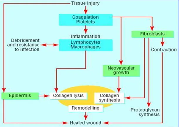
Fig 1 Phases of wound healing
During the proliferative phase, fibroblasts migrate into the wound and produce extracellular-matrix components, particularly collagen. Endothelial cells participate in angiogenesis, while keratinocytes migrate and proliferate to restore epidermal continuity. Wound contraction reduces the area that must be replaced and is strongly influenced by myofibroblasts, specialized contractile cells derived largely from fibroblast populations [2-4]. Remodeling subsequently reorganizes and strengthens the newly formed matrix. Although the wound may appear closed relatively early, matrix maturation can continue for months or longer.
Several local and systemic factors can disturb these events. Infection, impaired blood supply, excessive movement, foreign material and wound size may delay local repair, whereas advanced age, inadequate protein or micronutrient intake, systemic infection, glucocorticoid exposure and poorly controlled diabetes can impair healing [3,4]. The source thesis also highlights nutritional requirements for tissue repair, including adequate protein and selected micronutrients [4]. These considerations reinforce the need to evaluate candidate wound-healing agents in standardized models in which contraction and epithelialization can be quantified.
2.2 Analgesia and Nociception
Pain includes sensory-discriminative and affective-emotional dimensions. Nociception refers to neural processing of noxious stimuli and is not identical to the conscious experience of pain. Nociceptors are free nerve endings capable of responding to mechanical, thermal and chemical stimuli. Thermal models therefore provide controlled experimental stimuli for assessing changes in nociceptive response [32-34].
Opioid and non-opioid analgesics act through different mechanisms. Opioid receptors, including μ, δ and κ receptors, are G-protein-coupled receptors that reduce neuronal excitability and neurotransmitter release. Non-steroidal anti-inflammatory drugs primarily reduce prostaglandin synthesis through cyclooxygenase inhibition. Diclofenac, used as the reference drug in the present study, belongs to the NSAID class [32]. The analgesic activity observed with a test compound in a thermal model may involve central or peripheral pathways, but thermal latency alone cannot establish a specific molecular mechanism.
2.3 Sorbifolin as a pharmacological lead
The supplied phytochemical review describes Sorbifolin as a monomethoxy, trihydroxy flavone and reports its occurrence in several botanical sources [19-31]. Structural investigations have demonstrated the presence of sorbifolin or related glycosides in Sorbaria and Spathelia species, while later studies have identified it among flavonoids isolated from Pterogyne nitens and other plants [19-24,27-31]. These studies collectively demonstrate that Sorbifolin is chemically established, but they do not by themselves establish a wound-healing indication.
Several reported biological activities provide a hypothesis for further testing. Radical-scavenging activity is relevant because excessive reactive oxygen species can contribute to tissue injury and prolonged inflammation. Myeloperoxidase-related effects may be relevant to neutrophil-associated inflammatory responses. Other studies cited in the thesis suggest interactions with inflammatory signaling or protein targets, although these observations arise from different experimental systems and cannot be directly extrapolated to the wound model used here [24,25,29-31].
2.4 Rationale and study hypothesis
The study hypothesis was that Sorbifolin would enhance wound closure and epithelialization relative to untreated wounds and would increase thermal nociceptive latency relative to control animals. The wound-healing component was designed to determine whether repeated Sorbifolin treatment altered the macroscopic course of repair. The analgesic component was designed to determine whether a single oral dose produced measurable changes in thermal response. The combination of safety observation, wound assessment and two analgesic models provides a preliminary pharmacological profile, although it is not sufficient to define mechanism, long-term safety or clinical usefulness.
3. MATERIALS AND METHODS
3.1 Study design
This was a controlled preclinical experimental study using albino Wistar rats. The experimental plan in the supplied thesis comprised acute oral toxicity assessment, evaluation of wound healing using an open excision wound model, and evaluation of analgesic activity using tail immersion and hot plate tests. The overall design was intended to establish a preliminary safety margin and then examine pharmacological activity at the selected experimental dose.
3.2 Test compound
The test compound was Sorbifolin, identified in the source document as scutellarein 7-methyl ether with molecular formula C16H12O6. The manuscript retains the identity and terminology used in the supplied thesis. Details such as supplier, batch number, certificate of analysis, purity, storage conditions and analytical authentication were not clearly documented in the supplied material and should be inserted from laboratory records before journal submission.
3.3 Experimental animals and ethical considerations
The thesis states that adult Wistar albino rats of either sex, weighing approximately 180–250 g, were used for the pharmacological studies and were maintained under laboratory conditions at approximately 30 ± 2°C and 60–65% relative humidity with standard diet and water available ad libitum. The source document states that ethical committee clearance was obtained from the Institutional Animal Ethics Committee (IAEC) under CPCSEA-related requirements. However, the exact IAEC approval number is not populated in the supplied thesis. This information should be verified and inserted before submission.
Animals were divided into experimental groups, with six animals per group for the wound-healing and analgesic experiments. The thesis indicates random sampling for the acute toxicity study. The present manuscript does not infer additional randomization, blinding, allocation concealment or humane endpoints beyond those explicitly described in the source. These elements should be added if they were used experimentally.
3.4 Acute oral toxicity study
Acute toxicity was assessed using an approach described as OECD guideline 423, the acute toxic class method. The protocol uses sequential fixed-dose assessment and observation for mortality and overt toxicity. The source document describes overnight fasting with access to water, oral administration, observation at approximately 0.5, 1, 2 and 4 h after dosing, and subsequent daily observation for 14 days. Clinical observations included changes in skin and fur, eyes, mucous membranes, respiration, circulation, autonomic and central nervous system activity, motor behavior and signs such as salivation, tremors, convulsions, diarrhea, lethargy, sleep and coma.
The tabulated toxicity dataset records Sorbifolin at 200 mg/kg with three male and three female rats, with no mortality in either sex. Body weights were recorded before treatment and at the end of the observation period. Necropsy included external examination and opening of thoracic and abdominal cavities. Stomach, liver, heart and kidneys were examined histologically following fixation, paraffin embedding, sectioning at approximately 5 μm and hematoxylin-eosin staining.
Important source-data clarification: the supplied thesis contains an internal inconsistency. One narrative sentence refers to administration of 2000 mg/kg and another describes the isolated compound as being tested at one-tenth of that dose (200 mg/kg), while the actual mortality and body-weight tables are presented for 200 mg/kg. Because the reported dataset and dose selection are based on 200 mg/kg, this manuscript reports the tabulated 200 mg/kg toxicity dataset and does not reinterpret the conflicting 2000 mg/kg statement. The original laboratory record must be checked before publication.
3.5 Excision wound model
Wound-healing activity was assessed according to an open excision wound procedure based on the method attributed in the thesis to Morton and Malone [35], with wound measurement approaches referenced to subsequent experimental studies [36,37]. Animals were anesthetized using the procedure described in the source, dorsal hair was removed with a depilatory preparation, and a circular excision wound of approximately 500 mm² was created on the dorsal thoracic region using forceps and scissors. The wound was left open.
Wound area was recorded on post-wounding days 1, 4, 7, 10, 13 and 16. A transparent sheet was placed over the wound to trace its outline, and the traced area was quantified in square millimeters using graph paper. Wound contraction was followed as the reduction in wound area over time. Complete epithelialization was defined in the source as disappearance of the scab with no residual raw wound area. The time required to reach this endpoint was recorded as the epithelialization period.
3.6 Treatment groups for wound healing
The wound-healing experiment included three groups (n=6 per group): Group I, simple ointment base as control; Group II, Sorbifolin at 50 mg/kg as the test treatment; and Group III, 10% povidone-iodine cream as the reference topical treatment. The thesis contains a wording inconsistency in one sentence mentioning 200 and 400 mg/kg in relation to wound application, but all wound-healing results and the stated experimental design use 50 mg/kg. The manuscript therefore uses 50 mg/kg as the analyzed test dose.
Treatments were applied daily through day 16. Wound area was measured at the specified intervals, and percentage protection was reported for day 16. The source document cites Agrahari et al. for the calculation approach [38].
3.7 Analgesic activity: tail-immersion test
The tail-immersion test used Wistar albino rats divided into three groups of six animals. The source reports overnight fasting for 18 h. Group I received normal saline (5 mL/kg, orally), Group II received diclofenac sodium 10 mg/kg in normal saline, and Group III received Sorbifolin 50 mg/kg orally. The distal 5 cm of the tail was immersed in water maintained at 55°C and the latency to tail withdrawal was recorded. Measurements were made before treatment and at 30, 45, 60 and 90 min after treatment. A 15-s cutoff time was used to avoid thermal injury.
3.8 Analgesic activity: hot-plate test
The hot-plate test was conducted in Wistar albino rats divided into three groups of six. The source reports an animal weight range of 200–220 g for this experiment. Treatment groups were as described above. Animals were placed on a hot plate maintained at 55°C, and latency to paw licking or jumping was recorded. Measurements were made before treatment and at 30, 45, 60 and 90 min. A maximum response time of 45 s was specified to minimize tissue injury.
3.9 Statistical analysis
Results were expressed as mean ± SEM. The thesis states that data were analyzed using one-way analysis of variance followed by Dunnett's post-test, with six animals per group. A probability value of p<0.05 was considered statistically significant, while the wound-healing and analgesic result tables specifically mark selected comparisons at p<0.01 or p<0.001. Because the raw individual-level observations and complete ANOVA outputs were not provided, this manuscript reports the statistical outcomes exactly as documented rather than reconstructing test statistics or confidence intervals.
4. OUTCOME MEASURES AND DATA HANDLING
4.1 Primary wound-healing outcomes
The principal wound-healing outcome was wound area (mm²) at each scheduled post-wounding day. Serial wound-area measurements provide a direct macroscopic measure of contraction and closure. A second outcome was the reported percentage protection at day 16, together with the period of epithelialization in days. The source document reports these endpoints as summary values and does not provide individual animal measurements or the full calculation worksheet.
4.2 Analgesic outcomes
The principal analgesic outcome was latency to withdrawal or nociceptive behavior after thermal stimulation. For the tail-immersion test, latency was expressed in seconds and the thesis reported an inhibition percentage. For the hot-plate test, latency to paw licking or jumping was recorded and the source likewise reported an inhibition percentage. These measures indicate altered thermal nociceptive responsiveness but do not establish whether the effect is exclusively central, peripheral, anti-inflammatory, sedative or motor-related.
4.3 Interpretation framework
For wound healing, smaller wound area at later time points and shorter epithelialization period were interpreted as favorable responses. For analgesia, increased response latency relative to control was interpreted as analgesic activity. Diclofenac was used as a pharmacological reference rather than as a wound-healing comparator. Povidone-iodine was used as the topical reference in the wound model. The two standards therefore answer different comparative questions and should not be directly ranked against one another.
4.4 Reporting principles
The manuscript retains the numerical values in the thesis and avoids generating new experimental observations. Where the thesis uses terms such as 'percentage inhibition' or 'percentage protection', the terminology is retained but interpreted cautiously. In particular, the reported analgesic inhibition percentages are not independently recalculated because the source does not provide a single unambiguous formula and time-point dataset for the calculation. Likewise, the epithelialization endpoint is reported exactly as documented.
5. RESULTS
5.1 Acute oral toxicity
The acute oral toxicity experiment showed no mortality among the six animals documented at 200 mg/kg (three males and three females). The mortality table records zero deaths during the early observation periods and during the remainder of the 14-day observation period. No overt signs such as tremors, convulsions, excessive salivation, diarrhea, marked lethargy, sleep or coma were reported. Body weights increased modestly in all six animals between baseline and day 14, and no macroscopic abnormalities were detected at necropsy.
|
Animal |
Sex |
Dose (mg/kg) |
Before (g) |
After (g) |
Necropsy |
|
1 |
Male |
200 |
198 |
202 |
NAD |
|
2 |
Male |
200 |
187 |
191 |
NAD |
|
3 |
Male |
200 |
200 |
202 |
NAD |
|
4 |
Female |
200 |
193 |
196 |
NAD |
|
5 |
Female |
200 |
199 |
201 |
NAD |
|
6 |
Female |
200 |
201 |
205 |
NAD |
NAD, no abnormality detected. The source thesis reports the organ-weight findings for vehicle and Sorbifolin-treated animals. Mean heart weight was 1.15 ± 0.11 g in the vehicle group and 1.15 ± 0.19 g after Sorbifolin; spleen weight was 0.85 ± 0.10 and 0.90 ± 0.08 g; liver weight was 7.31 ± 0.04 and 7.39 ± 0.05 g; right kidney weight was 0.67 ± 0.04 and 0.64 ± 0.05 g; and left kidney weight was 0.62 ± 0.10 and 0.64 ± 0.06 g, respectively. Histological examination did not identify treatment-associated lesions in the examined organs.
|
Organ |
Vehicle: 1% Tween 80 |
Sorbifolin 200 mg/kg |
|
Heart (g) |
1.15 ± 0.11 |
1.15 ± 0.19 |
|
Spleen (g) |
0.85 ± 0.10 |
0.90 ± 0.08 |
|
Liver (g) |
7.31 ± 0.04 |
7.39 ± 0.05 |
|
Right kidney (g) |
0.67 ± 0.04 |
0.64 ± 0.05 |
|
Left kidney (g) |
0.62 ± 0.10 |
0.64 ± 0.06 |
Taken together, the documented observations support the thesis conclusion that no acute toxicity was evident at the tested 200 mg/kg dose. Because of the internal inconsistency between 200 and 2000 mg/kg in the narrative, the finding should be described as 'no observed acute toxicity at the tabulated 200 mg/kg dose' rather than as a definitive LD50 value.
5.2 Wound contraction
Serial measurement demonstrated a progressive decline in wound area in all groups, consistent with spontaneous repair. The control group decreased from 501 ± 10.3 mm² on day 1 to 184 ± 9.8 mm² on day 16. Sorbifolin produced a more rapid reduction, from 502 ± 4.7 mm² on day 1 to 71 ± 3.7 mm² on day 16. The povidone-iodine group decreased from 501 ± 8.2 mm² to 80 ± 5.8 mm² over the same period.
|
Group |
Day 1 |
Day 4 |
Day 7 |
Day 10 |
Day 13 |
Day 16 |
|
Control |
501 ± 10.3 |
478 ± 9.4 |
403 ± 7.6 |
346 ± 2.4 |
271 ± 9.4 |
184 ± 9.8 |
|
Sorbifolin 50 mg/kg |
502 ± 4.7 |
457 ± 6.4 |
337 ± 6.4* |
268 ± 3.7* |
143 ± 6.9* |
71 ± 3.7* |
|
Povidone-iodine 10% |
501 ± 8.2 |
401 ± 7.3* |
320 ± 9.4* |
245 ± 6.5* |
131 ± 7.3* |
80 ± 5.8* |
*p<0.001 versus control by one-way ANOVA with Dunnett's post-test, ; n=6.
At day 7, the Sorbifolin-treated wound area was 337 ± 6.4 mm² compared with 403 ± 7.6 mm² in control. By day 13, the corresponding values were 143 ± 6.9 and 271 ± 9.4 mm². At day 16, Sorbifolin-treated wounds remained smaller than control wounds, while the numerical value was slightly smaller than that reported for povidone-iodine. The magnitude of the day-16 reduction from the initial wound area was substantial in both active-treatment groups.
Using the tabulated means, the day-16 wound area represented approximately 14.1% of the initial area in the Sorbifolin group, 16.0% in the povidone-iodine group and 36.7% in control. These descriptive calculations are derived directly from the reported means and are included to facilitate interpretation; they are not a substitute for the thesis's stated percentage-protection endpoint.
5.3 Epithelialization
Sorbifolin also improved the epithelialization endpoint. The source reports a percentage protection of 97.49% and an epithelialization period of 15.24 days for Sorbifolin. The control group had 62.14% protection and required 24.36 days, whereas 10% povidone-iodine produced 96.86% protection and an epithelialization period of 15.52 days.
|
Group |
Treatment |
Percentage protection |
Period of epithelialization (days) |
|
I |
Control |
62.14 |
24.36 |
|
II |
Sorbifolin 50 mg/kg |
97.49 |
15.24 |
|
III |
Povidone-iodine 10% |
96.86 |
15.52 |
The Sorbifolin-treated group therefore showed the shortest reported epithelialization period among the three groups. The difference from control was approximately 9.12 days, while the difference from povidone-iodine was 0.28 days. The numerical similarity between Sorbifolin and povidone-iodine suggests broadly comparable performance in this endpoint, although formal direct statistical comparison between the two active groups was not reported.
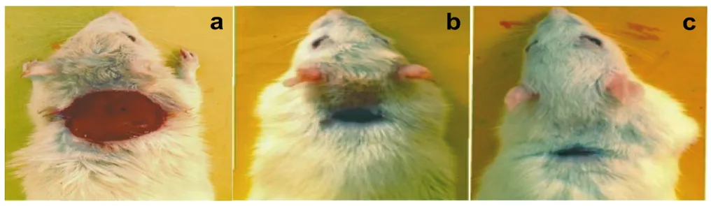
Figure 2: Effect of Sorbifolin on wound healing a. Control group; b. Sorbifolin group; c. Standard group
5.4 Analgesic activity: tail immersion
In the tail-immersion test, the control group showed little change in latency, from 2.21 ± 0.11 s before treatment to 2.12 ± 0.09 s after treatment. Diclofenac sodium increased latency from 2.33 ± 0.32 to 6.32 ± 0.44 s, corresponding to 64.2% inhibition. Sorbifolin increased latency from 2.22 ± 0.42 to 3.71 ± 0.24 s, corresponding to 41.4% inhibition. The source marks the post-treatment responses for both diclofenac and Sorbifolin as significant at p<0.01 versus control.
|
Group |
Pretreatment latency (s) |
Post-treatment latency (s) |
Reported inhibition (%) |
|
Control |
2.21 ± 0.11 |
2.12 ± 0.09 |
— |
|
Diclofenac sodium 10 mg/kg |
2.33 ± 0.32 |
6.32 ± 0.44** |
64.2 |
|
Sorbifolin 50 mg/kg |
2.22 ± 0.42 |
3.71 ± 0.24** |
41.4 |
**p<0.01 versus control by one-way ANOVA with Dunnett's post-test; n=6. The Sorbifolin response was lower than that of diclofenac, but the direction of effect was consistent with increased thermal response latency.
5.5 Analgesic activity: hot plate
The hot-plate findings showed a stronger Sorbifolin response than the tail-immersion model. Control latency changed minimally from 4.44 ± 0.38 to 4.42 ± 0.14 s. Diclofenac increased latency from 5.62 ± 0.93 to 10.92 ± 0.26 s, with a reported inhibition of 61.92%. Sorbifolin increased latency from 5.45 ± 0.27 to 9.92 ± 0.82 s, with a reported inhibition of 56.83%. Both active-treatment responses were marked as p<0.01 versus control.
|
Group |
Pretreatment latency (s) |
Post-treatment latency (s) |
Reported inhibition (%) |
|
Control |
4.44 ± 0.38 |
4.42 ± 0.14 |
— |
|
Diclofenac sodium 10 mg/kg |
5.62 ± 0.93 |
10.92 ± 0.26** |
61.92 |
|
Sorbifolin 50 mg/kg |
5.45 ± 0.27 |
9.92 ± 0.82** |
56.83 |
**p<0.01 versus control by one-way ANOVA with Dunnett's post-test; n=6. In this model, the post-treatment latency observed with Sorbifolin was approximately 91% of the diclofenac value, although direct statistical comparison between the two active groups was not reported.
6. DISCUSSION
The present study provides preliminary experimental evidence that Sorbifolin possesses two pharmacological properties relevant to tissue injury: enhancement of wound repair and attenuation of thermal nociceptive responses. The findings are notable because Sorbifolin has been studied primarily in phytochemical and other biological contexts, while direct investigation of its wound-healing and analgesic effects is comparatively limited in the supplied literature. The results should therefore be viewed as an initial pharmacological characterization rather than proof of therapeutic efficacy.
6.1 Wound contraction and epithelialization
The wound model demonstrated a consistent separation between control and active-treatment groups from the first week onward. Sorbifolin-treated wounds were smaller than control wounds at days 7, 10, 13 and 16, and the source reports p<0.001 for these comparisons. By day 16, the mean wound area in the Sorbifolin group was 71 mm², compared with 184 mm² in control. This corresponds to a substantial reduction in residual wound area and indicates accelerated macroscopic closure.
The observed effect is biologically plausible in the context of flavonoid pharmacology, but the present dataset does not directly measure fibroblast proliferation, collagen content, hydroxyproline, angiogenesis, inflammatory cytokines, oxidative stress markers or histological repair. Therefore, the thesis suggestion that Sorbifolin may promote fibroblast proliferation and collagen synthesis should be regarded as a mechanistic hypothesis rather than a demonstrated mechanism in this experiment. Liu et al. showed that another flavonoid could promote fibroblast migration and collagen synthesis in association with wound healing [18], and Kim et al. summarized multiple pathways through which plant-derived compounds may influence tissue repair [17]. These reports support the plausibility of a flavonoid-mediated effect without establishing identity of mechanism.
Sorbifolin performed numerically close to the reference topical treatment. At day 16, the Sorbifolin group had a mean residual wound area of 71 ± 3.7 mm², while povidone-iodine had 80 ± 5.8 mm². The reported epithelialization periods were similarly close, at 15.24 and 15.52 days. This is an important observation because it suggests that the effect was not limited to a single measurement of wound size. Nevertheless, povidone-iodine is primarily used for antiseptic purposes and the present experiment was not designed to determine whether Sorbifolin has equivalent antimicrobial activity. A direct head-to-head comparison should therefore be interpreted as a comparison of the measured wound endpoint rather than an assertion of therapeutic equivalence.
The wound-healing effect may reflect several overlapping biological processes. Reduction of excessive inflammation could create a more favorable environment for proliferation and matrix deposition, while antioxidant activity could reduce oxidative damage to cells participating in repair. The source literature reports radical-scavenging activity for flavones containing Sorbifolin [24], and broader studies of flavonoids describe anti-inflammatory and tissue-protective effects [17,18]. However, no antioxidant or inflammatory biomarker was measured in the present study, so the proposed relationship remains inferential.
6.2 Analgesic effects
Sorbifolin produced measurable increases in thermal nociceptive latency in both models. In the tail-immersion test, latency increased from 2.22 to 3.71 s, while in the hot-plate test it increased from 5.45 to 9.92 s. The reported inhibition values were 41.4% and 56.83%, respectively. These findings indicate that the analgesic response was reproducible across two thermal paradigms, although the magnitude differed between models.
Diclofenac sodium produced a larger effect in both assays, with reported inhibition values of 64.2% in tail immersion and 61.92% in the hot plate. This provides a useful benchmark: Sorbifolin demonstrated substantial activity but did not exceed the established reference drug. The finding is consistent with the cautious conclusion that Sorbifolin may have analgesic potential rather than being equivalent to a standard NSAID.
The mechanism of the analgesic response cannot be established from the present study. Thermal response tests can detect changes in nociceptive processing, but increased latency can theoretically arise from analgesia, altered arousal, motor impairment or other central effects. The source thesis proposes that Sorbifolin may influence cyclooxygenase or pain-related pathways based on the known activity of flavonoids. Diclofenac itself acts through cyclooxygenase inhibition [32]. Chen et al. described analgesic effects of scutellarin in a neuropathic pain model associated with NF-κB signaling modulation [33]. Because scutellarin and Sorbifolin are chemically related flavonoids, this literature provides a mechanistic hypothesis but not direct evidence that Sorbifolin acts through the same pathway.
6.3 Relationship between wound healing and analgesia
The concurrent wound-healing and analgesic findings are potentially important. Pain and inflammation can influence mobility, stress responses and patient adherence to wound-care procedures. A compound that can influence both tissue repair and nociception may have an integrated therapeutic profile. However, the present experiments were conducted as separate models and did not determine whether the analgesic effect contributed to the observed wound closure. Nor did they assess whether reduced pain behavior altered locomotion or wound manipulation.
A possible unifying hypothesis is modulation of inflammatory signaling. Inflammation is essential during the early phase of repair, but prolonged or excessive inflammation can delay resolution. Flavonoids may influence inflammatory mediators and redox pathways [17,18,33]. The source thesis also cites evidence that flavonoids can affect inflammatory signaling, including NF-κB-related processes [33]. Future work should therefore measure inflammatory cytokines, cyclooxygenase expression, prostaglandins, oxidative-stress markers, macrophage phenotype and tissue histology to determine whether these mechanisms contribute to the observed phenotype.
6.4 Safety findings
The acute toxicity experiment did not identify mortality, overt clinical toxicity or major macroscopic or histological abnormalities at the tabulated 200 mg/kg dose. Body weights increased in all documented animals. These observations are reassuring for preliminary experimentation but do not establish chronic safety, reproductive safety, genotoxicity, organ-specific pharmacokinetics or topical tolerability. Acute toxicity is only one component of a complete safety assessment.
Furthermore, the source document contains a discrepancy between a narrative reference to 2000 mg/kg and tables documenting 200 mg/kg. This is not a trivial editorial issue because the numerical interpretation of an acute toxic class study depends on the actual administered dose. The present manuscript deliberately reports the tabulated 200 mg/kg dataset and flags the discrepancy for verification. The final journal submission should reconcile this point using the original animal study records.
6.5 Comparison with previous literature
The phytochemical literature confirms that Sorbifolin is a structurally characterized natural flavone. Chan et al. reported the structure of sorbifolin from Spathelia sorbifolia, while Taylor et al. subsequently described synthetic work related to the compound [22,23]. Arisawa et al. identified sorbifolin as scutellarein-7-methyl ether in Sorbaria stellipila [21]. These studies provide a chemical foundation for considering Sorbifolin as a defined compound rather than an unidentified botanical extract.
The biological studies summarized in the thesis demonstrate that Sorbifolin can occur in chemically complex plant matrices and has been investigated in several pharmacological contexts. Fernandes et al. evaluated flavones from Pterogyne nitens for myeloperoxidase inhibition and radical scavenging [24]. Hernández-Bolio et al. reported that sorbifolin showed low anti-giardial activity relative to another flavonoid in their model and did not show cytotoxicity toward HEK-293 cells under the reported conditions [25]. Shimizu et al. observed antiviral effects against hepatitis C virus entry for flavonoids including Sorbifolin [29]. These findings illustrate the breadth of biological activity associated with the compound but also reinforce that activity is highly model-dependent.
The wound-healing findings in the present study are consistent with the broader literature describing flavonoids as multifunctional modulators of tissue repair. Liu et al. reported that a flavonoid isolated from Periploca sepium Bunge promoted fibroblast migration and collagen synthesis [18]. Kim et al. discussed plant-derived compounds that can influence inflammatory regulation, matrix formation and cell migration during wound repair [17]. The current findings extend this general concept by providing an in vivo wound-closure phenotype for Sorbifolin. Importantly, the study does not demonstrate the specific cellular pathways responsible.
The analgesic findings also fit within a wider flavonoid pharmacology framework. Chen et al. reported analgesic effects of scutellarin associated with modulation of NF-κB signaling in a neuropathic pain model [33]. The structural relationship between scutellarin-type flavonoids and Sorbifolin makes anti-inflammatory signaling a plausible area for investigation. Nevertheless, the tail-immersion and hot-plate results alone cannot determine whether Sorbifolin has anti-inflammatory, central analgesic, peripheral analgesic, or mixed activity.
6.6 Strengths of the study
The study has several strengths. First, the wound-healing assessment used serial measurements across multiple time points rather than a single endpoint. Second, epithelialization time was recorded in addition to wound area, providing a second indicator of repair. Third, two independent thermal nociception models were used for analgesic evaluation. Fourth, both wound and analgesic experiments included recognized reference treatments, allowing contextual interpretation of the magnitude of activity. Finally, the acute toxicity component provided an initial safety screen before pharmacological evaluation.
6.7 Limitations
The most important limitations are the small group size (n=6), absence of raw individual-level data, lack of detailed randomization/blinding information, incomplete documentation of compound purity and formulation, and absence of mechanistic biomarkers. The wound experiment did not report histopathological scoring of granulation tissue, collagen organization or angiogenesis. The analgesic experiments did not report all time-point values despite the methods specifying measurements at 30, 45, 60 and 90 min. Thus, the reported pre- and post-treatment summaries cannot establish a complete pharmacodynamic time course.
The study also contains source-document inconsistencies in acute toxicity dosing and wording regarding wound-treatment dose. These issues should be corrected by reference to the laboratory notebook before submission. In addition, the supplied thesis does not provide an exact IAEC approval number, supplier information, batch/purity information or a complete description of housing enrichment and humane endpoints. These are important reporting details for a contemporary animal study.
6.8 Implications for future research
Future investigations should include dose-ranging studies, topical versus systemic formulations, pharmacokinetic characterization, wound histology, hydroxyproline/collagen quantification, angiogenesis markers, inflammatory cytokines, oxidative-stress markers and microbiological assessment. Analgesic studies should incorporate additional inflammatory and chemical nociception models and examine the temporal profile of the response. Mechanistic experiments targeting cyclooxygenase, NF-κB, MAPK, Nrf2 or related pathways could help establish causality rather than relying on structural analogy.
Longer-term safety evaluation is also needed. Before clinical translation, reproducible pharmaceutical-grade Sorbifolin with defined purity and stability should be developed, and topical tolerability should be assessed. If wound healing remains the principal development objective, a standardized topical formulation may be particularly appropriate because local exposure can potentially be optimized while limiting systemic exposure. These are future research directions rather than conclusions established by the present experiment.
7. CONCLUSION
The present experimental investigation indicates that Sorbifolin has promising wound-healing and analgesic activities in rats. In the open excision wound model, Sorbifolin at the reported 50 mg/kg treatment produced faster wound contraction than control and yielded a day-16 wound area of 71 ± 3.7 mm², compared with 184 ± 9.8 mm² in control. It also produced a reported percentage protection of 97.49% and reduced the epithelialization period to 15.24 days. These values were numerically comparable to those obtained with 10% povidone-iodine.
In the analgesic experiments, Sorbifolin increased thermal response latency in both the tail-immersion and hot-plate tests. The reported inhibition values were 41.4% and 56.83%, respectively. Diclofenac sodium produced greater inhibition in both models, indicating that the Sorbifolin response, although substantial, was below the reference NSAID under the conditions tested.
The acute toxicity observations showed no mortality or overt toxicity in the tabulated 200 mg/kg dataset, supporting preliminary tolerability at the experimental dose. However, the internal dose discrepancy in the thesis must be reconciled before publication. Overall, the findings justify further investigation of Sorbifolin as a natural-product lead for wound repair and pain modulation, but mechanistic studies, dose optimization, formulation development, expanded toxicology and independent confirmatory experiments are required before any therapeutic claims can be made.
8. DECLARATIONS AND REPORTING STATEMENTS
Consent for publication: Not applicable to this animal study.
Data availability: The present manuscript reports the summary data available in the supplied thesis. Individual-level raw data were not provided in the source document.
Conflict of interest: To be completed by the authors.
Author contributions: To be completed by the authors according to the journal's taxonomy.
Acknowledgments: To be completed by the authors.
REFERENCES

K. Nikitha, Dr. C. Radhika, Dr. D. Jothieswari, Repurposing Sorbifolin for Wound Recovery and Analgesia: Investigating its Therapuetic Potential in Adult Albino Wistar Rats, Int. J. of Pharm. Sci., 2026, Vol 4, Issue 9, 2487-2503. https://doi.org/10.5281/zenodo.22877135
 10.5281/zenodo.22877135