View Article

Abstract

Diabetic retinopathy (DR) is a common microvascular complication of diabetes mellitus and is the leading cause of visual loss in the elderly. Hyperglycaemia and altered metabolic pathways lead to oxidative stress and the development of neurodegeneration in the initial stage of diabetic retinopathy. Diabetic retinopathy (DR) remains a leading cause of vision loss among adults with diabetes, necessitating a multifaceted approach to management. Current treatment strategies are tailored to disease severity and include strict metabolic control, intravitreal anti-VEGF injections, corticosteroid implants, laser photocoagulation, and surgical interventions such as vitrectomy for advanced cases. Despite availability of effective screening and treatment, patients often present late due to poor awareness and practices. Effective prevention relies heavily on patient awareness and adherence to regular ophthalmological screening, making the assessment of patient knowledge, attitudes, and practices a critical step in identifying barriers to care. This study aimed to assess the Knowledge, Attitude, and Practice (KAP) concerning diabetic retinopathy among a cohort of diabetic patients to identify gaps in patient education and preventative care, among diabetes mellitus outpatients at a primary health centre. A cross-sectional survey was conducted among 300 diabetic individuals (71.3% rural, 81.1% Type 2 diabetes). The results revealed significant knowledge deficits: only 42.5% of respondents had heard of DR, and a mere 26.8% could identify its correct cause. A majority (71.3%) were unaware that DR could be prevented or treated. Despite this, attitudes were largely positive, with 81.9% agreeing that DR is a serious complication and over 85?knowledging the importance of early detection. A striking attitude-practice gap was identified; while most endorsed the need for annual eye exams, 70.1% had never undergone a retinal screening (fundus examination), and 66.5% reported never visiting an eye doctor. This poor adherence to eye care contrasts sharply with high self-reported adherence to daily diabetes. Management, such as regular blood sugar monitoring (88.2%). Furthermore, 72.8% of participants reported never having received counseling on DR from a healthcare professional, suggesting a critical lapse in patient education.

Keywords

Diabetic retinopathy, Treatment Strategies, Anti-VEGF therapy, Laser photocoagulation, Vitrectomy, Eye injection, Fundus examination, Vision loss.

Introduction

EPIDEMIOLOGY

The economic burden of vision impairment and blindness due to diabetic retinopathy in globally is considerable. A global study in 2017 found that of the leading causes of vision impairment and blindness, the crude global prevalence (all ages) of diabetic retinopathy as a cause increased between 1990 and 2015, while more than460 million people live with diabetes, and this number is expected to rise to 700 million by 2045. Approximately one-third of individuals with diabetes have signs of DR, and about one in fifteen have vision-threating disease (proliferative DR or DME).

In India

A recent study from an urban population in Chennai, India estimated the age-standardised. The incidence VT-DR was higher (10.2%) among those who had diabetes for ≥15 years compared with those with 0–4 year’s duration (2%). Progression was associated with poor glycaemic control, systolic hypertension and anaemia.

CLINICAL FEATURES

DIABETIC MACULAR EDEMA (DME) retinal thickening and hard exudates near the macula, leading to central vision loss.

MICROANEURYSS (MA) small, balloon-like bulges in the tiny blood vessels of the retina.

NEOVASCULARIZATION (NV) The growth of new, abnormal blood vessel in the retina, a hallmark of proliferative diabetic retinopathy

SYMPTOMS

DR is characterized by blurred or fluctuating vision, Floaters, Dark or empty areas in vision, Poor night vision, Faded or washed-out colour, sudden vision loss. (Figure 1: symptoms)
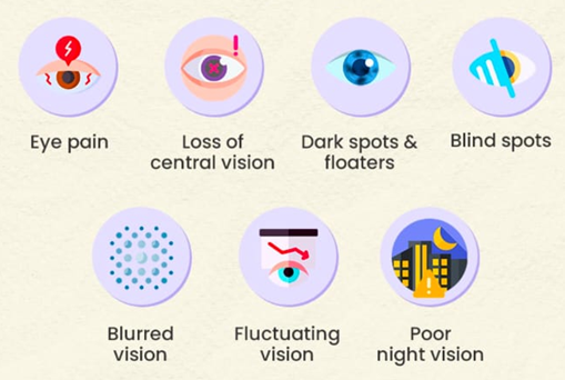
Figure 1: SYMPTOMS

RISK FACTORS

The development of diabetic retinopathy Strongly correlates with a longer duration of diabetes, greater hyperglycemia, and hypertension. Other factors such as High cholesterol and triglycerides, Male sex, Older age, Diabetic neuropathy, Nephropathy, Foot ulcers, Smoking, Anemia, Pregnancy, Family history, High salt intake, Glilatzone drugs.

LEVEL OF AWARENESS OF DIABETIC RETINOPATHY AMONG PATIENT, PROVIDERS IN INDIA

In India, levels of awareness about DR amongst DM in terms of their knowledge and the importance of annual eye examinations is low. Awareness amongst health care professionals is also not as good. There is limited evidence of the availability of services for patients with DR.

STAGES OF DIABETIC RETINOPATHY

This Classification system based on the ETDRS (Early Treatment Diabetic Retinopathy Study Research Group) can be found in the International Council of Ophthalmology (ICO) guidelines for diabetes eye care (International Council of Ophthalmology, 2017; Wong et al., 2018).

DR is divided into two broad stages: Non-proliferative DR (NPDR) and Proliferative DR (PDR). NPDR represents the earlier stages of the disease, characterized (progressively) by retinal Microaneurysm (MA), intra retinal hemorrhages, hard extrudes (HE), venous beading, and Intra microvascular Microaneurysms (IRMA)

PROLIFERATIVE DIABETIC RETIOPATHY (PDR):

Proliferative diabetic retinopathy (PDR) is the more advanced form of the disease, which is characterized by abnormal blood vessel growth (neovasculation) on the retina or optic disc.

NON-PROLIFERATIVE DIABETIC RETINOPATHY (NPDR):

Non-Proliferative diabetic retinopathy is further charaterizsed by three stages:

1. Mild NPDR: Characterized by microaneurysms

2. Moderate NPDR: Involves more microaneurysms, haemorrhages, cotton-wool spots and hard exudates.

3. Severe NPDR: Definite by the “4-2-1 rule”

  • Hemorrhages (4+retinal quadrants)
  • Venous bleeding (≥2 qadrants)
  • Intra retinal microvascular abnormalities [IRMA] ≥1 Quadrant

PATHOPHYSIOLOGY

The various metabolic mechanisms, pathologic processes, and systemic comorbidities involved in the development and progression of DR each provide important targets for disease-modifying therapeutics

As a complication of diabetes mellitus, hyperglycemia is one of the essential contributions to DR development. Several underlying mechanisms of elevated blood glucose leads to DR are identified, including the hexosamine pathway, advanced glycation end products accumulation, polyol pathway, protein kinase C pathway and poly polymerase activation.The metabolic dysregulations, together with oxidative and inflammatory imbalance, lead to the pathological presentation of DR. (figure 2: pathology of diabetic retinopathy)

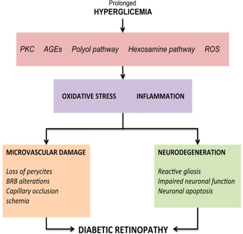
Figure 2: PATHOLOGY OF DIABETIC RETINOPATHY

Inflammation is also documented as a key component of DR pathogenesis via capillary damage and hypoxia induction, leading to increased VEGF expression and Neovascularization. Finally, endothelial proliferation driven by chronic microvascular ischemia results in the formation of visible intraretinal microvascular abnormalities (IRMA) and neovascularization (NV), with consequent vision-threatening complications such as Vitreous hemorrhage (VH) and tractional retinal detachment (TRD).

DIAGNOSIS OF DIABETIC RETINOPATHY

Early detection of DR is critical, as the disease is often asymptomatic in its initial stages and vision loss can be prevented or minimized with timely intervention. DR often develops without symptoms in its early stages. Vision loss typically occurs only when the disease has advanced or when complications such as diabetic macular edema (DME) or proliferative diabetic retinopathy (PDR) arise.

SCREENING RECOMMENDATIONS

  • Type 1 Diabetes: Begin screening 5 years after diagnosis or at puberty if diagnosed earlier.
  • Type 2 Diabetes: Begin screening at the time of diagnosis.
  • Pregnancy: Women with pre-existing diabetes should have an eye exam before conception and during the first trimester.
  • High-risk patients (long duration of diabetes, poor glycemic/lipid control, hypertension) require annual eye examinations, while those without evidence of DR may be screened every 2 years.

DIAGNOSITC TOOLS USED IN DIABETIC RETINOPATHY

A variety of diagnostic tools and imaging modalities are used to identify, monitor, and guide the treatment of DR.

  • Dilated Fundus Examination: The gold standard for DR screening and diagnosis is a comprehensive dilated fundus examination performed by an ophthalmologist or optometrist.
  • Stereoscopic examination of the posterior pole is essential for detecting diabetic macular enema (DME) and proliferative changes.
  • Fundus Photography: Fundus photography uses specialized cameras to capture detailed images of the retina.
  • Optical Coherence Tomography (OCT): OCT is a non-invasive imaging technique that provides high-resolution, cross-sectional images of the retina. Guides management, especially with anti-VEGF therapy.
  • Fluorescein Angiography: This imaging modality involves intravenous injection of fluorescein dye, which circulates through the retinal vasculature and is photographed using a special camera.
  • B-Scan Ultrasonography:B-scan ultrasound uses sound waves to create images of the eye’s internal structures.

Emerging Technologies

  • Tele ophthalmology: Remote screening using digital retinal images reviewed by offsite graders improves access and compliance.
  • Widefield Imaging: Captures larger areas of the retina, improving detection of peripheral lesions.
  • Artificial Intelligence (AI): Automated systems can assist in DR screening and grading, increasing efficiency and accessibility.

II. MANAGEMENT OF DIABETIC RETINOPATHY

1. MEDICAL MANAGEMENT

Effective management of DR begins with optimizing systemic health

  • Control of comorbidities – Hypertension, hyperlipidemia management slows retinal damage
  • Glycemic Control: Tight blood sugar management reduces onset & progression of DR. SGLT2 inhibitors show protective retinal effects by reducing vascular leakage and oxidative stress.
  • Blood Pressure Control: Hypertension accelerates DR progression. ACE inhibitors (e.g., Lisinopril) and β-blockers reduce risk, independent of blood pressure lowering.
  • Lipid Management:Statins and Fenofibrate (PPAR-α agonist) lower triglycerides and reduce DR progression.
  • Strict blood sugar control – reduces progression risk.
  • Regular monitoring – Fundus examination yearly (or more frequently in advanced cases)

2. OPHTHALMIC TREATMENT

  • Laser Photocoagulation
  • Conventional focal/grid laser seals microaneurysms, reduces VEGF drive, and prevents vision loss.
  • Modern PASCAL scanning lasers shorten treatment time and improve safety.
  • Panretinal Photocoagulation (PRP) Used in proliferative diabetic retinopathy (PDR) prevents neovascularization.

3. INTRAVITREAL INJECTIONS

  • Anti-VEGF Therapy First-line for diabetic macular edema (DME) and proliferative

DR. Agents: Ranibizumab, Aflibercept, Bevacizumab, Brolucizumab. Reduce vascular leakage, neovascularization, and improve visual acuity.

  • Intravitreal Corticosteroids Used when anti-VEGF response is poor.

Drugs: Dexamethasone (Ozurdex), Fluocinolone (Retisert), Triamcinolone. Suppress inflammation, VEGF expression, and reduce oedema.

4. SURGICAL OPTIONS

  • Vitrectomy (Retinal Surgery) Clears vitreous, relieves traction, improves visual outcomes.

Success rates: Anatomical (90–95%), visual improvement (60–80%).

  • Nutraceuticals & Antioxidants: Lutein, Zeaxanthin, Omega-3 fatty acids (DHA, EPA), Vitamins C & E. Delay DR progression via antioxidant, anti-inflammatory, and neuroprotective effects. Safe, adjunctive daily supplementation.

5. EMERGING THERAPIES

  • Gene Therapy: Targets retinal vasculopathy to stop DR progression.
  • Stem Cell Therapy: Aims at neuroprotection & vascular repair.
  • Nanotechnology: Sustained drug delivery, less frequent injections.
  • Integrin & Angiopoietin inhibitors: Novel vascular targets.
  • AI & Tele-Ophthalmology
  • Artificial intelligence aids in DR screening and grading.
  • Telemedicine improves access in remote areas.

III. OBJECTIVES

General Objective: To assess the knowledge, attitude, and practice regarding diabetic retinopathy among patients with diabetes mellitus.

Specific Objectives:

  • To evaluate the knowledge of diabetic patients about causes, risk factors, and prevention of diabetic retinopathy.
  • To assess their attitudes towards regular eye screening and treatment adherence.
  • To analyze the practices followed by patients related to blood sugar control and ophthalmic check-ups.
  • To educate the study population regarding Diabetic Retinopathy among diabetic patients
  • To categorize subject into Diabetic Retinopathy according to WHO guidelines.
  • To identify KAP level of subject using a validated questioner
  • To provide baseline data for future health education and awareness programs.
  • To explore associations between demographic/clinical variables and KAP levels

IV. MATERIALS AND METHODS

STUDY DESIGN: Cross-sectional descriptive study, Questionnaire-based KAP survey

STUDY SETTING AND DURATION: Conducted at

  • Dr.Gokul prakash, M.B.B.S, General, NO.142, NH77, next to Muruga hotel, Alampundi, Villupuram-604 151,  from July 2025 to August 2025
  • Dr.B.Mageshwaran, M.B.B.S, M.S General surgeon, B.M Hospital, Bus stop, No.755/A Min nagar, Thendral nagar, Vengikkal, Tiruvannamalai-606 604, from July 2025 to August 2025

STUDY POPULATION:

Patients aged ≥18 years with a confirmed diagnosis of type 1 or type 2 diabetes mellitus attending the outpatient clinic.

SAMPLE SIZE AND SAMPLING METHOD:

A total of 300 participants were selected using simple random sampling, based on a 95% confidence level and 5% margin of error.

INCLUSION CRITERIA:

Adults (≥18 years) with type 1 or type 2 diabetes. Willing to provide informed consent.

EXCLUSION CRITERIA:

Patients with cognitive impairment or severe illness. Individuals unable to communicate effectively.

DATA COLLECTION TOOL:

A structured, pre-tested questionnaire divided into four sections:

  • Personal information (name, age, gender, duration of diabetes, family history)
  • Knowledge (12 questions on DR causes, symptoms, complications, treatment)
  • Attitude (Belief about need for eye check-ups, treatment compliance, prevention)
  • Practice (eye check-up frequency, treatment adherence blood sugar control, compliance with treatment)

DATA COLLECTION PROCEDURE:

Face-to-face interviews conducted in the local language by trained data collectors after obtaining written informed consent. Each interview lasted 10–15 minutes.

DATA ANALYSIS:

  • Data entered in Microsoft Excel and analyzed using SPSS version [XX].
  • Descriptive statistics used for demographic and KAP scores.
  • KAP levels categorized as poor, moderate, or good.
  • Chi-square and t-tests applied to assess associations (significance at p < 0.05).

V. RESULTS

The study's findings reveal significant gaps in the knowledge and practices of diabetic patients regarding diabetic retinopathy (DR), despite a generally positive attitude towards eye care.

SOCIO-DEMOGRAPHIC CHARACTERISTICS

The study included 300 diabetic patients. The majority of participants (55%) were between 40 and 60 years of age

  • Location: The majority of respondents were from rural areas (71.3%), with the remaining 28.7% from urban areas.
  • ?Type of Diabetes: Type 2 diabetes was far more prevalent (81.1%) than Type 1 (18.9%).
  • ?Duration of Diabetes: A large portion of participants have had diabetes for less than 5 years (37.8%) or 5 to 10 years (38.6%). Fewer participants reported having diabetes for 10-15 years (14.2%) or longer.
  • ?Family History: The group was nearly split, with 51.6% reporting a family history of diabetes and 48.4% reporting none or being unsure.

KNOWLEDGE ASSESSMENT:

Overall knowledge about the specifics of diabetic retinopathy was found to be low, with a high frequency of "don't know" answers.

  • Awareness of DR: Less than half of the respondents (42.5%) had ever heard of diabetic retinopathy.
  • Cause of DR: Only 26.8% correctly identified "damages to blood vessel in retina due to high blood sugar" as the main cause. A significant number incorrectly cited "cataract" (40.2%) or didn't know (24.4%).
  • Consequences and Prevention: While 57.1% knew that DR can cause blindness, a concerning 42.9% answered "no" or "don't know". A large majority (71.3%) did not know if DR could be prevented or treated. Knowledge about the link between blood sugar control and preventing DR was poor, with 67.3% answering "No" or "don't know".
  • Risk Factors & Treatments: Awareness of other risk factors like high blood pressure, smoking, and cholesterol was also low. Similarly, knowledge about treatments like laser therapy or eye injections was minimal. (figure 3: chart representation)

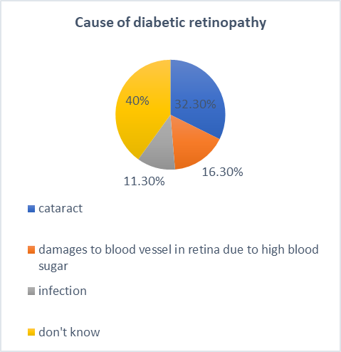
Figure 3: CHART REPRESENTATION

ATTITUDE ASSESSMENT:

Respondents generally displayed a positive and concerned attitude towards diabetic retinopathy, even when their specific knowledge was lacking.

  • Seriousness of DR: A significant majority (81.9%) either "agree" or "strongly agree" that DR is a serious complication of diabetes.
  • ?Importance of Early Detection: Overwhelmingly, respondents felt that early detection is "very important" (53.1%) or "important" (32.3%).
  • ?Necessity of Eye Exams: There is a strong consensus on the need for professional eye care.

?3.1% agree that visiting an ophthalmologist is necessary for all diabetic patients.

?70.1% believe a yearly eye examination is necessary.

PRACTICE ASSESSMENT:

Despite positive attitudes, the self-reported practices for preventing and screening for DR were alarmingly poor. This indicates a significant gap between belief and action.

  • Retinal Screening: A striking 70.1% of respondents have never undergone a retinal screening (fundus examination).
  • Eye Doctor Visits: The vast majority (66.5%) reported "never" visiting an eye doctor. Only 28% reported visiting once a year.
  • Diabetes Self-Management: In contrast to poor eye care practices, self-reported adherence to general diabetes management was high: 88.2% check their blood sugar regularly. 85.8% take their diabetes medicines as advised. 79.5% follow a recommended diabetic diet and lifestyle.
  • Healthcare Counseling: A critical finding is that 72.8% of participants reported having never received counseling or education about diabetic retinopathy from a healthcare professional.The positive attitude of the participants did not translate into consistent practice. (figure 4: chart representation)


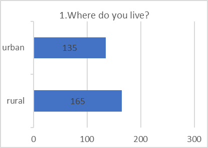
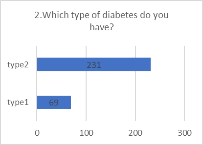
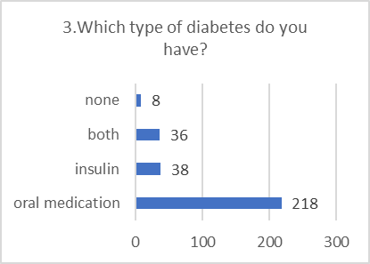
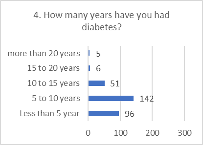
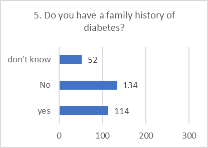
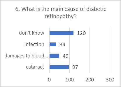
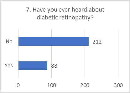
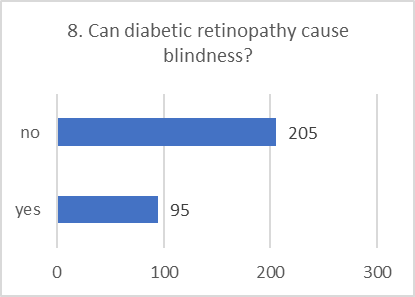
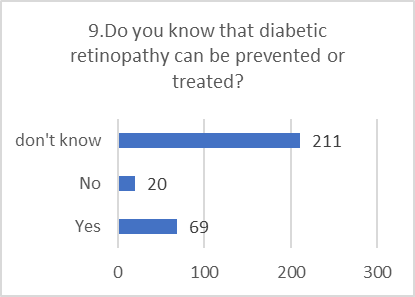
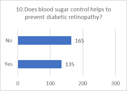
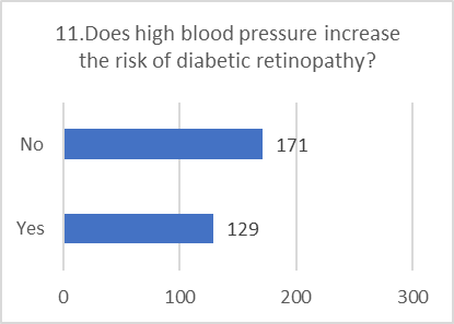
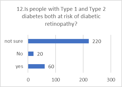
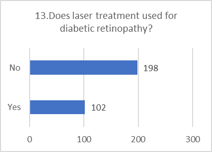
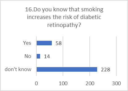
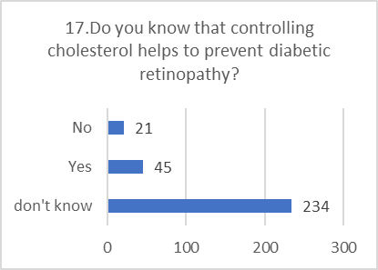
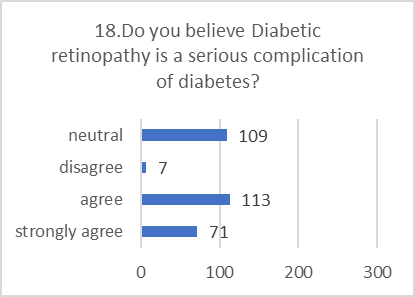
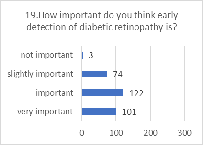
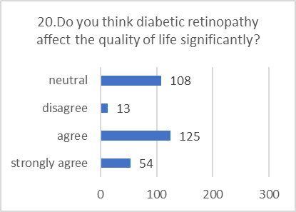
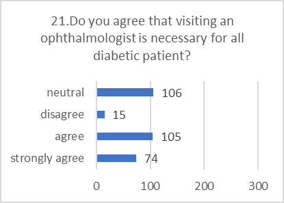
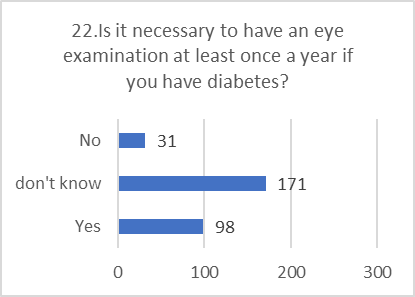
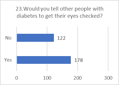
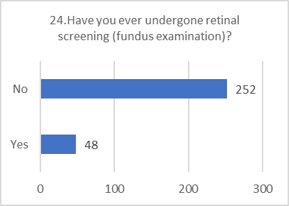
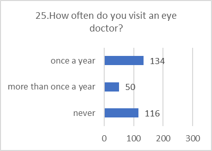
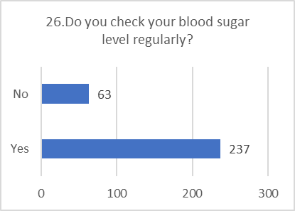
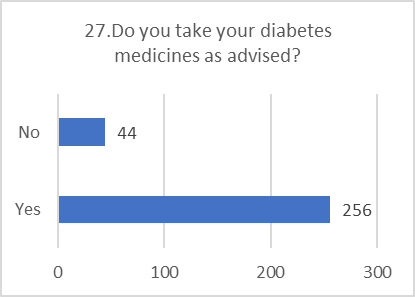
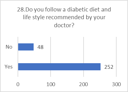
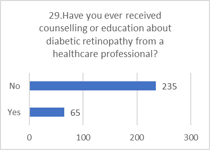
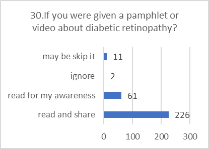
Figure 5: CHART REPRESENTATION

VI. DISCUSSION

The study reveals a critical disconnect in patient care for diabetic retinopathy. While patients demonstrate responsible behavior in managing their daily blood sugar, diet, and medication, they largely neglect the specific, sight-threatening complication of DR.

The Knowledge-Practice Deficit

The most significant finding is the wide gap between what patients believe and what they do. A vast majority agrees that DR is serious and that yearly eye exams are necessary. However, an almost identical majority has never had a retinal screening and never visits an eye doctor. This suggests that the primary barrier is not a lack of concern, but a combination of other factors.

Even among those who have "heard" of DR, specific knowledge is severely lacking. Most could not identify the correct cause, and the majority were unaware that controlling blood sugar helps prevent it. This indicates that awareness is superficial. Patients may have heard the term but do not understand the mechanism, the asymptomatic nature of early stages, or the direct link between their daily diabetes management and saving their sight.

?The Critical Role of Healthcare Providers

?The fact that nearly three-quarters of respondents have never been educated about DR by a healthcare professional is a major red flag. This points to a potential systemic failure in patient education. Patients are diligent in the practices their doctors emphasize (e.g., blood sugar checks, medication). The lack of practice in eye care is likely a direct reflection of a lack of emphasis and education from their healthcare providers.

VII. CONCLUSION

This study highlights a population of diabetic patients who are diligent in their general disease management but critically deficient in their knowledge and practice related to diabetic retinopathy. Their positive attitude and concern are not translating into the essential preventative action of regular eye screenings. This inaction is strongly associated with a lack of specific knowledge and a failure of the healthcare system to provide targeted education on this complication. There is an urgent need for targeted interventions to bridge this gap and improve eye care practices among individuals with diabetes.

VIII. LIMITATIONS

The study has certain limitations that should be considered when interpreting the results:

  • Limited Generalizability: As a single-center study with a relatively small sample size, the findings may not be representative of the entire diabetic population in other regions or healthcare settings.
  • Self-Reported Data: The data was collected through self-reported responses, which may introduce bias. Participants might have provided socially desirable answers, potentially overstating positive practices or understating negligence.

IX. RECOMMENDATIONS

Based on the study's conclusions, the following recommendations are proposed to reduce the burden of blindness from diabetic retinopathy:

  • Implement Health Education Programs: Develop and conduct targeted health education sessions for diabetic patients at both hospital and community levels to improve their understanding of DR.
  • Integrate DR Screening: Incorporate diabetic retinopathy screening as a standard and integral part of routine diabetes care to ensure regular check-ups.
  • Improve Accessibility: Work towards providing affordable and easily accessible eye care services for all diabetic patients, regardless of their financial status.
  • Enhance Collaboration: Foster stronger collaboration between diabetologists and ophthalmologists to create a more streamlined and effective referral and care system for patients.

Targeted Public Health Campaigns: Campaigns are needed, especially in rural areas, to move beyond simple awareness. They must clearly explain why annual screenings are essential even when vision is good and directly link blood sugar control to eye health.

REFERENCES

  1. International Diabetes Federation. IDF Diabetes Atlas, 10th ed [Internet]. Brussels, Belgium; 2021 [cited 2022 Oct 19]. Available from: https://diabetesatlas.org.
  2. Yau JWY, Rogers SL, Kawasaki R, et al. Global Prevalence and Major Risk Factors of Diabetic Retinopathy. Diabetes Care. 2012;35:556–564. [PubMed: 22301125]
  3. Fong DS, Aiello L, Gardner TW, et al. Retinopathy in diabetes. Diabetes Care. 2004;27Suppl 1:S84–87. [PubMed: 14693935]
  4. Köberlein J, Beifus K, Schaffert C, et al. The economic burden of visual impairment and blindness: a systematic review. BMJ Open. 2013;3:e003471.
  5. Moshfeghi AA, Lanitis T, Kropat G, et al. Social Cost of Blindness Due to AMD and Diabetic Retinopathy in the United States in 2020. Ophthalmic Surgery, Lasers and Imaging Retina. 2020;51:S6–S14.
  6. Lee R, Wong TY, Sabanayagam C. Epidemiology of diabetic retinopathy,diabetic macular edema and related vision loss. Eye Vision (London,England). 2015;2:17.
  7. American Optometric Association. Diabetic Retinopathy, St. Louis, MO: American Optometric Association; 2017. Available from:https://www.aoa.org/patients-and-public/eye-and-vision-problems/glossary-of-eye-and-vision-conditions/diabetic-retinopathy. Accessed April 15, 2019.
  8. Domingueti CP, Dusse LM, Carvalho M, de Sousa LP, Gomes KB, FernandesAP. Diabetes mellitus: the linkage between oxidative stress, inflammation,hypercoagulability and vascular complications. J Diabetes Complications. 2016;30(4):738–45.
  9. Li C, Miao X, Li F, Wang S, Liu Q, Wang Y, Sun J. Oxidative stress-relatedmechanisms and antioxidant therapy in diabetic retinopathy. Oxid Med Cell Longev. 2017;2017:9702820.
  10. Wu MY, Yiang GT, Lai TT, Li CJ. The oxidative stress and mitochondrialdysfunction during the pathogenesis of diabetic retinopathy. Oxid MedCell Longev. 2018;2018:3420187.
  11. "White JR. A brief history of the development of diabetes medications. Diabetes Spectr. 2014; 27:82–86. doi: 10.2337/diaspect.27.2.82. [DOI] [PMC free article] [PubMed] [Google Scholar]
  12. Quianzon CC, Cheikh IE. History of current non-insulin medications for diabetes mellitus. J Community Hosp Intern Med Perspect. 2012;2. 10.3402/jchimp. v3402i3403.19081. [DOI] [PMC free article] [PubMed]"
  13. https://pmc.ncbi.nlm.nih.gov/artic.White JR. A brief history of the development of diabetes medications. Diabetes Spectr. 2014; 27:82–86. doi: 10.2337/diaspect.27.2.82. [DOI] [PMC free article] [PubMed] [Google Scholar]
  14. Quianzon CC, Cheikh IE. History of current non-insulin medications for diabetes mellitus. J Community Hosp Intern Med Perspect. 2012;2. 10.3402/jchimp. v3402i3403.19081. [DOI] [PMC free article] [PubMed]
  15. Diabetic Retinopathy Study Research Group. Preliminary report on effects of photocoagulation therapy. Am J Ophthalmol 1976; 81:383–96. [DOI] [PubMed] [Google Scholar]
  16. ETDRS Research Group. Photocoagulation for diabetic macular edema. Arch Ophthalmol 1985; 103:1796–806. [PubMed] [Google Scholar]
  17. Davis S, Aldrich TH, Jones PF, Acheson A, Compton DL, Jain V, Ryan TE, Bruno J, Radziejewski C, Maisonpierre PC, Yancopoulos GD. Is/olation of angiopoietin-1, a ligand for the TIE2 receptor, by secretion-trap expression cloning. Cell. 1996; 87:1161–1169. doi: 10.1016/s0092-8674(00)81812-7. [DOI] [PubMed] [Google Scholar]
  18. Suri C, Jones PF, Patan S, Bartunkova S, Maisonpierre PC, Davis S, et al. Requisite role of angiopoietin-1, a ligand for the TIE2 receptor, during embryonic angiogenesis. Cell. 1996; 87:1171–1180. doi: 10.1016/s0092-8674(00)81813-9. [DOI] [PubMed] [Google Scholar]
  19. Speicher MA, Danis RP, Criswell M, Pratt L. Pharmacologic therapy for diabetic retinopathy. Expert OpinEmerg Drugs. 2003; 8:239–50. doi: 10.1517/14728214.8.1.239. [DOI] [PubMed] [Google Scholar]
  20. Ryan SJ. Retina. 4. St. Louis, Mo: Mosby Inc; 2004. [Google Scholar]
  21. Black, R.J.; Cross, M.; Haile, L.M.; Culbreth, G.T.; Steinmetz, J.D.; Hagins, H.; Kopec, J.A.; Brooks, P.M.; Woolf, A.D.; Ong, K.L.; et al. Global, Regional, and National Burden of Rheumatoid Arthritis, 1990–2020, and Projections to 2050: A Systematic Analysis of the Global Burden of Disease Study 2021. Lancet Rheumatol. 2023, 5, e594–e610. [CrossRef]
  22. Cheung, N.; Mitchell, P.; Wong, T.Y. Diabetic Retinopathy. Lancet 2010, 376, 124–136. [CrossRef] [PubMed]
  23. Simó-Servat, O.; Hernández, C.; Simó, R. Diabetic Retinopathy in the Context of Patients with Diabetes. Ophthalmic Res. 2019,62, 211–217. [CrossRef] [PubMed]
  24. Rasmussen RN, Matsumoto A, Arvin S, Yonehara K. Binocular Integration of Retinal Motion Information Underlies Optic Flow Processing by the Cortex. CurrBiol (2021) 31(6):1165–74. e6. doi: 10.1016/j.cub.2020.12.034
  25. Huang H. Pericyte-Endothelial Interactions in the Retinal Microvasculature.Int J MolSci (2020) 21(19):7413. doi: 10.3390/ijms21197413Yau JW, Rogers SL, Kawasaki R, et al. Global prevalence and major risk factors of diabetic retinopathy. Diabetes Care. 2012;35: 556–64. [PubMed: 22301125]
  26. Zhang X, Saaddine JB, Chou CF, et al. Prevalence of diabetic retinopathy in the United States, 2005–2008. JAMA. 2010; 304:649–56. [PubMed: 20699456]
  27. Klein R, Klein BE, Moss SE, et al. The Wisconsin epidemiologic study of diabetic retinopathy. III. Prevalence and risk of diabetic retinopathy when age at diagnosis is 30 or more years. Arch Ophthalmol. 1984; 102:527–32. [PubMed: 6367725]
  28. International Diabetes Federation: diabetes facts & figures. 2017. https://www.idf.org/aboutdiabetes/what-is-diabetes/facts-figures.html. Accessed 12 Jul 2019.
  29. Centers for Disease Control and Prevention. Prevalence of predia-betes. 2018.https://www.cdc.gov/diabetes/data/statistics-report/prevalence.html. Accessed 20 Jun 2019.
  30. Cheung N, Mitchell P, Wong TY. Diabetic retinopathy. Lancet.2010;377(9735):124–36.
  31. Cheung N, Mitchell P, Wong TY. Diabetic retinopathy. Lancet.2010;376(9735):124–36.
  32. NIH National Eye Institute. Diabetic eye disease, statistics and data [NEI]. 2010.Available at:  https://www.nei.nih.gov/eyedata/diabetic.asp. Accessed September 26, 2014.
  33. Early Treatment Diabetic Retinopathy Study Research Group. Photocoagulation for diabetic macular edema. Early Treatment Diabetic Retinopathy Study Report Number 1. Arch Ophthalmol 1985;103:1796–806.
  34. Van Leiden HA, Dekker JM, Moll AC, et al. Blood pressure, lipids, and obesity are associated with retinopathy: the Hoorn Study. Diabetes Care 2002;25(8): 1320–5.
  35. Klein R, Moss SE, Klein BE. Is gross proteinuria a risk factor for the incidence of proliferative diabetic retinopathy? Ophthalmology 1993;100(8):1140–6.
  36. Cruickshanks KJ, Ritter LL, Klein R, et al. The association of microalbuminuria with diabetic retinopathy. The Wisconsin Epidemiologic Study of Diabetic Retinopathy. Ophthalmology 1993; 100 (6): 862–7.
  37. Klein BE, Moss SE, Klein R. Effect of pregnancy on progression of diabetic retinopathy. Diabetes Care 1990;13(1):34–40.
  38. Writing Team for the Diabetes Control and Complications Trial/Epidemiology of Diabetes Interventions and Complications Research Group. Effect of intensive therapy on the microvascular complications of type 1 diabetes mellitus. JAMA 2002;287(19):2563–9.
  39. "Jackson, T. L. et al. Parsplana vitrectomy for diabetic macular edema: a systematic review, meta-analysis, and synthesis of safety literature. Retina 37 (5), 886–895 (2017)." https://www.nature.com/articles/s41598-025-93847-z#:~:text=Jackson%2C%20T.%20L.%20et%20al.%20Pars%20plana%20vitrectomy%20for%20diabetic%20macular%20edema%3A%20a%20systematic%20review%2C%20meta%2Danalysis%2C%20and%20synthesis%20of%20safety%20literature.%20Retina%2037%20(5)%2C%20886%E2%80%93895%20(2017)] Diabetic Retinopathy Clinical Research Network, Wells JA, Glassman AR, et al. Aflibercept, bevacizumab, or ranibizumab for diabetic macular edema. N Engl J Med. 2015;372:1193–203. [PubMed: 25692915]
  40. Jang C, Chen L, Rabinowitz JD. Metabolomics and Isotope Tracing. Cell(2018) 173(4):822–37. doi: 10.1016/j.cell.2018.03.055
  41. https://www.niddk.nih.gov/healthinformation/diabetes/overview/preventing-problems/diabetic-eye-disease 6. Diabetic Retinopathy - StatPearls - NCBI Bookshelf,
  42. https://www.mayoclinic.org/diseases-conditions/diabetic-retinopathy/diagnosis-treatment/drc-20371617
  43. Eye Health Annual Eye Exams - American Diabetes Association, https://diabetes.org/sites/default/files/2024-07/ADA24_Annual-Eye-Exam_AA_HEN_IN-7-23-24.pdf 14. Medical Management Of Glaucoma And Diabetic Retinopathy - North Texas Center for Sight,
  44. Retinal detachment - Diagnosis and treatment - Mayo Clinic, https://www.mayoclinic.org/diseases-conditions/retinal-detachment/diagnosis-treatment/drc-20351348 47. Vitreous hemorrhage – Causes, diagnosis, and management - PMC - PubMed Central,
  45. https://pmc.ncbi.nlm.nih.gov/articles/PMC10155538/ 48. Options available for vitreous hemorrhage in proliferative diabetic retinopathy - Healio,
  46. https://www.healio.com/news/ophthalmology/20220120/options-available-for-vitreous-hemorrhage-in-proliferative-diabetic-retinopathy 49. Gene Therapy Strategies for Diabetic Eye Diseases - ASGCT,
  47. https://www.asgct.org/publications/news/november-2021/diabetic-eye-disease-awareness-month 50. Updates on Gene Therapy for Diabetic Retinopathy - PMC - PubMed Central, https://pmc.ncbi.nlm.nih.gov/articles/PMC7228867/
  48. Future Directions in Diabetic Retinopathy Treatment: Stem Cell Therapy, Nanotechnology, and PPARα Modulation - PubMed, https://pubmed.ncbi.nlm.nih.gov/39941353/ 52. Future Directions in Diabetic Retinopathy Treatment: Stem Cell Therapy, Nanotechnology, and PPARα Modulation - MDPI,
  49. https://www.mdpi.com/20770383/14/3/683pmc.ncbi.nlm.nih.gov,https://pmc.ncbi.nlm.nih.gov/articles/PMC9115596/#:~:text=Gene%20therapy%20%E2%80%93%20targeting%20retinal%20vasculopathy,proved%20to%20stop%20DR%20progression.
  50. Eye Health Annual Eye Exams - American Diabetes Association,https://diabetes.org/sites/default/files/2024-07/ADA24_Annual-Eye-Exam_AA_HEN_IN-7-23-24.pdf 14. Medical Management Of Glaucoma And Diabetic Retinopathy - North Texas Center for Sight.

Reference

  1. International Diabetes Federation. IDF Diabetes Atlas, 10th ed [Internet]. Brussels, Belgium; 2021 [cited 2022 Oct 19]. Available from: https://diabetesatlas.org.
  2. Yau JWY, Rogers SL, Kawasaki R, et al. Global Prevalence and Major Risk Factors of Diabetic Retinopathy. Diabetes Care. 2012;35:556–564. [PubMed: 22301125]
  3. Fong DS, Aiello L, Gardner TW, et al. Retinopathy in diabetes. Diabetes Care. 2004;27Suppl 1:S84–87. [PubMed: 14693935]
  4. Köberlein J, Beifus K, Schaffert C, et al. The economic burden of visual impairment and blindness: a systematic review. BMJ Open. 2013;3:e003471.
  5. Moshfeghi AA, Lanitis T, Kropat G, et al. Social Cost of Blindness Due to AMD and Diabetic Retinopathy in the United States in 2020. Ophthalmic Surgery, Lasers and Imaging Retina. 2020;51:S6–S14.
  6. Lee R, Wong TY, Sabanayagam C. Epidemiology of diabetic retinopathy,diabetic macular edema and related vision loss. Eye Vision (London,England). 2015;2:17.
  7. American Optometric Association. Diabetic Retinopathy, St. Louis, MO: American Optometric Association; 2017. Available from:https://www.aoa.org/patients-and-public/eye-and-vision-problems/glossary-of-eye-and-vision-conditions/diabetic-retinopathy. Accessed April 15, 2019.
  8. Domingueti CP, Dusse LM, Carvalho M, de Sousa LP, Gomes KB, FernandesAP. Diabetes mellitus: the linkage between oxidative stress, inflammation,hypercoagulability and vascular complications. J Diabetes Complications. 2016;30(4):738–45.
  9. Li C, Miao X, Li F, Wang S, Liu Q, Wang Y, Sun J. Oxidative stress-relatedmechanisms and antioxidant therapy in diabetic retinopathy. Oxid Med Cell Longev. 2017;2017:9702820.
  10. Wu MY, Yiang GT, Lai TT, Li CJ. The oxidative stress and mitochondrialdysfunction during the pathogenesis of diabetic retinopathy. Oxid MedCell Longev. 2018;2018:3420187.
  11. "White JR. A brief history of the development of diabetes medications. Diabetes Spectr. 2014; 27:82–86. doi: 10.2337/diaspect.27.2.82. [DOI] [PMC free article] [PubMed] [Google Scholar]
  12. Quianzon CC, Cheikh IE. History of current non-insulin medications for diabetes mellitus. J Community Hosp Intern Med Perspect. 2012;2. 10.3402/jchimp. v3402i3403.19081. [DOI] [PMC free article] [PubMed]"
  13. https://pmc.ncbi.nlm.nih.gov/artic.White JR. A brief history of the development of diabetes medications. Diabetes Spectr. 2014; 27:82–86. doi: 10.2337/diaspect.27.2.82. [DOI] [PMC free article] [PubMed] [Google Scholar]
  14. Quianzon CC, Cheikh IE. History of current non-insulin medications for diabetes mellitus. J Community Hosp Intern Med Perspect. 2012;2. 10.3402/jchimp. v3402i3403.19081. [DOI] [PMC free article] [PubMed]
  15. Diabetic Retinopathy Study Research Group. Preliminary report on effects of photocoagulation therapy. Am J Ophthalmol 1976; 81:383–96. [DOI] [PubMed] [Google Scholar]
  16. ETDRS Research Group. Photocoagulation for diabetic macular edema. Arch Ophthalmol 1985; 103:1796–806. [PubMed] [Google Scholar]
  17. Davis S, Aldrich TH, Jones PF, Acheson A, Compton DL, Jain V, Ryan TE, Bruno J, Radziejewski C, Maisonpierre PC, Yancopoulos GD. Is/olation of angiopoietin-1, a ligand for the TIE2 receptor, by secretion-trap expression cloning. Cell. 1996; 87:1161–1169. doi: 10.1016/s0092-8674(00)81812-7. [DOI] [PubMed] [Google Scholar]
  18. Suri C, Jones PF, Patan S, Bartunkova S, Maisonpierre PC, Davis S, et al. Requisite role of angiopoietin-1, a ligand for the TIE2 receptor, during embryonic angiogenesis. Cell. 1996; 87:1171–1180. doi: 10.1016/s0092-8674(00)81813-9. [DOI] [PubMed] [Google Scholar]
  19. Speicher MA, Danis RP, Criswell M, Pratt L. Pharmacologic therapy for diabetic retinopathy. Expert OpinEmerg Drugs. 2003; 8:239–50. doi: 10.1517/14728214.8.1.239. [DOI] [PubMed] [Google Scholar]
  20. Ryan SJ. Retina. 4. St. Louis, Mo: Mosby Inc; 2004. [Google Scholar]
  21. Black, R.J.; Cross, M.; Haile, L.M.; Culbreth, G.T.; Steinmetz, J.D.; Hagins, H.; Kopec, J.A.; Brooks, P.M.; Woolf, A.D.; Ong, K.L.; et al. Global, Regional, and National Burden of Rheumatoid Arthritis, 1990–2020, and Projections to 2050: A Systematic Analysis of the Global Burden of Disease Study 2021. Lancet Rheumatol. 2023, 5, e594–e610. [CrossRef]
  22. Cheung, N.; Mitchell, P.; Wong, T.Y. Diabetic Retinopathy. Lancet 2010, 376, 124–136. [CrossRef] [PubMed]
  23. Simó-Servat, O.; Hernández, C.; Simó, R. Diabetic Retinopathy in the Context of Patients with Diabetes. Ophthalmic Res. 2019,62, 211–217. [CrossRef] [PubMed]
  24. Rasmussen RN, Matsumoto A, Arvin S, Yonehara K. Binocular Integration of Retinal Motion Information Underlies Optic Flow Processing by the Cortex. CurrBiol (2021) 31(6):1165–74. e6. doi: 10.1016/j.cub.2020.12.034
  25. Huang H. Pericyte-Endothelial Interactions in the Retinal Microvasculature.Int J MolSci (2020) 21(19):7413. doi: 10.3390/ijms21197413Yau JW, Rogers SL, Kawasaki R, et al. Global prevalence and major risk factors of diabetic retinopathy. Diabetes Care. 2012;35: 556–64. [PubMed: 22301125]
  26. Zhang X, Saaddine JB, Chou CF, et al. Prevalence of diabetic retinopathy in the United States, 2005–2008. JAMA. 2010; 304:649–56. [PubMed: 20699456]
  27. Klein R, Klein BE, Moss SE, et al. The Wisconsin epidemiologic study of diabetic retinopathy. III. Prevalence and risk of diabetic retinopathy when age at diagnosis is 30 or more years. Arch Ophthalmol. 1984; 102:527–32. [PubMed: 6367725]
  28. International Diabetes Federation: diabetes facts & figures. 2017. https://www.idf.org/aboutdiabetes/what-is-diabetes/facts-figures.html. Accessed 12 Jul 2019.
  29. Centers for Disease Control and Prevention. Prevalence of predia-betes. 2018.https://www.cdc.gov/diabetes/data/statistics-report/prevalence.html. Accessed 20 Jun 2019.
  30. Cheung N, Mitchell P, Wong TY. Diabetic retinopathy. Lancet.2010;377(9735):124–36.
  31. Cheung N, Mitchell P, Wong TY. Diabetic retinopathy. Lancet.2010;376(9735):124–36.
  32. NIH National Eye Institute. Diabetic eye disease, statistics and data [NEI]. 2010.Available at:  https://www.nei.nih.gov/eyedata/diabetic.asp. Accessed September 26, 2014.
  33. Early Treatment Diabetic Retinopathy Study Research Group. Photocoagulation for diabetic macular edema. Early Treatment Diabetic Retinopathy Study Report Number 1. Arch Ophthalmol 1985;103:1796–806.
  34. Van Leiden HA, Dekker JM, Moll AC, et al. Blood pressure, lipids, and obesity are associated with retinopathy: the Hoorn Study. Diabetes Care 2002;25(8): 1320–5.
  35. Klein R, Moss SE, Klein BE. Is gross proteinuria a risk factor for the incidence of proliferative diabetic retinopathy? Ophthalmology 1993;100(8):1140–6.
  36. Cruickshanks KJ, Ritter LL, Klein R, et al. The association of microalbuminuria with diabetic retinopathy. The Wisconsin Epidemiologic Study of Diabetic Retinopathy. Ophthalmology 1993; 100 (6): 862–7.
  37. Klein BE, Moss SE, Klein R. Effect of pregnancy on progression of diabetic retinopathy. Diabetes Care 1990;13(1):34–40.
  38. Writing Team for the Diabetes Control and Complications Trial/Epidemiology of Diabetes Interventions and Complications Research Group. Effect of intensive therapy on the microvascular complications of type 1 diabetes mellitus. JAMA 2002;287(19):2563–9.
  39. "Jackson, T. L. et al. Parsplana vitrectomy for diabetic macular edema: a systematic review, meta-analysis, and synthesis of safety literature. Retina 37 (5), 886–895 (2017)." https://www.nature.com/articles/s41598-025-93847-z#:~:text=Jackson%2C%20T.%20L.%20et%20al.%20Pars%20plana%20vitrectomy%20for%20diabetic%20macular%20edema%3A%20a%20systematic%20review%2C%20meta%2Danalysis%2C%20and%20synthesis%20of%20safety%20literature.%20Retina%2037%20(5)%2C%20886%E2%80%93895%20(2017)] Diabetic Retinopathy Clinical Research Network, Wells JA, Glassman AR, et al. Aflibercept, bevacizumab, or ranibizumab for diabetic macular edema. N Engl J Med. 2015;372:1193–203. [PubMed: 25692915]
  40. Jang C, Chen L, Rabinowitz JD. Metabolomics and Isotope Tracing. Cell(2018) 173(4):822–37. doi: 10.1016/j.cell.2018.03.055
  41. https://www.niddk.nih.gov/healthinformation/diabetes/overview/preventing-problems/diabetic-eye-disease 6. Diabetic Retinopathy - StatPearls - NCBI Bookshelf,
  42. https://www.mayoclinic.org/diseases-conditions/diabetic-retinopathy/diagnosis-treatment/drc-20371617
  43. Eye Health Annual Eye Exams - American Diabetes Association, https://diabetes.org/sites/default/files/2024-07/ADA24_Annual-Eye-Exam_AA_HEN_IN-7-23-24.pdf 14. Medical Management Of Glaucoma And Diabetic Retinopathy - North Texas Center for Sight,
  44. Retinal detachment - Diagnosis and treatment - Mayo Clinic, https://www.mayoclinic.org/diseases-conditions/retinal-detachment/diagnosis-treatment/drc-20351348 47. Vitreous hemorrhage – Causes, diagnosis, and management - PMC - PubMed Central,
  45. https://pmc.ncbi.nlm.nih.gov/articles/PMC10155538/ 48. Options available for vitreous hemorrhage in proliferative diabetic retinopathy - Healio,
  46. https://www.healio.com/news/ophthalmology/20220120/options-available-for-vitreous-hemorrhage-in-proliferative-diabetic-retinopathy 49. Gene Therapy Strategies for Diabetic Eye Diseases - ASGCT,
  47. https://www.asgct.org/publications/news/november-2021/diabetic-eye-disease-awareness-month 50. Updates on Gene Therapy for Diabetic Retinopathy - PMC - PubMed Central, https://pmc.ncbi.nlm.nih.gov/articles/PMC7228867/
  48. Future Directions in Diabetic Retinopathy Treatment: Stem Cell Therapy, Nanotechnology, and PPARα Modulation - PubMed, https://pubmed.ncbi.nlm.nih.gov/39941353/ 52. Future Directions in Diabetic Retinopathy Treatment: Stem Cell Therapy, Nanotechnology, and PPARα Modulation - MDPI,
  49. https://www.mdpi.com/20770383/14/3/683pmc.ncbi.nlm.nih.gov,https://pmc.ncbi.nlm.nih.gov/articles/PMC9115596/#:~:text=Gene%20therapy%20%E2%80%93%20targeting%20retinal%20vasculopathy,proved%20to%20stop%20DR%20progression.
  50. Eye Health Annual Eye Exams - American Diabetes Association,https://diabetes.org/sites/default/files/2024-07/ADA24_Annual-Eye-Exam_AA_HEN_IN-7-23-24.pdf 14. Medical Management Of Glaucoma And Diabetic Retinopathy - North Texas Center for Sight.

Photo
P. Praveen Kumar
Corresponding author

Arunai college of Pharmacy, Tiruvannamalai, Tamil Nadu 606603

Photo
S. K. Senthil Kumar
Co-author

Arunai college of Pharmacy, Tiruvannamalai, Tamil Nadu 606603

Photo
S. Tamil Priya
Co-author

Arunai college of Pharmacy, Tiruvannamalai, Tamil Nadu 606603

Photo
V. Tamilarasan
Co-author

Arunai college of Pharmacy, Tiruvannamalai, Tamil Nadu 606603

Photo
S. Thamizharasi
Co-author

Arunai college of Pharmacy, Tiruvannamalai, Tamil Nadu 606603

Photo
K. Vaishnudevi
Co-author

Arunai college of Pharmacy, Tiruvannamalai, Tamil Nadu 606603

P. Praveen Kumar, S. K. Senthil Kumar, S. Tamil Priya, V. Tamilarasan, S. Thamizharasi, K. Vaishnudevi, A Cross-Sectional Kap Study on Diabetic Retinopathy and its Treatment Among Diabetic Mellitus Patients, Int. J. of Pharm. Sci., 2025, Vol 3, Issue 9, 3695-3711. https://doi.org/10.5281/zenodo.17235897

More related articles
The Fundamental Aspect of Rabies...
Rushali Darade, Payal Dodamani, Akshata Patil, Tushar Shelke, ...
Electrospun Nanofibers as Advanced Platforms for S...
Dr. Nasheer S. Shaikh, Vaishnavi Karbhari, Shubham Chillarge, Vid...
Recent Advances In Drug Delivery System...
Nandkumar M. chaudhari, Amrita M. Singh, Nitin S. Bharud, Dhananj...
Analytical Method Development and Validation of UV Spectrophotometric for Levofl...
Dr. Kiran Kumar Buralla, Bhavani Chirra, Triveni Chodavarapu, Dommeti Divya SaiDurga, Dutchari Yamin...
Medical Surveys of an Antidiabetic Medication in village area of western Maharas...
Sohel Ilahi mujawar, Pritam salokhe, Nilesh Chougule, ...
Related Articles
A Comprehensive Review of Coleus aromaticus Encompassing its Botany, Phytochemis...
Bhavyashree K, Deeksha, Deepa K, Deepak, Charan D S, ...
UV Spectrophotometric Method Development and Validation for the Determination of...
Dipannita Naskar, Dr. Subhabrota Majumdar, Banhishikha Kar, ...
Microwave Assisted Organic Synthesis of Heterocyclic Compound...
Dr. N D Nizamuddin, Vittukuri Sanamanhal, ...
The Fundamental Aspect of Rabies...
Rushali Darade, Payal Dodamani, Akshata Patil, Tushar Shelke, ...
More related articles
The Fundamental Aspect of Rabies...
Rushali Darade, Payal Dodamani, Akshata Patil, Tushar Shelke, ...
Electrospun Nanofibers as Advanced Platforms for Skin Drug Delivery: Formulation...
Dr. Nasheer S. Shaikh, Vaishnavi Karbhari, Shubham Chillarge, Vidya Sirsat, ...
Recent Advances In Drug Delivery System...
Nandkumar M. chaudhari, Amrita M. Singh, Nitin S. Bharud, Dhananjay R. Chavhan, Rushikesh D. Kakade,...