
We use cookies to ensure our website works properly and to personalise your experience. Cookies policy
1,2,5Division of Fish Nutrition & Biochemistry, Faculty of Fisheries, SKUAST-Kashmir.
3,4Department of Pharmaceutical Sciences, University of Kashmir.
6Division of Aquatic Animal Health Management, Faculty of Fisheries, SKUAST-Kashmir.
7Division of Agri-statistics, Faculty of Horticulture, SKUAST-Kashmir.
8ICAR-CICFR, Bhimtal Uttarakhand, India.
9Division of Fish Genetics & Biotechnology, Faculty of Fisheries, SKUAST-Kashmir.
The Kashmir Valley, renowned for its breathtaking landscapes, is equally celebrated as a biological reservoir teeming with floristic diversity. Endowed with a temperate climatic regime, the region nurtures a wide range of medicinal plant species, many of which are valued for their therapeutic and pharmaceutical potential. Among these is Salix alba L., commonly known as White Willow, which has historically been utilized in ethnomedicine and continues to garner attention for its bioactive phytoconstituents. In this study, a crude extract of Salix alba L. was prepared using a hydroalcoholic extraction method. The process utilized a solvent system composed of ethanol and distilled water in a standardized ratio of 80:20 (v/v), ensuring optimal polarity balance for the extraction of both hydrophilic and lipophilic compounds. The plant material, comprising shade-dried leaves, was coarsely powdered using a traditional mortar and pestle and subsequently subjected to maceration in the solvent mixture within a macerator apparatus. The extraction was carried out at ambient temperature (25?±?2°C) for a duration of 72 hours with periodic agitation to enhance solute diffusion and solvent penetration. Following maceration, the resulting extract was filtered to separate the marc from the filtrate. To ensure complete removal of the ethanol component, a BUCHI Rotavapor R-300 rotary evaporator was employed under reduced pressure,facilitating the gentle evaporation of the volatile solvent without compromising phytochemical integrity. The concentrated extract was then carefully transferred to a China dish and placed in a desiccator to eliminate residual moisture and solvent traces. Subsequently, qualitative phytochemical screening was conducted to identify various classes of secondary metabolites, including flavonoids, alkaloids, terpenoids, phenols, glycosides, tannins, saponins, lignins, sterols, proteins, acidic compounds, and sugars. Each step of the protocol was executed with methodological precision, utilizing state-of-the-art analytical techniques to ensure reliability and reproducibility. This comprehensive extraction and profiling aim to underscore the pharmacogenetic value of Salix alba L. and contribute to the broader understanding of Kashmir’s phytochemical wealth.
Plants have long served as essential components of traditional medicine systems. Early scholars meticulously documented their observations and treatments based on natural remedies. For example, Al-Razi emphasized the importance of simplicity and safety in medical treatments, advocating for the use of nutrition over complex medications whenever possible (Tschanz, 1998; Tibi, 2006). Today, approximately 80% of the global population—particularly in developing nations—relies on herbal medicine as a primary healthcare source due to its cultural acceptance, cost-effectiveness, and accessibility (El-Seedi et al., 2013). Islamic teachings also reinforce this tradition. Prophet Muhammad (PBUH) stated, “There is no disease that Allah has created, except that He also has created its cure,” and encouraged lifelong pursuit of knowledge (Khan, 1997; Inhorn & Serour, 2011). Natural medicine remains central to both spiritual and empirical approaches to healing (Ahmad et al., 2009). In modern times, bacterial resistance to antibiotics has escalated due to genetic mutations, gene acquisition, and altered gene expression (Sultan et al., 2018). These resistant strains propagate rapidly, transferring resistance traits through horizontal gene transfer mechanisms such as transformation, transduction, and conjugation (Dubin et al., 2016; Munita & Arias, 2016). This alarming trend has led to a growing demand for alternative antimicrobial agents derived from natural sources. Medicinal plants are now globally recognized for their bioactive potential due to their affordability, biocompatibility, and reduced side effects. These phytochemicals, primarily secondary metabolites, exhibit broad therapeutic properties including antibacterial, antifungal, antipyretic, and diuretic effects (Gligori? et al., 2019; Mogashoa et al., 2019). In regions with limited access to modern healthcare, such as parts of the developing world, plant-based remedies offer a practical solution to prevalent health issues (Khunoana et al., 2019; Dafallah & Hossain, 2019). The Salix genus, particularly Salix alba L. (White Willow), comprises around 500 species found globally in wetland habitats. Its bark contains a range of bioactive compounds including salicylic acid, the natural precursor to aspirin, along with flavonoids, lignins, catechins, tannins, and essential vitamins and minerals (Bernardini et al., 2017; Forster et al., 2021). Willow bark has been historically used to treat pain, inflammation, and fever—applications supported by modern pharmacological studies. Willow bark preparations have shown therapeutic effects such as anti-inflammatory, antipyretic, and analgesic properties (Tawfeek et al., 2021; Ramos et al., 2019), largely attributed to their phenolic content (Pi?tczak et al., 2020; Gligori? et al., 2023). Recent research also suggests Salix alba exhibits potential in managing oxidative stress, cardiovascular diseases, and metabolic disorders, due to its antioxidant and vasodilatory properties (Pérez-Sánchez et al., 2020; Mahdi et al., 2022). Moreover, Salix alba extracts have demonstrated significant antibacterial activity against both Gram-positive and Gram-negative pathogens (Türedi et al., 2020), making it a viable candidate for combatting drug-resistant infections. Despite its pharmacological promise, Salix alba remains underexplored. Given the significance of efficient extraction techniques in determining the quality and efficacy of plant-based medicines, this study focuses on hydroalcoholic extraction, phytochemical screening and antibacterial analysis of Salix alba L. collected from the temperate valley of Kashmir. This study aims to contribute to the broader effort of discovering novel bioactive agents from traditionally valued medicinal plants.
MATERIALS AND METHODS
Collection and processing of plant samples
Plant specimens of Salix alba L. were systematically collected from diverse regions across Jammu and Kashmir, including the capital city Srinagar, the Ganderbal district in central Kashmir, and the Qazigund area in South Kashmir. Upon collection, the leaves were meticulously sorted to eliminate any dead, diseased, or decaying material, ensuring only healthy, viable plant parts were retained for further processing. The selected foliage was air-dried in the shade under controlled conditions for a specified duration to preserve its phytochemical integrity. Once sufficiently dried, the plant material was cut into smaller fragments and subsequently pulverized using a laboratory-grade pestle and mortar to obtain a coarse powder. This coarse preparation of S. alba L. served as the foundational material for both the hydroalcoholic extraction process and qualitative phytochemical analysis.
Identification of plant material
The botanical identity of the collected specimens was authenticated using the Flora of Jammu & Kashmir. Salix alba L., commonly referred to as White Willow, is a medium to large deciduous tree that typically reaches heights between 10 and 30 meters. The tree features a trunk diameter of up to 1 meter and often exhibits an irregular or leaning crown structure. Mature trees possess deeply fissured, grey-brown bark, characteristic of the species. The leaves of S. alba L. are notably lighter in appearance compared to other willow species. This is due to the presence of fine, silky white trichomes, particularly abundant on the leaf undersides. The leaves are narrowly lanceolate, ranging from 5–10 cm in length and 0.5–1.5 cm in width. Ecologically, like other members of the genus Salix, S. alba L. demonstrates a strong preference for moist, water-retentive environments. It is typically found thriving in poorly drained soils along the margins of rivers, lakes, and ponds.
Experimental Site
The experimental work was carried out in the Pharmacognosy Laboratory, Department of Pharmaceutical Sciences, University of Kashmir. The plant material underwent initial cleaning, followed by hydroalcoholic extraction using a solvent system composed of ethanol and distilled water in an 80:20 ratio. Subsequently, preliminary phytochemical screening and characterization of the extract were conducted at the Division of Pharmaceutical Sciences, University of Kashmir, using standard laboratory protocols.
Reagents and Equipment’s
All reagents used in the study were of analytical grade. Absolute ethanol was sourced from Changshu Hongheng Chemical Co. Ltd., Jiangshu Province, China, while distilled water was freshly prepared using the EASY-STILL MARK-2000 DDQ apparatus (Bhanu Scientific Instruments). The experimental procedures utilized a range of standard laboratory equipment, including a BUCHI Rotavapor R-300 for solvent evaporation, borosilicate glassware (1000 mL), a laboratory-grade pestle and mortar for sample preparation, and an analytical balance for precise measurements. Additional apparatus included desiccators, china dishes, measuring cylinders, beakers, test tubes, glass flasks, funnels, and Whatman filter paper. Various chemical reagents required for qualitative phytochemical analysis were also employed. All instruments were properly calibrated, and standard protocols were meticulously followed to ensure the reliability and reproducibility of the results.
Preparation of herbal plant extracts
The extraction of Salix alba was carried out using the maceration technique. Initially, the plant material was shade-dried for an adequate period to preserve its phytochemical constituents. Once fully dried, the crude drug was coarsely powdered using a laboratory-grade pestle and mortar. A total of 100 grams of the powdered sample was accurately weighed using an analytical balance with a 10,000 g capacity. The maceration process was performed in a 1000 mL borosilicate glass container using a hydroalcoholic solvent mixture composed of ethanol and distilled water in an 80:20 ratio. The plant material was completely immersed in the solvent system and subjected to periodic agitation for 72 hours at room temperature. After maceration, the extract was filtered and concentrated using the BUCHI Rotavapor R-300 under reduced pressure. To ensure maximum recovery of phytoconstituents, the crude drug was re-extracted four additional times using the same hydroalcoholic solvent. The final pooled extract was collected in a china dish and placed on a water bath to facilitate complete evaporation of residual ethanol.
Phytochemical Screening of Salix alba L. ethanolic extract
Phytochemical screening is a fundamental analytical procedure employed to identify and categorize naturally occurring chemical constituents—known as phytochemicals—within plant extracts. This process is critical in the early stages of drug discovery, as it helps uncover bioactive compounds with potential therapeutic applications. A comprehensive series of qualitative phytochemical tests was carried out on the hydroalcoholic extract of Salix alba to identify its bioactive constituents. These tests confirmed the presence of several important phytochemicals, including alkaloids, flavonoids, tannins, phenols, saponins, glycosides, terpenoids, steroids, and proteins. The detection of these compounds highlights the therapeutic potential of Salix alba and supports its traditional use in herbal and pharmacological applications.
Qualitative determination of Chemical Constituency
Qualitative phytochemical screening is a preliminary approach used to detect the presence or absence of various secondary metabolites in plant extracts. Standardized biochemical assays were employed to achieve this objective. The following tests were performed on Salix alba extract:
Table: Phytochemical Screening Tests
|
Phytochemical Group |
Test Name |
Detailed Procedure |
Positive Result |
Reference |
|
Flavonoids |
Shinoda Test |
A few magnesium turnings are added to the ethanolic extract, followed by the careful addition of concentrated hydrochloric acid drop by drop. |
Orange-red (flavones), red to pink (flavonoids), pink to magenta (flavanones) |
Sri Adelila Sari et al., 2020 |
|
Conc. H?SO? Test |
A measured amount of the plant extract was transferred into a test tube, followed by the gentle addition of a few drops of concentrated sulfuric acid along the inner wall. |
Yellow to orange coloration |
Ramya et al., 2019 |
|
|
Alkaloids
|
Wagner’s Test |
A small portion of the extract is treated with 1 mL of diluted hydrochloric acid, followed by the careful addition of Wagner’s reagent. The mixture is gently shaken. |
Reddish-brown precipitate |
Yakubu Rufai et al., 2016 |
|
Picric Acid Test |
A small amount of the extract is placed in a test tube, followed by the addition of a few drops of saturated picric acid solution. |
Yellow crystalline precipitate |
Goveas S. W. et al., 2014 |
|
|
Terpenoids |
Salkowski Test |
A measured amount of the extract is mixed with chloroform. Concentrated sulfuric acid is added along the inner wall of the tube to form a separate layer. |
Reddish-brown coloration at the interface |
Ayoola et al., 2008 |
|
Phenols |
Ferric Chloride Test |
A small quantity of the ethanolic extract is transferred into a test tube. A few drops of 5% ferric chloride solution are added and the mixture is gently shaken. |
Bluish-green or bluish-black coloration |
Rufai et al., 2016 |
|
Glycosides |
Keller–Killiani Test |
A small quantity of alcoholic extract is transferred to a test tube. Add 1.5 mL glacial acetic acid, a few drops of 5% FeCl?, and layer concentrated H?SO?. |
Bluish-green color at the interface |
Ayoola et al., 2008 |
|
Baljet Test |
A small quantity of the extract is placed in a test tube, followed by a few drops of Baljet’s reagent (picric acid + NaOH). |
Yellow to orange coloration |
Ramya et al., 2019 |
|
|
Tannins |
Vanillin–HCl Test |
A small quantity of the plant sample is placed in a test tube, followed by freshly prepared vanillin reagent and concentrated HCl. The mixture is gently shaken. |
Bright red or crimson coloration |
Ramya et al., 2019 |
|
Saponins |
Foam Test |
A small quantity of extract is mixed with distilled water in a test tube, shaken vigorously, and left undisturbed for 30 minutes. |
Persistent and stable froth (honeycomb-like) |
Banso and Adeyemo, 2006 |
|
Lignins |
Labet Test |
A small quantity of the plant extract is placed in a test tube, followed by the addition of a few drops of gallic acid solution. |
Olive green coloration |
Kumar AK et al., 2016 |
|
Acidic Compounds
|
Methyl Orange Test |
A small amount of extract is placed in a test tube, and a few drops of methyl orange indicator are added. |
Yellow (neutral to alkaline), orange (mild acid), red (strong acid) |
Cho EJ et al., 2003 |
|
Litmus Test |
Blue litmus turns red in acidic conditions; red litmus turns blue in alkaline conditions when dipped in the extract. |
Color change indicates pH |
Cho EJ et al., 2003 |
|
|
Sugars |
Molisch’s Test |
A small quantity of extract is placed in a test tube, followed by a few drops of Molisch’s reagent. Concentrated H?SO? is added carefully along the side of the tube. |
Purple or violet ring at the interface |
El-Khateeb, 2020 |
|
Sugars |
Barfoed’s Test |
Add 2 mL of extract to a test tube, add a drop of Barfoed’s reagent, heat in boiling water bath for 3 mins, then cool. |
Brick-red precipitate (monosaccharides) |
El-Khateeb, 2020; Katoch et al., 2011 |
|
Sterols |
Libermann–Burchard Test |
A sample is treated with acetic anhydride followed by concentrated sulfuric acid. |
Blue-green coloration at the interface |
Godlewska et al., 2022 |
|
Proteins |
Biuret Test |
Add Biuret reagent (copper sulfate in alkaline medium) to the extract. |
Color change from blue to violet |
Boyer, 2000 |
RESULTS
Hydroalcoholic extraction of Salix alba
The hydroalcoholic extraction of Salix alba L. using an ethanol and distilled water mixture in an 80:20 ratio proved to be efficient, yielding a substantial quantity of extract. The process was enhanced by employing shade drying, which preserved the integrity of thermolabile bioactive compounds. Sequential maceration and multiple washings ensured maximum recovery of phytoconstituents. The clarity and concentration of the final extract indicate the method's effectiveness in isolating key secondary metabolites.

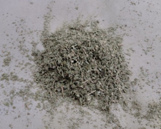
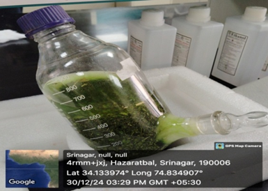
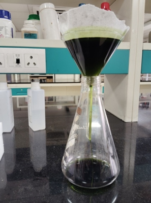
Figure 4. Filtration of plant material after 72 hours using Borosilicate glass conical flask, funnel & filter paper.
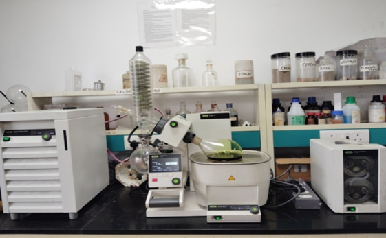
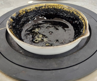
Figure 6. Final extract of Salix alba L. in the China Dish placed on REMI Water Bath for removal of any additional solvent to get a clarified extract.
A high-quality and substantial yield of Salix alba L. leaf extract was successfully obtained using the hydroalcoholic extraction technique, employing ethanol and distilled water in an optimized ratio of 80:20. The plant material was shade-dried to preserve thermolabile phytoconstituents and then coarsely powdered to facilitate maximum solvent penetration. The extraction process was carried out in multiple successive washings to ensure complete recovery of soluble bioactive compounds. Residual ethanol from the pooled extracts was efficiently evaporated using BUCHI Rotavapor R-300 under reduced pressure, yielding a concentrated, phytochemical-rich crude extract. The consistency, clarity, and quantity of the final extract confirm the effectiveness of the selected extraction method, rendering it suitable for further phytochemical screening and pharmacological evaluation.
Qualitative Phytochemical Analysis of Salix alba L.
Qualitative phytochemical analysis was conducted to determine the presence or absence of key secondary metabolites in the ethanolic extract of Salix alba L. leaves. Standard biochemical assays were employed to screen for a range of phytoconstituents, including flavonoids, alkaloids, terpenoids, phenols, glycosides, tannins, saponins, lignins, organic acids, carbohydrates, sterols, and proteins. The outcomes of these tests are summarized in Table 1. The analysis revealed that all the aforementioned phytochemicals were present in the hydroalcoholic leaf extract of S. alba L., suggesting the plant’s rich phytochemical profile and potential therapeutic value.
Quantitative Phytochemical Analysis
Test for Flavonoids
Shinoda test:
Upon addition of a few magnesium filings followed by dropwise addition of concentrated hydrochloric acid to the ethanolic extract of Salix alba L., the appearance of a red coloration was observed, indicating a positive result for the presence of flavonoids.
Concentrated H2SO4 test /Acid test for flavanoids:
Upon careful addition of concentrated sulfuric acid along the inner wall of the test tube containing a small quantity of Salix alba L. ethanolic extract, a distinct yellow coloration was observed, indicating a positive result for the presence of flavonoids.
Test for Alkaloids
Wagners test:
Upon performing Wagner’s test on the ethanolic extract of Salix alba L., 1 mL of diluted hydrochloric acid was added to a small quantity of the extract, followed by the addition of Wagner’s reagent (a solution of iodine and potassium iodide in distilled water). The mixture was shaken thoroughly to ensure complete interaction. A distinct reddish-brown precipitate was formed, which is indicative of the presence of alkaloids. This positive result confirms that Salix alba L. leaves contain alkaloidal constituents, contributing to its potential pharmacological activities.
Picric acid test
Picric acid test was performed on the ethanolic extract of Salix alba L. by adding a few drops of saturated picric acid solution to a small quantity of the extract in a test tube. The contents were mixed thoroughly and observed for any color change. The appearance of a yellow crystalline precipitate confirmed the presence of alkaloids in the plant extract, supporting the outcome of previous alkaloid detection tests.
Test for Terpenoids
Terpenoid test
To assess the presence of terpenoids, the ethanolic leaf extract of Salix alba L. was subjected to the Salkowski test. A measured volume of the extract was mixed with chloroform, followed by the careful addition of concentrated sulfuric acid along the inner wall of the test tube. The development of a distinct reddish-brown coloration at the interface was indicative of a positive result, confirming the presence of terpenoid constituents in the extract.
Test for Phenols
Ferric chloride test
The presence of phenolic compounds in the ethanolic extract of Salix alba L. was confirmed by the ferric chloride test. Upon the addition of a few drops of 5% FeCl? solution to the extract, a characteristic bluish-green coloration was observed. This color change indicates a positive reaction, confirming the presence of phenolic constituents in the plant extract.
Test for Glycosides
Keller Killiani test
To detect the presence of glycosides, a small quantity of the alcoholic extract of Salix alba L. was placed in a test tube. Subsequently, 1.5 mL of glacial acetic acid was added, followed by a few drops of 5% ferric chloride solution. Concentrated sulfuric acid was then carefully introduced along the inner wall of the test tube. The appearance of a bluish-green coloration at the interface indicated a positive result, confirming the presence of glycosidic compounds in the extract.
Baljet test
The presence of glycosides in the extract of Salix alba L. was assessed using the Baljet test. A small volume of the plant extract was treated with a few drops of Baljet’s reagent in a test tube. The development of an orange coloration indicated a positive reaction, confirming the presence of glycosidic compounds in the extract.
Test for Tannins
Vanillin-hydrochloric acid test
To detect tannins, a small quantity of the leaf extract of Salix alba L. was placed in a test tube. A few drops of vanillin solution were added, followed by the addition of concentrated hydrochloric acid. The appearance of a bright red coloration indicated a positive result, confirming the presence of tannins in the plant extract.
Test for Saponins
Foam test
A measured amount of the alcoholic extract of Salix alba L. was placed in a test tube, and a few drops of distilled water were added. The mixture was vigorously shaken for several minutes. The formation of a stable, honeycomb-like froth indicated a positive reaction, confirming the presence of saponins in the plant extract.
Test for lignins
Labet test
A small volume of the ethanolic extract of Salix alba L. was transferred into a test tube, and a few drops of gallic acid solution were added. Upon mixing, a distinct olive green coloration was observed. This color change indicated a positive result for the presence of lignin in the plant extract.
Test for acidic compounds
Methyl Orange Test
To evaluate the pH of the ethanolic extract of Salix alba L., a small amount of the extract was placed in a test tube, followed by the addition of a few drops of methyl orange indicator. The resulting color change to orange-red suggested a mildly acidic nature of the extract.
Litmus Test
The pH nature of the leaf extract of Salix alba L. was assessed using litmus paper. When blue litmus paper was introduced to the extract, a color change to red was observed, indicating acidic properties. In contrast, red litmus paper showed no color change upon contact with the extract, further confirming its acidic nature.
Test for sugars
Barefoed’s Test
An analysis for monosaccharides was conducted by mixing 2 mL of Salix alba L. extract with a drop of Barfoed’s reagent in a test tube. The mixture was heated gently for approximately three minutes and then allowed to cool at room temperature. The appearance of a brick-red precipitate confirmed the presence of monosaccharides in the extract.
Test for Sterols
Liberman Buchard test
The presence of sterols in the leaf extract of Salix alba L. was evaluated using the Libermann-Burchard test. A small quantity of the extract was treated with acetic anhydride, followed by the careful addition of concentrated sulfuric acid. A blue-green coloration developed at the interface, indicating a positive reaction. This color change is characteristic of sterols, resulting from their interaction with the reagents, particularly at the hydroxyl group.
Table 2: Qualitative Phytochemical Screening of Hydroalcoholic Leaf Extract of Salix alba L.
|
S. No. |
Phytochemical Group |
Test Employed |
Reagents Used |
Observed Result |
Phytochemical Presence |
|
1 |
Flavonoids |
Shinoda Test |
Magnesium ribbon + Concentrated HCl |
Appearance of reddish coloration |
Confirmed |
|
2 |
Concentrated H?SO? Test |
Concentrated sulfuric acid |
Development of yellow coloration |
Confirmed |
|
|
3 |
Alkaloids |
Wagner’s Test |
Dilute HCl + Wagner’s reagent (Iodine + Potassium iodide) |
Reddish-brown precipitate |
Confirmed |
|
4 |
Picric Acid Test |
Saturated picric acid solution |
Yellow crystalline precipitate |
Confirmed |
|
|
5 |
Terpenoids |
Salkowski Reaction |
Chloroform + Concentrated H?SO? |
Reddish-brown ring at the interface |
Confirmed |
|
6 |
Phenolic Compounds |
Ferric Chloride Test |
5% Ferric chloride aqueous solution |
Bluish-green coloration |
Confirmed |
|
7 |
Glycosides |
Keller–Killiani Test |
Glacial acetic acid + Ferric chloride + Concentrated H?SO? (layered carefully) |
Bluish-green ring at the interface |
Confirmed |
|
8 |
Baljet’s Test |
Baljet’s reagent (Sodium picrate in NaOH) |
Formation of orange coloration |
Confirmed |
|
|
9 |
Tannins |
Vanillin–HCl Test |
Vanillin solution + Concentrated HCl |
Bright red coloration |
Confirmed |
|
10 |
Saponins |
Foam Test |
Distilled water + vigorous shaking |
Persistent, honeycomb-like froth |
Confirmed |
|
11 |
Lignins |
Lignin (Läbet) Test |
Gallic acid solution |
Olive-green coloration |
Confirmed |
|
12 |
Organic Acids |
Methyl Orange Indicator Test |
Methyl orange reagent |
Orange-red coloration |
Mildly acidic |
|
13 |
Litmus Test |
Blue and red litmus papers |
Blue paper turned red; red paper unchanged |
Acidic |
|
|
14 |
Monosaccharides |
Barfoed’s Test |
Barfoed’s reagent + gentle boiling |
Brick-red precipitate |
Confirmed |
|
15 |
Sterols |
Liberman–Burchard Test |
Acetic anhydride + Concentrated H?SO? |
Blue-green coloration |
Confirmed |
CONCLUSION
The comprehensive phytochemical investigation of Salix alba L. leaf extract using hydroalcoholic extraction has revealed a diverse and rich profile of secondary metabolites, highlighting its potential pharmacological significance. The 80:20 ethanol-water extraction method proved to be efficient in preserving and concentrating bioactive constituents, as evidenced by the clarity, yield, and phytochemical richness of the final extract. Qualitative analyses demonstrated the presence of several important classes of phytochemicals, including flavonoids, alkaloids, terpenoids, phenols, glycosides, tannins, saponins, lignins, acidic compounds, monosaccharides, and sterols. Flavonoids and phenols, known for their antioxidant and anti-inflammatory properties, were strongly indicated by both the Shinoda and ferric chloride tests. Alkaloids were confirmed by Wagner’s and picric acid tests, supporting the plant’s potential use in neuroprotective and antimicrobial therapies. The presence of glycosides and tannins, detected through Keller-Killiani, Baljet, and vanillin-HCl tests, suggests possible cardioprotective and astringent effects. Terpenoids and sterols, indicated by the Salkowski and Libermann-Burchard tests, further underscore the plant’s therapeutic relevance, especially in anti-inflammatory and cholesterol-lowering applications. Additionally, the foam and Labet tests confirmed saponins and lignins, which contribute to immune-modulating and structural functions, respectively. Tests for acidity (methyl orange and litmus) indicated the mildly acidic nature of the extract, which may influence its solubility and biological interactions. Furthermore, the presence of monosaccharides, detected via Barfoed’s test, supports the nutritional and metabolic relevance of the extract. Overall, these findings substantiate the traditional medicinal use of Salix alba L. and provide a strong foundation for further pharmacological and biochemical studies. The presence of multiple bioactive compounds positions Salix alba as a promising candidate for the development of plant-based therapeutics, meriting deeper investigation into its mechanisms of action, dosage standardization, and clinical efficacy.
REFERENCES

Umar Rasool Parry*, Oyas A. Asimi, Weekar Younus Raja, Iflah Hassan, Mir Ishfaq Nazir, Syed Shariq Nazir Qadiri, Imran Khan, Pankaj Kumar, Irfan Ahmad Khan, Phytochemical Profiling and Hydroalcoholic Extraction of White Willow (Salix alba L.) from Kashmir’s Temperate Bioclimatic Zone Highlighting Its Ethnopharmaceutical Potential, Int. J. of Pharm. Sci., 2025, Vol 3, Issue 7, 2671-2684. https://doi.org/10.5281/zenodo.16149137
 10.5281/zenodo.16149137