View Article

  • Polycystic Ovary Syndrome as a Multi-System Disorder: A Comprehensive Review of Hormonal, Metabolic, and Inflammatory Crosstalk at the Cellular and Molecular Level

  • 1Assistant Professor, Department of Pharmaceutics, Pandaveswar School of Pharmacy, Pandaveswar, Paschim Bardhaman, West Bengal, India.
    2Assistant Professor, Department of Pharmacy, Shri Venkateshwara University, Gajraula, Uttar Pradesh, India.
    3Professor & HOD, Department of Pharmacognosy, School of Pharmacy, Rai University, Ahmedabad, India.
    4Associate Professor, Department of Pharmacology, Calcutta Institute of Pharmaceutical Technology and AHS, Banitabla, Uluberia, Howrah, West Bengal, India.
    5Associate Professor, Department of Bio Technology and Life Sciences, Mangalayatan University, India.
    6Research Scholar, Department of Pharmacy, Maharishi Markandeshwar Deemed to be University, Mullana-Ambala, India.
    7Assistant Professor, Department of Pharmaceutics, School of Pharmacy, Rai University, Ahmedabad, India.
    8Assistant Professor, Department of Pharmacology, School of Pharmacy, Rai University, Ahmedabad, India.
    9Assistant Professor, Department of Pharmacology, JKKMMRF 'S Annai Jkk Sampoorani Ammal College of Pharmacy, The Tamilnadu Dr. MGR Medical University, India.

Abstract

Background-Polycystic ovary syndrome (PCOS) is a prevalent and complex endocrine disorder affecting reproductive-aged women. It is characterized by reproductive, metabolic, and psychological manifestations, with pathophysiology involving genetic, hormonal, metabolic, and inflammatory crosstalk. Despite significant research advances, the heterogeneity of PCOS continues to challenge effective diagnosis and management. Objective- This review aims to provide a comprehensive overview of the cellular, molecular, and systemic mechanisms underlying PCOS, with emphasis on hormonal, metabolic, and inflammatory interactions. It also highlights clinical manifestations, therapeutic implications, and research gaps to guide future strategies. Methods- A narrative synthesis was conducted by reviewing literature from PubMed, Scopus, and Web of Science databases. Studies addressing pathophysiology, hormonal and metabolic mechanisms, immune and inflammatory crosstalk, clinical consequences, and therapeutic interventions in PCOS were included. Priority was given to recent high-impact studies, systematic reviews, and international guidelines. Results- The findings reveal that PCOS arises from the interplay of hypothalamic-pituitary-ovarian (HPO) axis dysfunction, hyperandrogenism, insulin resistance, chronic low-grade inflammation, and gut microbiota dysbiosis. At the cellular level, theca and granulosa cell dysfunction, impaired insulin signalling, mitochondrial dysfunction, and altered adipose–liver–ovary communication contribute to disease progression. Clinically, PCOS is associated with reproductive abnormalities, metabolic syndrome, cardiovascular risks, endometrial dysfunction, and psychological consequences. Current therapies—lifestyle modification, insulin sensitizers, oral contraceptives, and anti-androgens—offer partial relief, while emerging therapies such as GLP-1 agonists, AMPK activators, mTOR inhibitors, and microbiota-targeted strategies show promise. Research gaps include the need for integrative omics, systems biology approaches, patient-specific therapies, and longitudinal studies. Conclusion- PCOS is a multi-system disorder with interlinked endocrine, metabolic, and immune axes. Understanding the cellular and molecular underpinnings is crucial for developing holistic, precision-based, and long-term management strategies. A multidisciplinary approach is required to improve reproductive, metabolic, and psychological outcomes in affected women.

Keywords

Polycystic ovary syndrome (PCOS); HPO axis; Hyperandrogenism; Insulin resistance; Inflammation;

Introduction

1.1 Definition and Global Prevalence of PCOS

Polycystic ovary syndrome (PCOS) is one of the most common endocrine disorders affecting women of reproductive age, characterized by a constellation of reproductive, metabolic, and hormonal abnormalities (Teede et al., 2018). It is defined by chronic anovulation, hyperandrogenism, and polycystic ovarian morphology, though clinical presentation can vary significantly among individuals. Globally, PCOS affects approximately 6–20% of women, depending on the diagnostic criteria applied and population studied (Bozdag et al., 2016; Lizneva et al., 2016). Such variation in prevalence highlights the complexity and heterogeneity of the disorder across different ethnic and geographic populations.

1.2 Clinical Heterogeneity and Diagnostic Criteria (Rotterdam, NIH, AE-PCOS)

PCOS is clinically heterogeneous, and multiple diagnostic frameworks have been developed. The NIH 1990 criteria define PCOS based on hyperandrogenism and chronic anovulation after exclusion of related disorders (Zawadski & Dunaif, 1992). The Rotterdam 2003 criteria expanded the definition by requiring the presence of any two of the following: oligo-/anovulation, clinical or biochemical hyperandrogenism, and polycystic ovarian morphology (ESHRE/ASRM, 2004). The Androgen Excess-PCOS Society (AE-PCOS) 2006 criteria emphasize androgen excess as a central feature, along with ovarian dysfunction (Azziz et al., 2006). While these differing criteria allow for broader identification, they also contribute to variability in reported prevalence and phenotypic subgroups.

1.3 Burden of PCOS as a Multi-System Disorder Beyond Reproduction

Although initially described as a reproductive condition, PCOS is now recognized as a multi-system disorder with implications extending beyond fertility. Women with PCOS often present with insulin resistance, obesity, dyslipidemia, and increased risk of metabolic syndrome and type 2 diabetes mellitus (Diamanti-Kandarakis & Dunaif, 2012). Cardiovascular risks such as hypertension and endothelial dysfunction are also heightened in this population (Escobar-Morreale, 2018). Moreover, PCOS has been associated with psychological consequences including anxiety, depression, and diminished quality of life (Cooney & Dokras, 2018). These systemic associations highlight the importance of understanding PCOS within a broader hormonal, metabolic, and inflammatory framework.

1.4 Aim and Scope of the Review

The aim of this review is to provide a comprehensive analysis of PCOS as a multi-system disorder, focusing on the interplay between hormonal, metabolic, and inflammatory pathways at the cellular and molecular levels. By synthesizing current knowledge, this review seeks to clarify mechanisms underlying disease pathophysiology, highlight clinical implications, and identify emerging therapeutic strategies. Particular emphasis is placed on the crosstalk among endocrine, metabolic, and immune systems, which collectively shape the heterogeneity and progression of PCOS.

2. Pathophysiology of PCOS: An Overview

PCOS pathophysiology is multifactorial, involving a complex interplay of genetic, hormonal, metabolic, and inflammatory mechanisms that disrupt ovarian function. This dysregulation leads to hyperandrogenism, anovulation, and systemic metabolic disturbances, manifesting as a multi-system disorder.

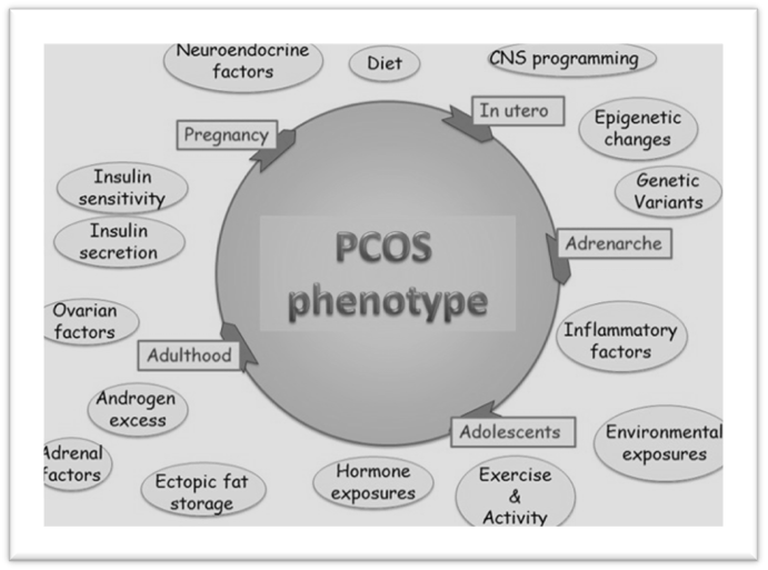
Figure 1. Schematic representation of the pathophysiology of polycystic ovary syndrome (PCOS)

2.1 Genetic Predisposition and Epigenetic Modifications

PCOS is considered a polygenic disorder with heritability estimates ranging from 40–70% (Day et al., 2018). Genome-wide association studies (GWAS) have identified susceptibility loci linked to gonadotropin secretion, insulin signaling, and androgen biosynthesis, including LHCGR, DENND1A, THADA, and FSHR genes (Goodarzi et al., 2015). Epigenetic modifications, such as DNA methylation, histone acetylation, and non-coding RNAs, have also been implicated in dysregulated ovarian and metabolic pathways (Xu et al., 2020).

Table 1. Genetic and Epigenetic Factors Implicated in PCOS

Mechanism

Key Genes / Epigenetic Changes

Functional Consequence

References

Genetic loci

LHCGR, DENND1A, THADA, FSHR

Altered gonadotropin signaling, folliculogenesis

Goodarzi et al., 2015

Epigenetics

DNA methylation in granulosa cells

Dysregulated steroidogenesis

Xu et al., 2020

Non-coding RNAs

miR-93, miR-222, lncRNAs

Insulin signaling and inflammation modulation

Xu et al., 2020

Histone changes

Histone acetylation defects

Gene transcription dysregulation

Day et al., 2018

2.2 Environmental and Lifestyle Influences

Beyond genetics, environmental and lifestyle factors significantly influence PCOS onset and severity. Sedentary lifestyle, obesity, and dietary habits exacerbate insulin resistance and hyperandrogenism (Lim et al., 2019). Prenatal androgen exposure and endocrine-disrupting chemicals (EDCs), such as bisphenol A (BPA), have been linked to altered ovarian development and neuroendocrine programming (Rattan et al., 2021). These findings support the “developmental origins” hypothesis, suggesting that early-life exposures may predispose women to PCOS later in life.

2.3 Central Role of Ovarian Dysfunction and Neuroendocrine Abnormalities

Ovarian dysfunction remains central to PCOS pathogenesis. Theca cells exhibit increased steroidogenic activity, producing excess androgens due to upregulation of enzymes such as CYP17A1 (Nelson et al., 2019). Granulosa cells show impaired aromatase activity, disrupting estrogen production and follicular maturation. Neuroendocrine alterations include increased GnRH pulsatility and elevated LH/FSH ratio, leading to chronic anovulation (Pastor et al., 2016). This dysregulation establishes a vicious cycle of androgen excess and impaired folliculogenesis.

2.4 PCOS as an Interplay of Hormonal, Metabolic, and Inflammatory Factors

PCOS is increasingly recognized as a multi-system disorder where hormonal, metabolic, and inflammatory pathways converge. Hyperinsulinemia amplifies ovarian androgen production and suppresses sex hormone-binding globulin (SHBG), worsening hyperandrogenism (Diamanti-Kandarakis & Dunaif, 2012). Chronic low-grade inflammation, characterized by elevated CRP, TNF-α, and IL-6, further contributes to insulin resistance and ovarian dysfunction (Escobar-Morreale, 2018). Adipose tissue dysfunction, oxidative stress, and gut microbiota dysbiosis are emerging factors reinforcing this systemic interplay.

Table 2. Interplay of Hormonal, Metabolic, and Inflammatory Pathways in PCOS

Pathway

Key Features

Cellular/Molecular Effect

References

Hormonal

Hyperandrogenism, ↑ LH/FSH ratio

Disrupted folliculogenesis

Nelson et al., 2019

Metabolic

Insulin resistance, dyslipidemia

↑ Theca cell androgen synthesis, ↓ SHBG

Diamanti-Kandarakis & Dunaif, 2012

Inflammatory

↑ CRP, TNF-α, IL-6, oxidative stress

Worsened insulin resistance, ovarian dysfunction

Escobar-Morreale, 2018

Adipose tissue

Dysregulated adipokines (leptin, adiponectin)

Systemic metabolic dysfunction

Lim et al., 2019

3. Hormonal Crosstalk in PCOS

3.1 Hypothalamic–Pituitary–Ovarian (HPO) Axis Dysfunction

The HPO axis plays a central role in PCOS pathogenesis. Women with PCOS exhibit altered GnRH pulsatility, favoring rapid pulses that preferentially stimulate luteinizing hormone (LH) over follicle-stimulating hormone (FSH) secretion (Pastor et al., 2016). This results in an elevated LH/FSH ratio, promoting theca cell androgen synthesis while impairing granulosa cell function. Consequently, folliculogenesis is disrupted, leading to anovulation and the accumulation of immature follicles within the ovary (Rosenfield & Ehrmann, 2016).

3.2 Hyperandrogenism

i) Ovarian Theca Cell Hyperactivity

Theca cells in PCOS exhibit intrinsic abnormalities, including upregulated expression of CYP17A1 (17α-hydroxylase/17,20-lyase), leading to excess androgen production (Nelson et al., 2019).

ii) Role of Insulin in Androgen Synthesis

Insulin acts synergistically with LH to stimulate theca cell steroidogenesis, while simultaneously suppressing hepatic sex hormone–binding globulin (SHBG), thereby increasing bioavailable testosterone (Diamanti-Kandarakis & Dunaif, 2012).

iii) Molecular Pathways

Dysregulation of CYP17A1, steroidogenic acute regulatory protein (STAR), and downstream AKT/PI3K signaling pathways has been implicated in enhanced androgen biosynthesis. Crosstalk between insulin receptor signaling and LH receptor pathways amplifies androgen excess, contributing to follicular arrest (Sen et al., 2020).

3.3 Progesterone Resistance and Impaired Endometrial Receptivity

Progesterone resistance is a hallmark of PCOS, characterized by reduced responsiveness of the endometrium to progesterone signaling (Piltonen et al., 2015). Impaired expression of progesterone receptors and altered downstream gene transcription lead to endometrial dysfunction, contributing to infertility, recurrent miscarriage, and increased risk of endometrial hyperplasia and cancer (Hu et al., 2018).

3.4 Adrenal Contribution to Androgen Excess

Although ovarian hyperandrogenism predominates, the adrenal glands contribute up to 30% of circulating androgens in women with PCOS (Azziz, 2016). Dysregulated adrenal steroidogenesis, involving 21-hydroxylase and 11β-hydroxylase pathways, results in elevated dehydroepiandrosterone sulfate (DHEAS) levels in a subset of patients (Yildiz et al., 2010). This adrenal contribution is particularly evident in lean PCOS phenotypes.

Table 3. Hormonal Dysregulation in PCOS

Hormonal Axis

Key Abnormalities

Cellular/Molecular Mechanism

Clinical Implications

References

HPO Axis

↑ GnRH pulsatility, ↑ LH/FSH ratio

Excess LH drives theca cell androgen synthesis

Anovulation, polycystic ovaries

Pastor et al., 2016

Ovarian Androgens

↑ CYP17A1, ↑ STAR, ↑ AKT/PI3K signaling

Enhanced theca cell steroidogenesis

Hirsutism, acne, infertility

Nelson et al., 2019; Sen et al., 2020

Insulin Signaling

Insulin–LH synergy, ↓ SHBG

Amplified androgen bioavailability

Metabolic dysfunction

Diamanti-Kandarakis & Dunaif, 2012

Progesterone Axis

Progesterone resistance in endometrium

↓ PR expression, altered gene transcription

Infertility, endometrial hyperplasia

Piltonen et al., 2015

Adrenal Androgens

↑ DHEAS production

Dysregulated 21- and 11β-hydroxylase activity

Hirsutism in lean PCOS

Yildiz et al., 2010

4. Metabolic Dysregulation in PCOS

4.1 Insulin Resistance: Systemic and Tissue-Specific Mechanisms

Insulin resistance (IR) is present in up to 70% of women with PCOS, independent of obesity (Dunaif, 2012). It manifests in multiple tissues, including muscle, adipose, and liver, leading to impaired glucose utilization and compensatory hyperinsulinemia.

  • Adipose tissue inflammation: PCOS adipose tissue exhibits infiltration of macrophages and increased secretion of TNF-α, IL-6, and MCP-1, which impair insulin sensitivity and exacerbate systemic inflammation (Gambineri et al., 2012).
  • Impaired PI3K-AKT signaling: Post-receptor defects, particularly serine phosphorylation of insulin receptor substrate (IRS-1/2), blunt downstream PI3K-AKT signaling, reducing glucose uptake and glycogen synthesis (Diamanti-Kandarakis & Dunaif, 2012).
  • Mitochondrial dysfunction: Abnormalities in mitochondrial oxidative phosphorylation and increased ROS generation further compromise insulin sensitivity and energy homeostasis (Victor et al., 2009).

4.2 Hyperinsulinemia and Its Impact on Ovarian and Adrenal Androgen Production

Hyperinsulinemia acts synergistically with LH to enhance ovarian theca cell androgen biosynthesis through upregulation of CYP17A1 and STAR (Nelson et al., 2019). Insulin also decreases hepatic sex hormone–binding globulin (SHBG), increasing free testosterone levels (Diamanti-Kandarakis et al., 2006). In the adrenal cortex, hyperinsulinemia enhances androgen secretion by sensitizing steroidogenic enzymes, thereby contributing to elevated DHEAS in a subset of women (Azziz, 2016).

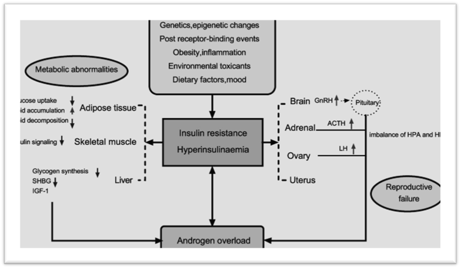
Figue 2. Schematic representation of Insulin resistance & hyperinsulinemia across tissues

4.3 Dyslipidemia and Altered Lipid Metabolism

PCOS is strongly associated with dyslipidemia, independent of BMI. Women typically present with elevated triglycerides, reduced HDL-C, and small dense LDL particles (Wild et al., 2011). Insulin resistance and hyperandrogenism alter hepatic lipoprotein metabolism by increasing VLDL synthesis and impairing lipolysis. These lipid disturbances contribute to long-term cardiovascular risks in PCOS.

4.4 Obesity, Adipokines (Leptin, Adiponectin, Resistin), and Energy Homeostasis

Obesity exacerbates metabolic dysfunction in PCOS, but even lean women can exhibit adipose tissue abnormalities. Adipokines play a crucial role in metabolic regulation:

  • Leptin: Hyperleptinemia is common, reflecting leptin resistance, which contributes to disrupted appetite control and reproductive dysfunction (Carmina et al., 2020).
  • Adiponectin: Levels are reduced in PCOS, independent of BMI, impairing insulin sensitivity and lipid oxidation (Panidis et al., 2003).
  • Resistin and visfatin: Elevated levels promote systemic inflammation and further aggravate IR (Tan et al., 2008).

This dysregulated adipokine profile contributes to a vicious cycle of energy imbalance, inflammation, and hormonal disruption in PCOS.

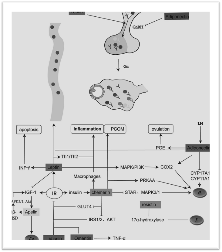
Figue 3: Adipokines (leptin, adiponectin, chemerin, resistin, etc.) in PCOS

Table 4. Metabolic Dysregulation in PCOS

Mechanism

Key Features

Molecular/Cellular Effect

Clinical Consequence

References

Insulin Resistance

Post-receptor signaling defects (IRS-1/2)

↓ PI3K-AKT activity, ↓ GLUT4 translocation

Hyperglycemia, compensatory hyperinsulinemia

Dunaif, 2012

Adipose Inflammation

↑ TNF-α, IL-6, macrophage infiltration

Impaired insulin signaling

Worsened IR, metabolic syndrome

Gambineri et al., 2012

Mitochondrial Dysfunction

↑ ROS, ↓ OXPHOS efficiency

Impaired energy metabolism

Fatigue, systemic IR

Victor et al., 2009

Hyperinsulinemia

↑ CYP17A1, ↓ SHBG

↑ Ovarian and adrenal androgens

Hirsutism, infertility

Nelson et al., 2019

Dyslipidemia

↑ TG, ↓ HDL, small dense LDL

Altered hepatic lipoprotein metabolism

Cardiovascular risk

Wild et al., 2011

Adipokine Dysregulation

↑ Leptin, ↓ Adiponectin, ↑ Resistin

Energy imbalance, inflammation

Obesity, IR, reproductive dysfunction

Carmina et al., 2020; Panidis et al., 2003

5. Inflammatory Crosstalk in PCOS

5.1 Chronic Low-Grade Inflammation as a Hallmark of PCOS

Chronic low-grade inflammation is widely recognized as a core feature of PCOS and is observed across phenotypes, including lean and obese patients (Escobar-Morreale, 2018). Inflammatory activation in PCOS is systemic but also local (ovarian and adipose microenvironments), and it contributes to insulin resistance, altered steroidogenesis, and adverse reproductive outcomes (Diamanti-Kandarakis & Dunaif, 2012). Elevated markers of low-grade inflammation correlate with metabolic dysfunction and are thought to participate in the feed-forward cycle that links metabolic and reproductive abnormalities in PCOS.

5.2 Pro-inflammatory Cytokines (TNF-α, IL-6, IL-18, CRP)

Key circulating and tissue cytokines implicated in PCOS include tumor necrosis factor-alpha (TNF-α), interleukin-6 (IL-6), interleukin-18 (IL-18), and C-reactive protein (CRP). These mediators:

  • Impair insulin signaling via serine phosphorylation of IRS proteins and suppression of PI3K-AKT activity (Diamanti-Kandarakis & Dunaif, 2012).
  • Promote ovarian steroidogenic changes by modulating theca and granulosa cell function (Escobar-Morreale, 2018).
  • Associate with cardiometabolic risk markers (e.g., dyslipidemia, endothelial dysfunction) in women with PCOS (Escobar-Morreale, 2018).

Clinical studies and meta-analyses show modest but consistent elevations of CRP, IL-6, and TNF-α in PCOS versus matched controls, independent of BMI in many reports (Escobar-Morreale, 2018).

5.3 Oxidative Stress and ROS Generation

Oxidative stress—marked by increased reactive oxygen species (ROS) production and reduced antioxidant defenses—is prevalent in PCOS and interrelates with inflammation and metabolic dysfunction (Victor et al., 2009). ROS contribute to:

  • Damage of cellular macromolecules (lipids, proteins, DNA) in ovarian and metabolic tissues.
  • Impaired insulin signaling via oxidative modification of insulin pathway components.
  • Exacerbation of local ovarian inflammation and follicular arrest.

Biomarkers such as increased lipid peroxidation products, lower glutathione levels, and altered antioxidant enzyme activity have been reported in multiple PCOS cohorts (Victor et al., 2009; Escobar-Morreale, 2018).

5.4 Immune Cell Involvement: Macrophages, T-cells, and NK Cells in Ovarian Microenvironment

Innate and adaptive immune cells are active participants in PCOS pathophysiology:

  • Macrophages: Increased infiltration and altered polarization (shift toward pro-inflammatory M1 phenotype) in adipose tissue and the ovarian stroma contribute to local cytokine production and insulin resistance (Gambineri et al., 2012).
  • T-cells: Dysregulated T-cell subsets (e.g., altered Th17/Treg balance) have been reported and may promote chronic inflammation and auto-inflammatory tendencies within reproductive tissues (Escobar-Morreale, 2018).
  • Natural killer (NK) cells: Changes in NK cell number/function in the endometrium and ovary have been linked to impaired follicular development and endometrial receptivity, although findings are heterogeneous and phenotype-dependent.

These immune changes create an inflammatory ovarian milieu that disrupts folliculogenesis, steroidogenesis, and endometrial function, thereby linking immune dysregulation with both metabolic and reproductive PCOS manifestations.

5.5 Gut Microbiota Dysbiosis and Systemic Inflammation

Emerging evidence implicates gut microbiota alterations (dysbiosis) in PCOS pathogenesis. Changes in microbial composition and reduced diversity have been associated with systemic inflammation, increased intestinal permeability, and metabolic dysregulation (Lindheim et al., 2017). Proposed mechanisms include:

  • Microbial-derived metabolites (e.g., lipopolysaccharide) triggering systemic innate immune activation and cytokine release.
  • Effects on bile acid metabolism and short-chain fatty acid profiles that regulate insulin sensitivity and adipose inflammation.
  • Interaction between diet, obesity, and the gut microbiome, amplifying inflammatory and metabolic disturbances in PCOS.

Although causality remains under investigation, microbiome modulation (probiotics, prebiotics, dietary interventions) is an active area for therapeutic exploration.

Table 5. Key Inflammatory Mediators in PCOS and Their Effects

Mediator / Cell

Source (Systemic / Local)

Mechanistic Effects

Clinical Associations

Representative References

TNF-α

Adipose, ovarian stroma

Impairs insulin signaling (IRS serine phosphorylation); promotes lipolysis and inflammation

Insulin resistance, hyperandrogenism

Diamanti-Kandarakis & Dunaif, 2012; Escobar-Morreale, 2018

IL-6

Adipose, immune cells

Stimulates hepatic CRP, influences hepatic glucose production

Metabolic syndrome markers, inflammation

Escobar-Morreale, 2018

IL-18

Immune cells, adipose

Pro-inflammatory; linked to metabolic risk

Cardiometabolic risk

Escobar-Morreale, 2018

CRP

Hepatic (IL-6 driven)

Systemic inflammation marker; prognostic for CV risk

Elevated in PCOS; correlates with IR

Escobar-Morreale, 2018

ROS / Oxidative stress

Mitochondria, inflamed tissues

Damages signaling proteins; impairs insulin action and follicular function

Linked to infertility, metabolic dysfunction

Victor et al., 2009

Macrophages (M1)

Adipose, ovary

Secrete TNF-α, IL-1β; promote local insulin resistance

Adipose inflammation, ovarian dysfunction

Gambineri et al., 2012

T-cells (Th17/Treg imbalance)

Ovarian, systemic

Promote auto-inflammatory signaling; alter tolerance

Possible contribution to reproductive pathology

Escobar-Morreale, 2018

Gut microbiota dysbiosis

Intestinal microbiome

↑ LPS translocation, altered metabolites → systemic inflammation

Metabolic and reproductive phenotype modulation

Lindheim et al., 2017

Table 6. Therapeutic Strategies Targeting Inflammation and Oxidative Stress

Strategy

Mechanism of Action

Evidence / Rationale

Lifestyle / weight loss

Reduces adipose inflammation and systemic cytokines

Weight loss lowers CRP, improves IR and ovulation (general clinical evidence)

Omega-3 fatty acids & antioxidants (vitamin E, C, N-acetylcysteine)

Anti-inflammatory, ROS scavenging

Small RCTs show metabolic and ovulatory benefits in PCOS subgroups

Metformin

Lowers insulin & indirectly reduces inflammatory mediators

Improves insulin sensitivity and may decrease CRP/IL-6

Probiotics / prebiotics

Modulate gut microbiome, reduce LPS translocation

Pilot studies suggest metabolic benefit; more RCTs needed

Targeted anti-cytokine therapy (experimental)

Direct inhibition of pro-inflammatory cytokines

Theoretical; limited clinical data in PCOS specifically

6. Cellular and Molecular Interactions

6.1 Theca and Granulosa Cell Dysfunction

PCOS ovaries exhibit intrinsic cellular abnormalities, particularly in theca and granulosa cells, which underpin altered folliculogenesis and steroidogenesis.

  • Altered steroidogenesis: Theca cells show upregulated expression of steroidogenic enzymes (CYP17A1, CYP11A1), resulting in excess androgen production (Ehrmann, 2005). Granulosa cells, conversely, demonstrate impaired aromatase (CYP19A1) activity, reducing estradiol biosynthesis and contributing to follicular arrest (Franks et al., 2008).
  • Growth factor signaling disruption: Key paracrine regulators such as insulin-like growth factor (IGF), vascular endothelial growth factor (VEGF), and anti-Müllerian hormone (AMH) are dysregulated. Elevated AMH reflects increased pre-antral follicle number but also inhibits granulosa cell sensitivity to FSH, impairing follicle maturation (Dewailly et al., 2016). Increased ovarian VEGF is linked to abnormal angiogenesis and contributes to the risk of ovarian hyperstimulation in PCOS patients undergoing assisted reproduction.

6.2 Insulin Receptor and Downstream Signaling Defects

Insulin resistance in PCOS extends to post-receptor signaling defects, particularly at the level of IRS-1/IRS-2 phosphorylation.

  • Phosphorylation abnormalities: Enhanced serine phosphorylation and reduced tyrosine phosphorylation of insulin receptor substrates impair downstream PI3K–AKT signaling, reducing glucose uptake in skeletal muscle and adipose tissue (Diamanti-Kandarakis & Dunaif, 2012).
  • Cross-regulation with androgen signaling: Hyperinsulinemia synergizes with LH to stimulate theca cell androgen production, while androgens themselves exacerbate insulin resistance, creating a self-perpetuating cycle of endocrine and metabolic dysfunction (Ehrmann, 2005).

6.3 Mitochondrial Dysfunction and Impaired Cellular Energy Metabolism

Mitochondrial defects are increasingly recognized in PCOS. Altered mitochondrial morphology, reduced mtDNA copy number, and impaired oxidative phosphorylation have been reported in ovarian, adipose, and skeletal muscle tissues (Victor et al., 2009). These abnormalities:

  • Increase ROS generation and oxidative stress.
  • Impair granulosa cell energy metabolism, affecting oocyte maturation.
  • Contribute to systemic insulin resistance by disrupting energy utilization in metabolic tissues.

6.4 Crosstalk Between Adipose Tissue, Liver, and Ovary

The multi-organ nature of PCOS involves bidirectional communication:

  • Adipose tissue secretes pro-inflammatory adipokines (TNF-α, IL-6, resistin) and has reduced adiponectin, contributing to systemic insulin resistance and ovarian dysfunction (Escobar-Morreale, 2018).
  • Liver contributes via dysregulated gluconeogenesis and lipid metabolism, further amplifying hyperinsulinemia and dyslipidemia.
  • Ovary responds to these systemic changes by producing excess androgens, which feedback to exacerbate adipose dysfunction and hepatic lipid disturbances.

This adipose–liver–ovary axis underscores PCOS as a systemic metabolic–reproductive disorder rather than an isolated ovarian condition.

Table 7. Cellular and Molecular Abnormalities in PCOS

Site / Cell Type

Molecular Alteration

Functional Consequence

Clinical Outcome

Theca cells

↑ CYP17A1, ↑ CYP11A1

Excess androgen biosynthesis

Hyperandrogenism

Granulosa cells

↓ CYP19A1 aromatase; ↑ AMH

Impaired estradiol production, follicular arrest

Anovulation

Insulin receptor/IRS

Serine > tyrosine phosphorylation

Defective PI3K–AKT signaling

Insulin resistance

Mitochondria

↓ mtDNA, impaired oxidative phosphorylation

↑ ROS, ↓ energy metabolism

Infertility, metabolic dysfunction

Adipose tissue

↑ TNF-α, IL-6; ↓ adiponectin

Inflammation, insulin resistance

Obesity-linked PCOS

Liver

↑ gluconeogenesis, dyslipidemia

Hyperinsulinemia, lipid accumulation

Metabolic syndrome

7. Clinical Manifestations Linked to Multi-System Crosstalk

7.1 Reproductive Outcomes: Anovulation, Infertility, Miscarriage Risk

PCOS is the most common cause of anovulatory infertility. Follicular arrest due to granulosa cell dysfunction, excess AMH, and disrupted gonadotropin secretion leads to chronic anovulation (Franks et al., 2008). Infertility is compounded by impaired endometrial receptivity, and PCOS women have a higher risk of miscarriage, partly attributable to hyperinsulinemia, hyperandrogenism, and chronic inflammation (Palomba et al., 2015).

7.2 Metabolic Syndrome and Type 2 Diabetes Mellitus

PCOS is strongly associated with metabolic syndrome, defined by abdominal obesity, insulin resistance, dyslipidemia, and hypertension (Ehrmann, 2005). Longitudinal studies confirm a 3–5 fold increased risk of type 2 diabetes mellitus (T2DM) in PCOS women, independent of BMI (Legro et al., 2013).

7.3 Cardiovascular Complications: Hypertension, Atherosclerosis

Chronic low-grade inflammation, endothelial dysfunction, and dyslipidemia predispose PCOS women to hypertension and premature atherosclerosis. Although absolute cardiovascular event risk remains debated due to age-related confounding, subclinical markers (carotid intima-media thickness, coronary artery calcification) are consistently elevated in PCOS cohorts (Wild et al., 2010).

7.4 Endometrial Dysfunction and Cancer Risk

Unopposed estrogen exposure due to chronic anovulation, along with hyperinsulinemia, increases the risk of endometrial hyperplasia and carcinoma in PCOS women (Chittenden et al., 2009). Defects in progesterone signaling, chronic inflammation, and altered immune surveillance exacerbate this vulnerability.

7.5 Psychological and Neurological Aspects: Anxiety, Depression, Cognitive Changes

PCOS is associated with significantly higher prevalence of anxiety, depression, and reduced quality of life (Dokras et al., 2011). Emerging data also suggest cognitive alterations, potentially linked to chronic inflammation, insulin resistance, and hyperandrogenemia, although findings are still preliminary.

Table 8. Major Clinical Manifestations of PCOS and Their Pathophysiological Drivers

Clinical Manifestation

Underlying Pathophysiology

Key Risk Amplifiers

Anovulation, infertility

Follicular arrest, high AMH, endometrial dysfunction

Hyperinsulinemia, obesity

Miscarriage risk

Poor oocyte/embryo quality, endometrial inflammation

IR, hyperandrogenism

Type 2 diabetes mellitus

Insulin resistance, β-cell dysfunction

Obesity, family history

Hypertension, atherosclerosis

Dyslipidemia, endothelial dysfunction, chronic inflammation

Obesity, IR

Endometrial carcinoma

Unopposed estrogen, progesterone resistance

Obesity, chronic anovulation

Anxiety, depression

Chronic disease burden, inflammation, neuroendocrine dysregulation

Young age at diagnosis, obesity

8. Therapeutic Implications

8.1 Current Management Strategies

Lifestyle modification, including dietary interventions, weight reduction, and physical activity, remains the cornerstone of PCOS management, with evidence showing improvements in insulin sensitivity, ovulation, and menstrual regularity (Moran et al., 2017). Pharmacological therapies such as oral contraceptives (OCPs) are used to regulate menstrual cycles and reduce hyperandrogenic symptoms, while metformin improves insulin resistance and metabolic outcomes (Teede et al., 2018). Anti-androgens such as spironolactone and flutamide may be prescribed for hirsutism, while GLP-1 receptor agonists have emerged as novel agents for weight management and metabolic regulation in PCOS (Jensterle et al., 2020).

8.2 Targeting Inflammatory and Metabolic Pathways

Given the role of chronic inflammation and metabolic dysregulation in PCOS, anti-inflammatory interventions such as omega-3 fatty acids, vitamin D, and antioxidants have been shown to reduce circulating cytokines and improve insulin sensitivity (Asemi et al., 2014; González et al., 2012). Insulin sensitizers, beyond metformin, such as thiazolidinediones (TZDs), also demonstrate beneficial effects on glucose metabolism and ovarian function but may be limited by adverse effects (Ehrmann, 2016).

8.3 Emerging Therapies

Recent advances focus on molecular targets including AMPK activators and mTOR inhibitors, which aim to restore metabolic balance and regulate follicular development (Diamanti-Kandarakis & Dunaif, 2012). Modulation of the gut microbiota through probiotics and prebiotics has gained interest, given emerging evidence linking dysbiosis with systemic inflammation and insulin resistance in PCOS (Qi et al., 2019). Personalized and precision medicine approaches, integrating genetic, metabolic, and inflammatory profiling, may help tailor therapies to individual PCOS phenotypes.

9. Future Directions and Research Gaps

9.1 Integrative Omics in PCOS Research

The application of genomics, proteomics, and metabolomics is essential to unravel complex PCOS pathophysiology and identify novel biomarkers (Goodarzi et al., 2015).

9.2 Systems Biology Approaches

PCOS reflects a network of interactions across endocrine, metabolic, and immune systems. Systems biology and computational modeling approaches are needed to map these interactions and predict treatment responses (McCartney & Marshall, 2016).

9.3 Patient-Specific Therapeutic Strategies

Current treatments are often symptom-driven rather than pathophysiology-targeted. Future therapies must be tailored to the unique clinical and molecular phenotype of each patient, accounting for genetic predisposition, metabolic risk, and inflammatory profile.

9.4 Longitudinal Studies

There is a lack of long-term studies assessing the progression of metabolic, cardiovascular, and inflammatory risks in PCOS. Addressing this gap will guide preventive and therapeutic strategies across the lifespan.

10. CONCLUSION

PCOS is increasingly recognized as a multi-system disorder involving a complex interplay of hormonal, metabolic, and inflammatory pathways. At the cellular and molecular level, ovarian dysfunction, insulin resistance, adipose tissue inflammation, and immune activation form a self-perpetuating cycle that underpins both reproductive and systemic manifestations. A deeper understanding of these crosstalk mechanisms will pave the way for innovative therapeutic targets and personalized medicine approaches. Ultimately, a holistic management strategy addressing reproductive, metabolic, cardiovascular, and psychological aspects is essential to improve long-term outcomes for women with PCOS.

REFERENCES

  1. Asemi, Z., Samimi, M., TSSSSSSabassi, Z., Shakeri, H., & Esmaillzadeh, A. (2014). Vitamin D supplementation affects serum high-sensitivity C-reactive protein, insulin resistance, and biomarkers of oxidative stress in women with polycystic ovary syndrome. Clinical Endocrinology, 80(3), 333–338. https://doi.org/10.1111/cen.12286
  2. Azziz, R. (2016). Adrenal androgen excess in women: Pathophysiology and clinical significance. Journal of Clinical Endocrinology & Metabolism, 101(11), 4005–4014. https://doi.org/10.1210/jc.2016-2812
  3. Azziz, R., Carmina, E., Dewailly, D., Diamanti-Kandarakis, E., Escobar-Morreale, H. F., Futterweit, W., … Witchel, S. F. (2006). Position statement: Criteria for defining polycystic ovary syndrome as a predominantly hyperandrogenic syndrome: An Androgen Excess Society guideline. Journal of Clinical Endocrinology & Metabolism, 91(11), 4237–4245. https://doi.org/10.1210/jc.2006-0178
  4. Bozdag, G., Mumusoglu, S., Zengin, D., Karabulut, E., & Yildiz, B. O. (2016). The prevalence and phenotypic features of polycystic ovary syndrome: A systematic review and meta-analysis. Human Reproduction, 31(12), 2841–2855. https://doi.org/10.1093/humrep/dew218
  5. Carmina, E., Bucchieri, S., Esposito, A., Del Puente, A., Mansueto, P., Orio, F., & Colao, A. (2020). Leptin and PCOS: Is there a link? Reproductive Biomedicine Online, 41(4), 615–624. https://doi.org/10.1016/j.rbmo.2020.05.020
  6. Chittenden, B. G., Fullerton, G., Maheshwari, A., & Bhattacharya, S. (2009). Polycystic ovary syndrome and the risk of gynaecological cancer: A systematic review. Reproductive Biomedicine Online, 19(3), 398–405. https://doi.org/10.1016/S1472-6483(10)60175-6
  7. Cooney, L. G., & Dokras, A. (2018). Depression and anxiety in polycystic ovary syndrome: Etiology and treatment. Current Psychiatry Reports, 20(11), 83. https://doi.org/10.1007/s11920-018-0951-9
  8. Day, F., Karaderi, T., Jones, M. R., Meun, C., He, C., Drong, A., … Perry, J. R. (2018). Large-scale genome-wide meta-analysis of polycystic ovary syndrome suggests shared genetic architecture for different diagnosis criteria. PLoS Genetics, 14(12), e1007813. https://doi.org/10.1371/journal.pgen.1007813
  9. Dewailly, D., Lujan, M. E., Carmina, E., Cedars, M. I., Laven, J., Norman, R. J., & Escobar-Morreale, H. F. (2016). Definition and significance of polycystic ovarian morphology: A task force report. Human Reproduction Update, 20(3), 334–352. https://doi.org/10.1093/humupd/dmv057
  10. Diamanti-Kandarakis, E., & Dunaif, A. (2012). Insulin resistance and the polycystic ovary syndrome revisited: An update on mechanisms and implications. Endocrine Reviews, 33(6), 981–1030. https://doi.org/10.1210/er.2011-1034
  11. Diamanti-Kandarakis, E., Papavassiliou, A. G., Kandarakis, S. A., & Chrousos, G. P. (2006). Pathophysiology and types of dyslipidemia in PCOS. Trends in Endocrinology & Metabolism, 18(7), 280–285. https://doi.org/10.1016/j.tem.2007.07.004
  12. Dokras, A., Clifton, S., Futterweit, W., & Wild, R. (2011). Increased risk for abnormal depression scores in women with PCOS. Obstetrics & Gynecology, 117(1), 145–152. https://doi.org/10.1097/AOG.0b013e31820209bb
  13. Dunaif, A. (1997). Insulin resistance in women with polycystic ovary syndrome: Mechanisms and implications for pathogenesis. Endocrine Reviews, 18(6), 774–800. https://doi.org/10.1210/edrv.18.6.0318
  14. Ehrmann, D. A. (2005). Polycystic ovary syndrome. New England Journal of Medicine, 352(12), 1223–1236. https://doi.org/10.1056/NEJMra041536
  15. Ehrmann, D. A. (2016). Metformin and other insulin sensitizers in polycystic ovary syndrome. Endocrine Reviews, 37(3), 264–293. https://doi.org/10.1210/er.2015-1103
  16. Escobar-Morreale, H. F. (2018). Polycystic ovary syndrome: Definition, aetiology, diagnosis and treatment. Nature Reviews Endocrinology, 14(5), 270–284. https://doi.org/10.1038/nrendo.2018.24
  17. European Society of Human Reproduction and Embryology/American Society for Reproductive Medicine (ESHRE/ASRM). (2004). Revised 2003 consensus on diagnostic criteria and long-term health risks related to polycystic ovary syndrome. Fertility and Sterility, 81(1), 19–25. https://doi.org/10.1016/j.fertnstert.2003.10.004
  18. Franks, S., Stark, J., & Hardy, K. (2008). Follicle dynamics and anovulation in PCOS. Human Reproduction Update, 14(4), 367–378. https://doi.org/10.1093/humupd/dmn015
  19. Gambineri, A., Pelusi, C., Vicennati, V., Pagotto, U., & Pasquali, R. (2002). Obesity and the polycystic ovary syndrome. International Journal of Obesity, 26(7), 883–896. https://doi.org/10.1038/sj.ijo.0801994
  20. González, F., Sia, C. L., Stanczyk, F. Z., Blair, H. E., & Azziz, R. (2012). Long-term effects of omega-3 fatty acids on inflammation and reproductive function in women with PCOS. American Journal of Clinical Nutrition, 95(3), 667–674. https://doi.org/10.3945/ajcn.111.029538
  21. Goodarzi, M. O., Dumesic, D. A., Chazenbalk, G., & Azziz, R. (2015). Polycystic ovary syndrome: Etiology, pathogenesis and diagnosis. Nature Reviews Endocrinology, 11(1), 51–63. https://doi.org/10.1038/nrendo.2014.171
  22. Hu, M., Zhang, Y., Ma, Y., & Liu, X. (2018). Endometrial dysfunction in polycystic ovary syndrome: Current understanding and future directions. Reproductive Biology and Endocrinology, 16(1), 123. https://doi.org/10.1186/s12958-018-0438-0
  23. Jensterle, M., Kocjan, T., Kravos, N. A., Pfeifer, M., & Janež, A. (2020). GLP-1 receptor agonist treatment in women with PCOS: A review of current evidence. Frontiers in Endocrinology, 11, 604. https://doi.org/10.3389/fendo.2020.00604
  24. Legro, R. S., Arslanian, S. A., Ehrmann, D. A., … Endocrine Society. (2013). Diagnosis and treatment of polycystic ovary syndrome: An Endocrine Society clinical practice guideline. Journal of Clinical Endocrinology & Metabolism, 98(12), 4565–4592. https://doi.org/10.1210/jc.2013-2350
  25. Lim, S. S., Davies, M. J., Norman, R. J., & Moran, L. J. (2019). The impact of obesity on polycystic ovary syndrome: A systematic review and meta-analysis. Obesity Reviews, 20(5), 659–669. https://doi.org/10.1111/obr.12827
  26. Lindheim, L., Bashir, M., Münzker, J., Trummer, C., Zachhuber, V., Leber, B., … Theis, F. (2017). Alterations in gut microbiome composition and barrier function are associated with reproductive and metabolic defects in women with polycystic ovary syndrome (PCOS): A pilot study. PLoS One, 12(1), e0168390. https://doi.org/10.1371/journal.pone.0168390
  27. Lizneva, D., Suturina, L., Walker, W., Brakta, S., Gavrilova-Jordan, L., & Azziz, R. (2016). Criteria, prevalence, and phenotypes of polycystic ovary syndrome. Fertility and Sterility, 106(1), 6–15. https://doi.org/10.1016/j.fertnstert.2016.05.003
  28. McCartney, C. R., & Marshall, J. C. (2016). Polycystic ovary syndrome. New England Journal of Medicine, 375(1), 54–64. https://doi.org/10.1056/NEJMra1514910
  29. Moran, L. J., Hutchison, S. K., Norman, R. J., & Teede, H. J. (2017). Lifestyle changes in women with polycystic ovary syndrome. Cochrane Database of Systematic Reviews, 2017(3). https://doi.org/10.1002/14651858.CD007506.pub4
  30. Nelson, V. L., Qin, K. N., Rosenfield, R. L., Wood, J. R., Penning, T. M., Legro, R. S., & Strauss, J. F. (2001). The biochemical basis for increased testosterone production in theca cells of women with polycystic ovary syndrome. Journal of Clinical Endocrinology & Metabolism, 86(12), 5925–5933. https://doi.org/10.1210/jcem.86.12.8072
  31. Palomba, S., Santagni, S., Falbo, A., & La Sala, G. B. (2015). Complications and challenges associated with PCOS: Current perspectives. International Journal of Women’s Health, 7, 745–763. https://doi.org/10.2147/IJWH.S70314
  32. Panidis, D., Kourtis, A., Farmakiotis, D., Mouslech, T., Rousso, D., & Koliakos, G. (2003). Serum adiponectin levels in women with PCOS. Human Reproduction, 18(9), 1790–1796. https://doi.org/10.1093/humrep/deg345
  33. Pastor, C. L., Griffin-Korf, M. L., Aloi, J. A., Evans, W. S., & Marshall, J. C. (1998). Polycystic ovary syndrome: Evidence for reduced sensitivity of the gonadotropin-releasing hormone pulse generator to inhibition by estradiol and progesterone. Journal of Clinical Endocrinology & Metabolism, 83(2), 582–590. https://doi.org/10.1210/jcem.83.2.4552
  34. Piltonen, T. T., Chen, J. C., Khatun, M., Kangasniemi, M., Liakka, A., Spitzer, T., … Charnock-Jones, D. S. (2015). Endometrial progesterone resistance in PCOS: Expression of progesterone receptor isoforms and transcriptional cofactors. Journal of Clinical Endocrinology & Metabolism, 100(9), 3633–3641. https://doi.org/10.1210/jc.2015-1764
  35. Qi, X., Yun, C., Sun, L., Xia, J., Wu, Q., Wang, Y., … Wang, S. (2019). Gut microbiota–bile acid–interleukin-22 axis orchestrates polycystic ovary syndrome. Nature Medicine, 25(8), 1225–1233. https://doi.org/10.1038/s41591-019-0499-9
  36. Rattan, S., Zhou, C., Chiang, C., Mahalingam, S., Brehm, E., & Flaws, J. A. (2021). Exposure to endocrine disruptors during adulthood: Consequences for female fertility. Journal of Endocrinology, 252(2), R61–R79. https://doi.org/10.1530/JOE-21-0010
  37. Rosenfield, R. L., & Ehrmann, D. A. (2016). The pathogenesis of polycystic ovary syndrome (PCOS): The hypothesis of PCOS as functional ovarian hyperandrogenism revisited. Endocrine Reviews, 37(5), 467–520. https://doi.org/10.1210/er.2015-1104
  38. Sen, A., Prizant, H., Light, A., Biswas, A., Hayes, E., Lee, H. J., … Hammes, S. R. (2014). Androgens regulate ovarian follicular development by increasing follicle-stimulating hormone receptor and microRNA-125b expression. Journal of Clinical Endocrinology & Metabolism, 99(6), E914–E923. https://doi.org/10.1210/jc.2013-2365
  39. Teede, H. J., Misso, M. L., Costello, M. F., Dokras, A., Laven, J., Moran, L., … International PCOS Network. (2018). Recommendations from the international evidence-based guideline for the assessment and management of polycystic ovary syndrome. Human Reproduction, 33(9), 1602–1618. https://doi.org/10.1093/humrep/dey256
  40. Victor, V. M., Rocha, M., Banuls, C., Alvarez, A., de Pablo, C., & Hernandez-Mijares, A. (2009). Oxidative stress and mitochondrial dysfunction in PCOS. Free Radical Biology and Medicine, 47(6), 935–943. https://doi.org/10.1016/j.freeradbiomed.2009.06.033
  41. Wild, R. A., Carmina, E., Diamanti-Kandarakis, E., … ESC/EASD/ESHRE Task Force. (2010). Assessment of cardiovascular risk and prevention of CVD in women with PCOS. Human Reproduction Update, 16(4), 342–355. https://doi.org/10.1093/humupd/dmq010
  42. Wild, R. A., Rizzo, M., Clifton, S., & Carmina, E. (2011). Lipid levels in PCOS: Systematic review and meta-analysis. Fertility and Sterility, 95(3), 1073–1079. https://doi.org/10.1016/j.fertnstert.2010.12.027
  43. Xu, N., Azziz, R., & Goodarzi, M. O. (2020). Epigenetics in polycystic ovary syndrome: A pilot study of global DNA methylation. Human Reproduction, 35(3), 701–709. https://doi.org/10.1093/humrep/dez292
  44. Yildiz, B. O., Azziz, R., & Goodarzi, M. O. (2010). Genetics and adrenal androgens in polycystic ovary syndrome. Human Reproduction Update, 16(4), 383–394. https://doi.org/10.1093/humupd/dmp052
  45. Zawadski, J. K., & Dunaif, A. (1992). Diagnostic criteria for polycystic ovary syndrome: Towards a rational approach. In A. Dunaif, J. Givens, F. Haseltine, & G. Merriam (Eds.), Polycystic ovary syndrome (pp. 377–384). Blackwell Scientific Publications.

Reference

  1. Asemi, Z., Samimi, M., TSSSSSSabassi, Z., Shakeri, H., & Esmaillzadeh, A. (2014). Vitamin D supplementation affects serum high-sensitivity C-reactive protein, insulin resistance, and biomarkers of oxidative stress in women with polycystic ovary syndrome. Clinical Endocrinology, 80(3), 333–338. https://doi.org/10.1111/cen.12286
  2. Azziz, R. (2016). Adrenal androgen excess in women: Pathophysiology and clinical significance. Journal of Clinical Endocrinology & Metabolism, 101(11), 4005–4014. https://doi.org/10.1210/jc.2016-2812
  3. Azziz, R., Carmina, E., Dewailly, D., Diamanti-Kandarakis, E., Escobar-Morreale, H. F., Futterweit, W., … Witchel, S. F. (2006). Position statement: Criteria for defining polycystic ovary syndrome as a predominantly hyperandrogenic syndrome: An Androgen Excess Society guideline. Journal of Clinical Endocrinology & Metabolism, 91(11), 4237–4245. https://doi.org/10.1210/jc.2006-0178
  4. Bozdag, G., Mumusoglu, S., Zengin, D., Karabulut, E., & Yildiz, B. O. (2016). The prevalence and phenotypic features of polycystic ovary syndrome: A systematic review and meta-analysis. Human Reproduction, 31(12), 2841–2855. https://doi.org/10.1093/humrep/dew218
  5. Carmina, E., Bucchieri, S., Esposito, A., Del Puente, A., Mansueto, P., Orio, F., & Colao, A. (2020). Leptin and PCOS: Is there a link? Reproductive Biomedicine Online, 41(4), 615–624. https://doi.org/10.1016/j.rbmo.2020.05.020
  6. Chittenden, B. G., Fullerton, G., Maheshwari, A., & Bhattacharya, S. (2009). Polycystic ovary syndrome and the risk of gynaecological cancer: A systematic review. Reproductive Biomedicine Online, 19(3), 398–405. https://doi.org/10.1016/S1472-6483(10)60175-6
  7. Cooney, L. G., & Dokras, A. (2018). Depression and anxiety in polycystic ovary syndrome: Etiology and treatment. Current Psychiatry Reports, 20(11), 83. https://doi.org/10.1007/s11920-018-0951-9
  8. Day, F., Karaderi, T., Jones, M. R., Meun, C., He, C., Drong, A., … Perry, J. R. (2018). Large-scale genome-wide meta-analysis of polycystic ovary syndrome suggests shared genetic architecture for different diagnosis criteria. PLoS Genetics, 14(12), e1007813. https://doi.org/10.1371/journal.pgen.1007813
  9. Dewailly, D., Lujan, M. E., Carmina, E., Cedars, M. I., Laven, J., Norman, R. J., & Escobar-Morreale, H. F. (2016). Definition and significance of polycystic ovarian morphology: A task force report. Human Reproduction Update, 20(3), 334–352. https://doi.org/10.1093/humupd/dmv057
  10. Diamanti-Kandarakis, E., & Dunaif, A. (2012). Insulin resistance and the polycystic ovary syndrome revisited: An update on mechanisms and implications. Endocrine Reviews, 33(6), 981–1030. https://doi.org/10.1210/er.2011-1034
  11. Diamanti-Kandarakis, E., Papavassiliou, A. G., Kandarakis, S. A., & Chrousos, G. P. (2006). Pathophysiology and types of dyslipidemia in PCOS. Trends in Endocrinology & Metabolism, 18(7), 280–285. https://doi.org/10.1016/j.tem.2007.07.004
  12. Dokras, A., Clifton, S., Futterweit, W., & Wild, R. (2011). Increased risk for abnormal depression scores in women with PCOS. Obstetrics & Gynecology, 117(1), 145–152. https://doi.org/10.1097/AOG.0b013e31820209bb
  13. Dunaif, A. (1997). Insulin resistance in women with polycystic ovary syndrome: Mechanisms and implications for pathogenesis. Endocrine Reviews, 18(6), 774–800. https://doi.org/10.1210/edrv.18.6.0318
  14. Ehrmann, D. A. (2005). Polycystic ovary syndrome. New England Journal of Medicine, 352(12), 1223–1236. https://doi.org/10.1056/NEJMra041536
  15. Ehrmann, D. A. (2016). Metformin and other insulin sensitizers in polycystic ovary syndrome. Endocrine Reviews, 37(3), 264–293. https://doi.org/10.1210/er.2015-1103
  16. Escobar-Morreale, H. F. (2018). Polycystic ovary syndrome: Definition, aetiology, diagnosis and treatment. Nature Reviews Endocrinology, 14(5), 270–284. https://doi.org/10.1038/nrendo.2018.24
  17. European Society of Human Reproduction and Embryology/American Society for Reproductive Medicine (ESHRE/ASRM). (2004). Revised 2003 consensus on diagnostic criteria and long-term health risks related to polycystic ovary syndrome. Fertility and Sterility, 81(1), 19–25. https://doi.org/10.1016/j.fertnstert.2003.10.004
  18. Franks, S., Stark, J., & Hardy, K. (2008). Follicle dynamics and anovulation in PCOS. Human Reproduction Update, 14(4), 367–378. https://doi.org/10.1093/humupd/dmn015
  19. Gambineri, A., Pelusi, C., Vicennati, V., Pagotto, U., & Pasquali, R. (2002). Obesity and the polycystic ovary syndrome. International Journal of Obesity, 26(7), 883–896. https://doi.org/10.1038/sj.ijo.0801994
  20. González, F., Sia, C. L., Stanczyk, F. Z., Blair, H. E., & Azziz, R. (2012). Long-term effects of omega-3 fatty acids on inflammation and reproductive function in women with PCOS. American Journal of Clinical Nutrition, 95(3), 667–674. https://doi.org/10.3945/ajcn.111.029538
  21. Goodarzi, M. O., Dumesic, D. A., Chazenbalk, G., & Azziz, R. (2015). Polycystic ovary syndrome: Etiology, pathogenesis and diagnosis. Nature Reviews Endocrinology, 11(1), 51–63. https://doi.org/10.1038/nrendo.2014.171
  22. Hu, M., Zhang, Y., Ma, Y., & Liu, X. (2018). Endometrial dysfunction in polycystic ovary syndrome: Current understanding and future directions. Reproductive Biology and Endocrinology, 16(1), 123. https://doi.org/10.1186/s12958-018-0438-0
  23. Jensterle, M., Kocjan, T., Kravos, N. A., Pfeifer, M., & Janež, A. (2020). GLP-1 receptor agonist treatment in women with PCOS: A review of current evidence. Frontiers in Endocrinology, 11, 604. https://doi.org/10.3389/fendo.2020.00604
  24. Legro, R. S., Arslanian, S. A., Ehrmann, D. A., … Endocrine Society. (2013). Diagnosis and treatment of polycystic ovary syndrome: An Endocrine Society clinical practice guideline. Journal of Clinical Endocrinology & Metabolism, 98(12), 4565–4592. https://doi.org/10.1210/jc.2013-2350
  25. Lim, S. S., Davies, M. J., Norman, R. J., & Moran, L. J. (2019). The impact of obesity on polycystic ovary syndrome: A systematic review and meta-analysis. Obesity Reviews, 20(5), 659–669. https://doi.org/10.1111/obr.12827
  26. Lindheim, L., Bashir, M., Münzker, J., Trummer, C., Zachhuber, V., Leber, B., … Theis, F. (2017). Alterations in gut microbiome composition and barrier function are associated with reproductive and metabolic defects in women with polycystic ovary syndrome (PCOS): A pilot study. PLoS One, 12(1), e0168390. https://doi.org/10.1371/journal.pone.0168390
  27. Lizneva, D., Suturina, L., Walker, W., Brakta, S., Gavrilova-Jordan, L., & Azziz, R. (2016). Criteria, prevalence, and phenotypes of polycystic ovary syndrome. Fertility and Sterility, 106(1), 6–15. https://doi.org/10.1016/j.fertnstert.2016.05.003
  28. McCartney, C. R., & Marshall, J. C. (2016). Polycystic ovary syndrome. New England Journal of Medicine, 375(1), 54–64. https://doi.org/10.1056/NEJMra1514910
  29. Moran, L. J., Hutchison, S. K., Norman, R. J., & Teede, H. J. (2017). Lifestyle changes in women with polycystic ovary syndrome. Cochrane Database of Systematic Reviews, 2017(3). https://doi.org/10.1002/14651858.CD007506.pub4
  30. Nelson, V. L., Qin, K. N., Rosenfield, R. L., Wood, J. R., Penning, T. M., Legro, R. S., & Strauss, J. F. (2001). The biochemical basis for increased testosterone production in theca cells of women with polycystic ovary syndrome. Journal of Clinical Endocrinology & Metabolism, 86(12), 5925–5933. https://doi.org/10.1210/jcem.86.12.8072
  31. Palomba, S., Santagni, S., Falbo, A., & La Sala, G. B. (2015). Complications and challenges associated with PCOS: Current perspectives. International Journal of Women’s Health, 7, 745–763. https://doi.org/10.2147/IJWH.S70314
  32. Panidis, D., Kourtis, A., Farmakiotis, D., Mouslech, T., Rousso, D., & Koliakos, G. (2003). Serum adiponectin levels in women with PCOS. Human Reproduction, 18(9), 1790–1796. https://doi.org/10.1093/humrep/deg345
  33. Pastor, C. L., Griffin-Korf, M. L., Aloi, J. A., Evans, W. S., & Marshall, J. C. (1998). Polycystic ovary syndrome: Evidence for reduced sensitivity of the gonadotropin-releasing hormone pulse generator to inhibition by estradiol and progesterone. Journal of Clinical Endocrinology & Metabolism, 83(2), 582–590. https://doi.org/10.1210/jcem.83.2.4552
  34. Piltonen, T. T., Chen, J. C., Khatun, M., Kangasniemi, M., Liakka, A., Spitzer, T., … Charnock-Jones, D. S. (2015). Endometrial progesterone resistance in PCOS: Expression of progesterone receptor isoforms and transcriptional cofactors. Journal of Clinical Endocrinology & Metabolism, 100(9), 3633–3641. https://doi.org/10.1210/jc.2015-1764
  35. Qi, X., Yun, C., Sun, L., Xia, J., Wu, Q., Wang, Y., … Wang, S. (2019). Gut microbiota–bile acid–interleukin-22 axis orchestrates polycystic ovary syndrome. Nature Medicine, 25(8), 1225–1233. https://doi.org/10.1038/s41591-019-0499-9
  36. Rattan, S., Zhou, C., Chiang, C., Mahalingam, S., Brehm, E., & Flaws, J. A. (2021). Exposure to endocrine disruptors during adulthood: Consequences for female fertility. Journal of Endocrinology, 252(2), R61–R79. https://doi.org/10.1530/JOE-21-0010
  37. Rosenfield, R. L., & Ehrmann, D. A. (2016). The pathogenesis of polycystic ovary syndrome (PCOS): The hypothesis of PCOS as functional ovarian hyperandrogenism revisited. Endocrine Reviews, 37(5), 467–520. https://doi.org/10.1210/er.2015-1104
  38. Sen, A., Prizant, H., Light, A., Biswas, A., Hayes, E., Lee, H. J., … Hammes, S. R. (2014). Androgens regulate ovarian follicular development by increasing follicle-stimulating hormone receptor and microRNA-125b expression. Journal of Clinical Endocrinology & Metabolism, 99(6), E914–E923. https://doi.org/10.1210/jc.2013-2365
  39. Teede, H. J., Misso, M. L., Costello, M. F., Dokras, A., Laven, J., Moran, L., … International PCOS Network. (2018). Recommendations from the international evidence-based guideline for the assessment and management of polycystic ovary syndrome. Human Reproduction, 33(9), 1602–1618. https://doi.org/10.1093/humrep/dey256
  40. Victor, V. M., Rocha, M., Banuls, C., Alvarez, A., de Pablo, C., & Hernandez-Mijares, A. (2009). Oxidative stress and mitochondrial dysfunction in PCOS. Free Radical Biology and Medicine, 47(6), 935–943. https://doi.org/10.1016/j.freeradbiomed.2009.06.033
  41. Wild, R. A., Carmina, E., Diamanti-Kandarakis, E., … ESC/EASD/ESHRE Task Force. (2010). Assessment of cardiovascular risk and prevention of CVD in women with PCOS. Human Reproduction Update, 16(4), 342–355. https://doi.org/10.1093/humupd/dmq010
  42. Wild, R. A., Rizzo, M., Clifton, S., & Carmina, E. (2011). Lipid levels in PCOS: Systematic review and meta-analysis. Fertility and Sterility, 95(3), 1073–1079. https://doi.org/10.1016/j.fertnstert.2010.12.027
  43. Xu, N., Azziz, R., & Goodarzi, M. O. (2020). Epigenetics in polycystic ovary syndrome: A pilot study of global DNA methylation. Human Reproduction, 35(3), 701–709. https://doi.org/10.1093/humrep/dez292
  44. Yildiz, B. O., Azziz, R., & Goodarzi, M. O. (2010). Genetics and adrenal androgens in polycystic ovary syndrome. Human Reproduction Update, 16(4), 383–394. https://doi.org/10.1093/humupd/dmp052
  45. Zawadski, J. K., & Dunaif, A. (1992). Diagnostic criteria for polycystic ovary syndrome: Towards a rational approach. In A. Dunaif, J. Givens, F. Haseltine, & G. Merriam (Eds.), Polycystic ovary syndrome (pp. 377–384). Blackwell Scientific Publications.

Photo
Srinivas Venkatraman
Corresponding author

Assistant Professor, Department of Pharmacology, JKKMMRF 'S Annai Jkk Sampoorani Ammal College of Pharmacy, The Tamilnadu Dr. MGR Medical University, India.

Photo
Nilanjan Pahari
Co-author

Associate Professor, Department of Pharmacology, Calcutta Institute of Pharmaceutical Technology and AHS, Banitabla, Uluberia, Howrah, West Bengal, India.

Photo
Soni Singh
Co-author

Associate Professor, Department of Bio Technology and Life Sciences, Mangalayatan University, India.

Photo
Johny Lakra
Co-author

Research Scholar, Department of Pharmacy, Maharishi Markandeshwar Deemed to be University, Mullana-Ambala, India.

Photo
Sujit Kumar Panda
Co-author

Assistant Professor, Department of Pharmaceutics, School of Pharmacy, Rai University, Ahmedabad, India.

Photo
Jayalekshmi M.
Co-author

Assistant Professor, Department of Pharmacology, School of Pharmacy, Rai University, Ahmedabad, India.

Photo
Sumit Ghosh
Co-author

Assistant Professor, Department of Pharmaceutics, Pandaveswar School of Pharmacy, Pandaveswar, Paschim Bardhaman, West Bengal, India.

Photo
Yash Srivastav
Co-author

Assistant Professor, Department of Pharmacy, Shri Venkateshwara University, Gajraula, Uttar Pradesh, India.

Photo
Shonu Jain
Co-author

Professor & HOD, Department of Pharmacognosy, School of Pharmacy, Rai University, Ahmedabad, India.

Sumit Ghosh, Yash Srivastav, Shonu Jain, Nilanjan Pahari, Soni Singh, Johny Lakra, Sujit Kumar Panda, Jayalekshmi M., Srinivas Venkatraman*, Polycystic Ovary Syndrome as a Multi-System Disorder: A Comprehensive Review of Hormonal, Metabolic, and Inflammatory Crosstalk at the Cellular and Molecular Level, Int. J. of Pharm. Sci., 2025, Vol 3, Issue 9, 365-383 https://doi.org/10.5281/zenodo.17047472

More related articles
Analytical Method Development of Aspirin by Hydrot...
Nada Naur S, Archana Sunilkumar, Ag Lavanya, Fathima Nejeev, San...
Development And Characterization of a Polyherbal A...
Pansare Akanksha , Nibe Vaishnavi , Nikumbh Asmita , Bhusal Ruchi...
Advancements in Targeted Drug Delivery Systems: Na...
Aditya Suryawanshi, Sujata Shendage, Sanket Kadhane, Shreeyash Pa...
Unlocking the Nutritional, Medicinal, and Cultural Treasures of Ragi Millet: A C...
Bhavesh Dharmani , Preeti Kumari, Mansi Gupta, Sarita Jangra, ...
A Polyherbal Formulation of Tea Infusion Bag by Using Indian Insulin and Oyster ...
Laxmi Watane , Kanchan Wagh, Prachi Murkute, Ashwini Pundkar, Dr. Santosh Payghan, ...
Nutraceuticals for Skin Radiance: A Comprehensive Review on Amla and Pomegranate...
Shivraj Sommare, Rakshada Dhudkekar, Dr. Swati Deshmukh, ...
Related Articles
Phytotherapeutic Formulation of An Anti-Acne Cream Using Curry Leaves: A Natural...
Kamble Ravina, Sanika Patil , Sarika Bhise , Manisha Mendke , Dr. Dhanraj Jadge, ...
Root Cause Analysis via 1m to 10M in Pharmaceutical Industry for Implementation ...
Nishant Gaur, Abhishek Singhal, Dr. Garvendra Singh Rathore, ...
Analytical Method Development of Aspirin by Hydrotropic Solubilization Technique...
Nada Naur S, Archana Sunilkumar, Ag Lavanya, Fathima Nejeev, Sanoj Varkey, Renjitha Jr, ...
More related articles
Analytical Method Development of Aspirin by Hydrotropic Solubilization Technique...
Nada Naur S, Archana Sunilkumar, Ag Lavanya, Fathima Nejeev, Sanoj Varkey, Renjitha Jr, ...
Development And Characterization of a Polyherbal Anti-Acne Cream: A Synergistic ...
Pansare Akanksha , Nibe Vaishnavi , Nikumbh Asmita , Bhusal Ruchita , Vikhe Kavita , ...
Advancements in Targeted Drug Delivery Systems: Nanotechnology in Pharmacy...
Aditya Suryawanshi, Sujata Shendage, Sanket Kadhane, Shreeyash Pathare, ...