

We use cookies to ensure our website works properly and to personalise your experience. Cookies policy
Global College of Pharmaceutical Technology, Nadia, West Bengal, India.
Type 2 diabetes mellitus is a chronic metabolic disorder with insulin resistance and impaired glucose homeostasis. PPAR-Gamma is a key regulator of glucose and lipid metabolism and a well-established therapeutic target. The present study is to synthesize and identify new thiazolidinedione derivatives as a potential PPAR-Gamma agonist by In Silico methods. A number of compounds were designed and molecular docking studies were conducted using CB-DOCK2 to assess their binding affinity and interaction with target receptor. Also pharmacokinetic and toxicity properties of compounds were determined using QSAR analysis and ADMET profiling by ChemMaster software. Docking results showed that some derivatives exhibited strong binding affinities and favourable interactions with major active site residues of the receptor. QSAR analysis confirmed structural elements involved in biological activity. ADMET predictions suggested acceptable pharmacokinetic properties with low toxicity profiles for top ranked compounds. The results suggest that the identified thiazolidinedione derivatives can be promising candidates for further development as PPAR-Gamma agonists in the treatment of Type 2 Diabetes Mellitus. Nevertheless, further experimental confirmation through in vitro and in vivo studies is essential to verify the therapeutic potential of these compounds.
Diabetes Mellitus (DM) is one of the oldest known metabolic disorders, with descriptions dating back to ancient Egyptian manuscripts about 3000 years ago [1]. The classification of diabetes into type 1 and type 2 was clearly defined in the early 20th Century, improving understanding of disease mechanisms and management [2]. Type 2 Diabetes Mellitus (non-insulin dependent diabetes) is the most common form of Diabetes Mellitus and is characterised by hyperglycemia, insulin resistance and relative insulin deficiency [3]. Type 2 Diabetes Mellitus results from a combination of factors and the interactions between genetic predisposition, environmental influences and lifestyle related behavioural factors are complex [4]. Individuals with Type 2 Diabetes Mellitus are at risk of short-term and long-term complications, including cardiovascular diseases, neuropathy, nephropathy and retinopathy, which contribute substantially to morbidity and mortality globally. The disease can be left undiagnosed for years, especially in growing countries where access to healthcare is restricted [4].
EXISTING TREATMENTS OF TYPE 2 DIABETES MELLITUS:
The management of type 2 diabetes mellitus is mainly aimed at maintaining optimal glycemic control and preventing long-term complications. Lifestyle modification involving dietary changes, weight reduction and increased physical activity are essential in improving insulin sensitivity and are usually the initial treatment [5].
Pharmacological therapy is initiated when lifestyle interventions alone are not sufficient. The currently available pharmacological treatments for type 2 Diabetes Mellitus are summarized below:
Table 1: Existing therapeutic agents used in the management of type 2 Diabetes Mellitus [5]
|
DRUG CLASS |
EXAMPLE DRUG(S) |
MECHANISM OF ACTION |
|
BIGUANIDES |
Metformin |
↓ Hepatic glucose production ↑ Insulin sensitivity |
|
SULFONYLUREAS |
Glipizide Glibenclamide |
Stimulate pancreatic insulin secretion |
|
THIAZOLIDINEDIONES (TZD) |
Pioglitazone, Rosiglitazone |
Activate PPAR-Gamma ↑ Insulin sensitivity in adipose tissue and muscles |
|
DPP-4 INHIBITORS |
Sitagliptin Linagliptin |
↑ Incretin Hormones ↑ Insulin Release |
|
SGLT2 INHIBITORS |
Dapagliflozin Canagliflozin |
↑ Glucose excretion via urine |
|
GLP-1 RECEPTOR AGONISTS |
Liraglutide Exenatide |
↑ Insulin , ↓ Glucagon, Slow gastric emptying |
|
INSULIN THERAPY |
Human Insulin, analogues |
Direct glucose lowering |
Despite the availability of multiple therapeutic options, limitations such as side effects, drug resistance and incomplete glycemic control necessitate the development of novel therapeutic agents.
RATIONALE FOR NOVEL THERAPEUTIC APPROACHES:
Type 2 Diabetes Mellitus has many therapeutic options, but effective long-term management has become a challenge due to limitations such as adverse effects, high cost and less patient compliance. Metformin is one of the frequently used drugs that are usually well tolerated but may cause gastrointestinal disturbances [6]. In addition, Sulfonylureas are connected with the hazards of hypoglycemia and gaining of physical weight which can negatively impact patient outcomes [7].
Furthermore, newer agents such as SGLT2 inhibitors and GLP1 receptor agonists are useful but expensive and can cause side-effects such as urinary tract infection or gastrointestinal upset [5]. These limitations underline the requirement for safer, more effective and affordable therapeutic alternatives. The Peroxisome Proliferator Activated Receptor Gamma (PPAR-Gamma) is a nuclear receptor. It plays a key role in the regulation cum direction of glucose metabolism, lipid homeostasis and insulin sensitivity. It is a hopeful beneficial target in the administration of Type 2 Diabetes Mellitus. PPAR-Gamma activation improves insulin sensitivity in peripheral tissue and as such is an attractive drug target [8].
Computational approaches such as molecular docking have emerged as powerful tools in modern drug discovery, enabling the identification and optimization of potential drug candidates by predicting their binding affinity and interaction with target proteins. Therefore, the present study aims to evaluate potential ligands targeting PPAR-Gamma using In Silico docking techniques to identify novel compounds with improved efficacy and reduced side effects.
MATERIALS AND METHODOLOGY [9-13]:
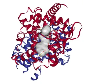
The Peroxisome Proliferator Activated Receptor Gamma (PPAR-Gamma) was selected as target for the study. The crystal structure of the ligand-binding domain (LDB) was retrieved from the RCSB Protein Data Bank (PDB) using the PDB accession code 1KNU [14]. This structure was selected because it represents the receptor in an active conformation complexed with a high affinity agonist.
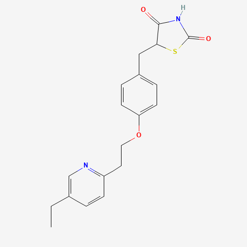
Standard reference ligands, such as Pioglitazone, were retrieved from the PubChem database [15]. A series of Thizolidinedione (TZD) derivatives were designed and prepared for docking using standard energy-minimization protocols to reach their local minima [16].
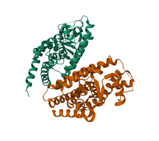
Figure 1: Human Peroxisome Proliferator Activated Receptor Gamma ligand-binding domain
(1KNU)
Table 2: Standard Compound
|
Sl. No. |
Standard Compound |
Structure |
|
1 |
Pioglitazone |
|
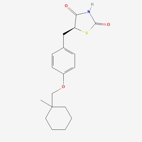
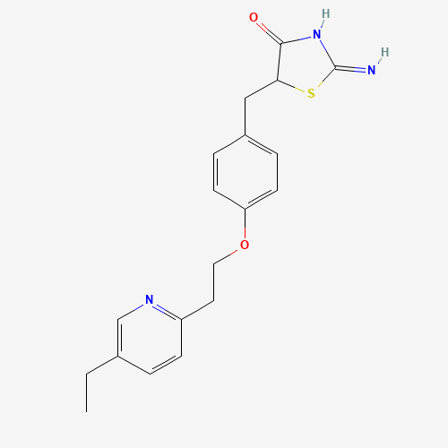
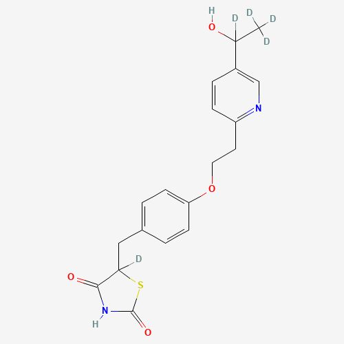
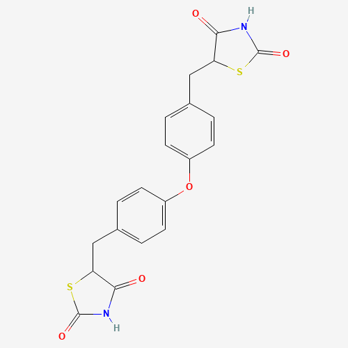
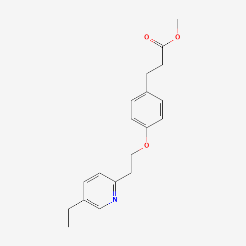
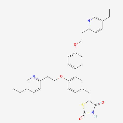
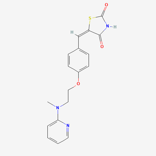
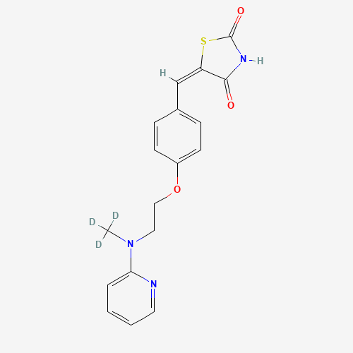
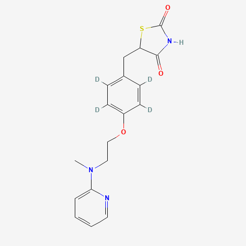
Table 3: List of Test Samples
|
Sl. No. |
Test Samples |
Structure |
|
1 |
TEST SAMPLE 1 |
|
|
2 |
TEST SAMPLE 2 |

|
|
3 |
TEST SAMPLE 3 |
|
|
4 |
TEST SAMPLE 4 |
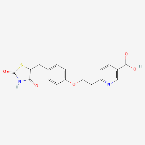
|
|
5 |
TEST SAMPLE 5 |
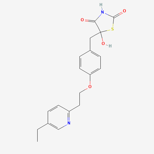
|
|
6 |
TEST SAMPLE 6 |
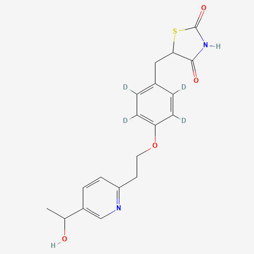
|
|
7 |
TEST SAMPLE 7 |
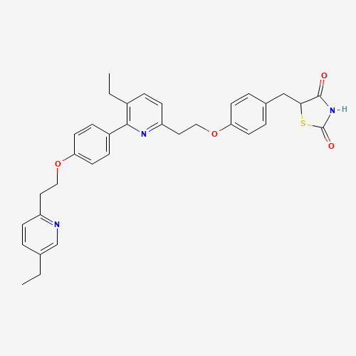
|
|
8 |
TEST SAMPLE 8 |
|
|
9 |
TEST SAMPLE 9 |
|
|
10 |
TEST SAMPLE 10 |
|
|
11 |
TEST SAMPLE 11 |
|
|
12 |
TEST SAMPLE 12 |
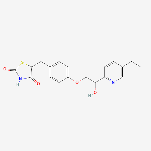
|
|
13 |
TEST SAMPLE 13 |
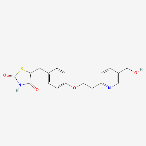
|
|
14 |
TEST SAMPLE 14 |
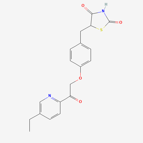
|
|
15 |
TEST SAMPLE 15 |
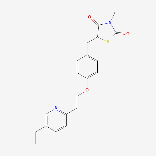
|
|
16 |
TEST SAMPLE 16 |
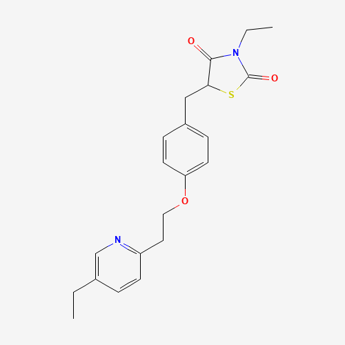
|
|
17 |
TEST SAMPLE 17 |
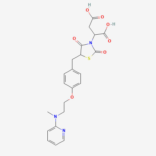
|
|
18 |
TEST SAMPLE 18 |
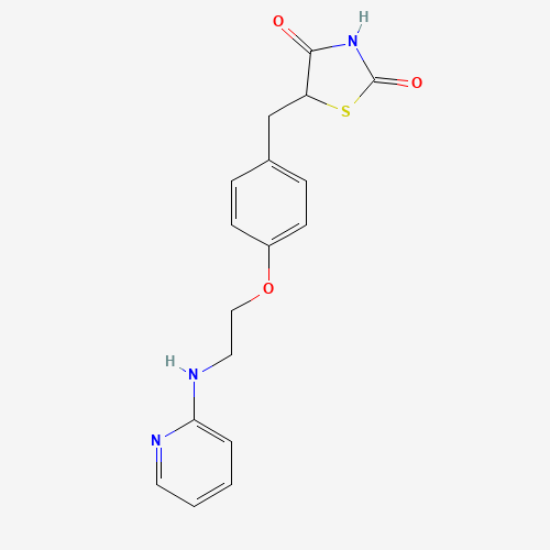
|
|
19 |
TEST SAMPLE 19 |
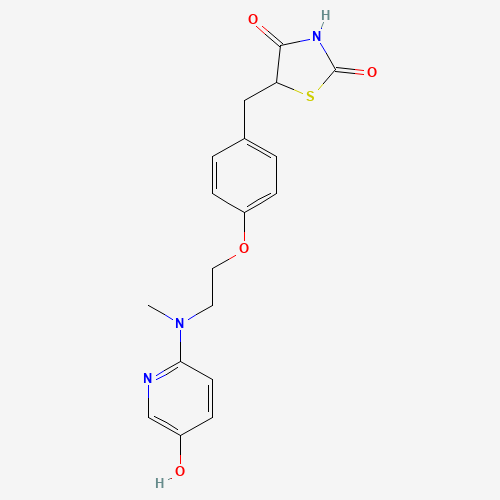
|
|
20 |
TEST SAMPLE 20 |
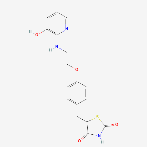
|
|
21 |
TEST SAMPLE 21 |
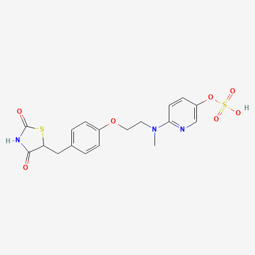
|
|
22 |
TEST SAMPLE 22 |
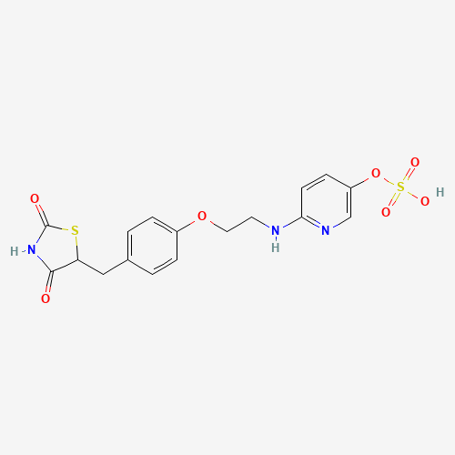
|
|
23 |
TEST SAMPLE 23 |
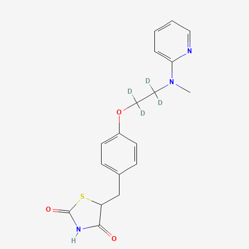
|
|
24 |
TEST SAMPLE 24 |
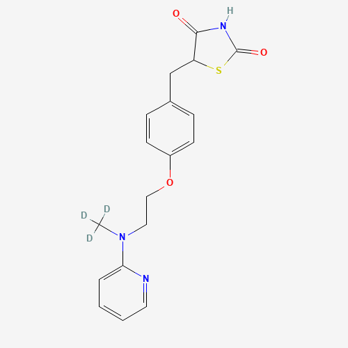
|
|
25 |
TEST SAMPLE 25 |
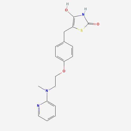
|
|
26 |
TEST SAMPLE 26 |
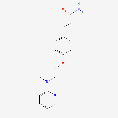
|
|
27 |
TEST SAMPLE 27 |
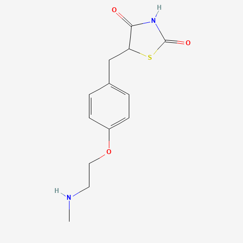
|
|
28 |
TEST SAMPLE 28 |
|
|
29 |
TEST SAMPLE 29 |
|
|
30 |
TEST SAMPLE 30 |
|
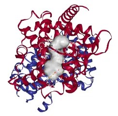
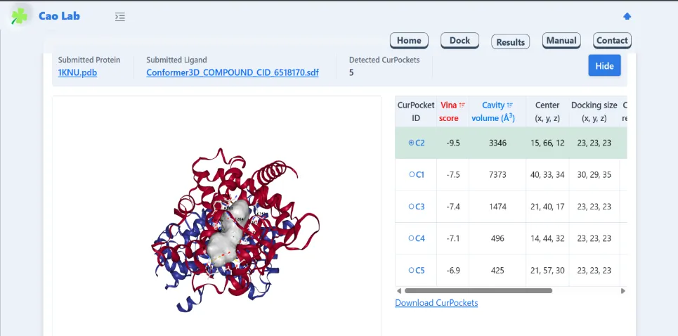
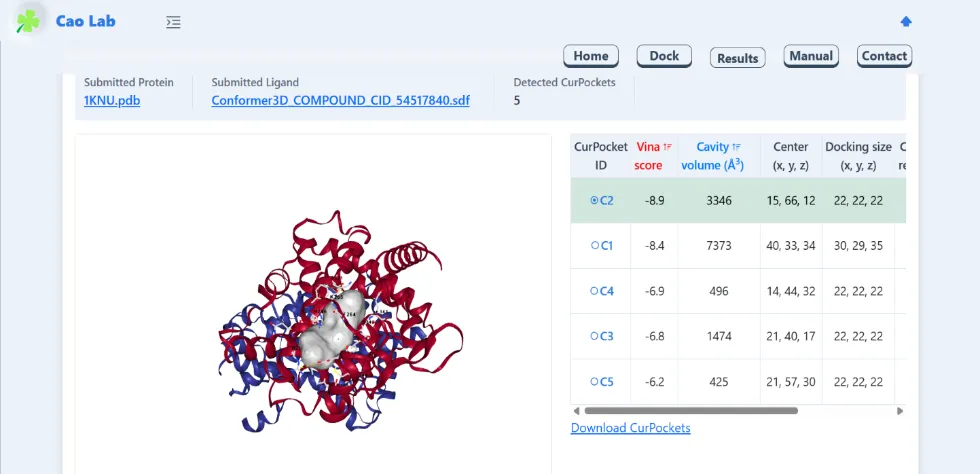
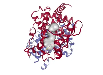
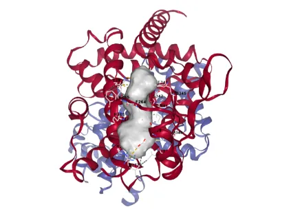
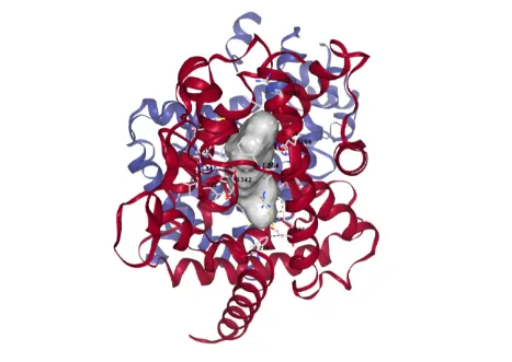
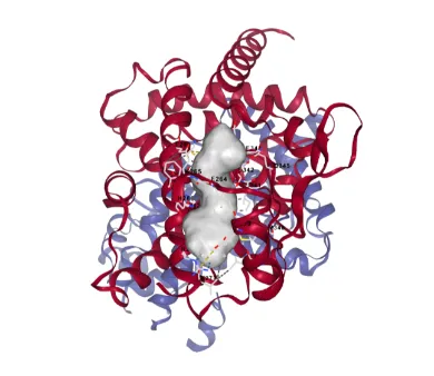
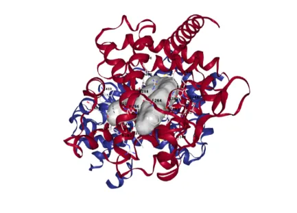
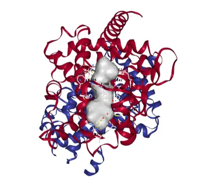
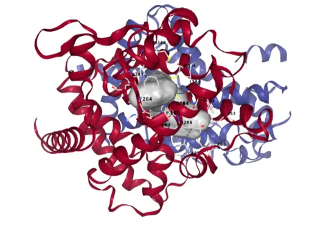
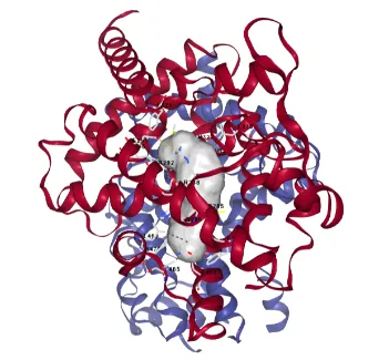
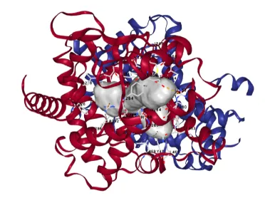
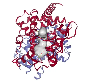
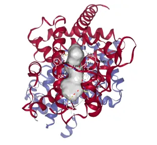
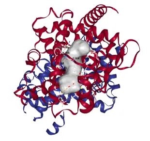
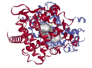
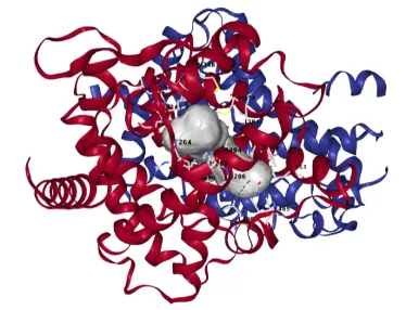
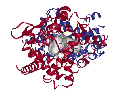
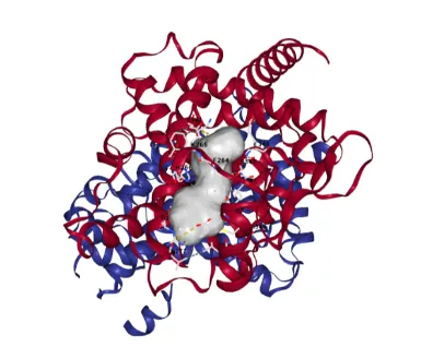
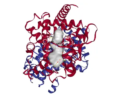
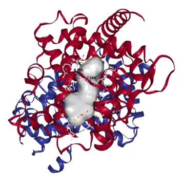
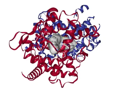
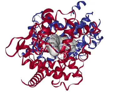
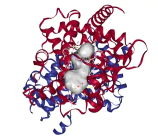
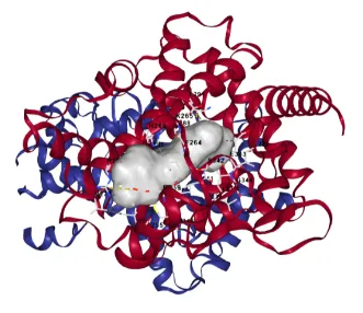
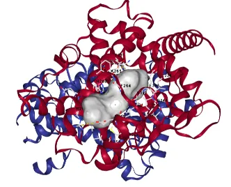
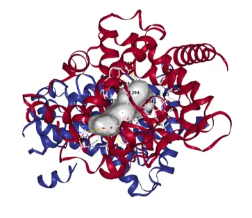
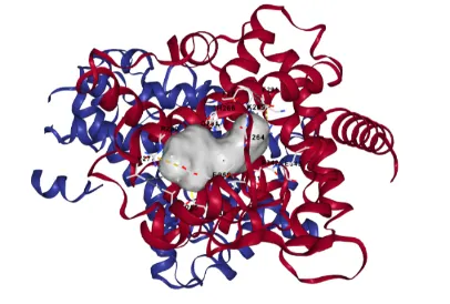
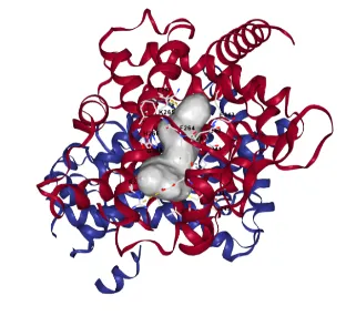
Molecular docking was performed using CB-DOCK2 server to evaluate the binding affinity of the 30 designed derivatives [17]. The CB-DOCK2 algorithm was used for blind docking, which involves automatic cavity detection followed by docking with the AutoDock Vina engine. The compounds were selected based on their binding energy (Vina score) and their ability to mimic the orientation of the co-crystallized agonist found in the 1KNU structure.
Table 4: Receptor Activated Site (Coordinates)
|
Receptor |
X |
Y |
Z |
|
Human Peroxisome Proliferator Activated Receptor Gamma ligand -binding domain (1KNU) |
15 |
66 |
12 |
Table 5: Molecular Docking analysis of Pioglitazone with the PPAR-Gamma Receptor (1KNU)
|
Sl. No. |
Name of the Standard Compound |
Docking Result [Binding Energy (kcal/mol)] |
Molecular Docking |
|
1
|
Pioglitazone |
-9.4 |
|
Table 6: Molecular docking results of the sample compounds against the PPAR-Gamma Receptor (1KNU)
|
Sl. No. |
Name of the Test Sample |
Docking Result [Binding Energy (kcal/mol)] |
Molecular Docking |
|
1
|
Test Sample 1 |
-9.5 |
|
|
2
|
Test Sample 2 |
-8.9 |
|
|
3
|
Test Sample 3 |
-9.5 |
|
|
4
|
Test Sample 4 |
-9.5 |
|
|
5
|
Test Sample 5 |
-8.6 |
|
|
6
|
Test Sample 6 |
-9.6 |
|
|
7
|
Test Sample 7 |
-11.1 |
|
|
8
|
Test Sample 8 |
−9.5 |
|
|
9
|
Test Sample 9 |
−10.8 |
|
|
10
|
Test Sample 10 |
−8.1 |
|
|
11
|
Test Sample 11 |
−11.5 |
|
|
12
|
Test Sample 12 |
−9.5 |
|
|
13
|
Test Sample 13 |
−9.4 |
|
|
14
|
Test Sample 14 |
−9.4 |
|
|
15
|
Test Sample 15 |
−9.4 |
|
|
16
|
Test Sample 16 |
−9.3 |
|
|
17
|
Test Sample 17 |
−9.1 |
|
|
18
|
Test Sample 18 |
−9.3 |
|
|
19
|
Test Sample 19 |
−9.5 |
|
|
20
|
Test Sample 20 |
−9.0 |
|
|
21
|
Test Sample 21 |
−9.5 |
|
|
22
|
Test Sample 22 |
−9.2 |
|
|
23
|
Test Sample 23 |
−9.1 |
|
|
24
|
Test Sample 24 |
−9.4 |
|
|
25
|
Test Sample 25 |
−9.0 |
|
|
26
|
Test Sample 26 |
−8.1 |
|
|
27
|
Test Sample 27 |
−7.7 |
|
|
28
|
Test Sample 28 |
−9.1 |

|
|
29
|
Test Sample 29 |
−8.8 |
|
|
30
|
Test Sample 30 |
−9.3 |
|
The selected 30 derivatives were subjected to Quantitative Structure Activity Relationship (QSAR) analysis using ChemMaster software [19].
Physicochemical descriptors including Molecular Weight, LogP, Hydrogen Bond Donor, Hydrogen Bond Acceptor, Molar Refractivity (MR), Rotatable Bonds were calculated for each compound.
The dataset was classified into training and test sets. Multiple Linear Regression (MLR) was used to generate the predictive model. The model’s reliability was assessed through the coefficient of determination (R2) and error metrices (RMSE, MAE) [20].
RESULTS AND DISCUSSION:
The 30 designed thiazolidinedione (TZD) derivatives were initially screened for their pharmacokinetic potential using Lipinski’s Rule of Five (RoF). High oral bioavailability is a prerequisite for effective Type 2 Diabetes Mellitus (T2 DM) treatments [21].
Table 7: Drug-likeness evaluation of the designed TZD derivatives
|
Sl. No. |
Molecular Formula |
Molecular Weight |
Log P < 5 |
H-Bond Donor <5 |
H-Bond Acceptor
|
Molar Refractivity |
Rotatable Bonds |
Lipinski’s Rule Compliance |
|
1 |
C18H23NO3S |
333.453 |
3.9299 |
1 |
4 |
91.7507 |
6 |
YES |
|
2 |
C18H23NO3S |
333.453 |
4.082 |
2 |
4 |
92.5395 |
7 |
YES |
|
3 |
C19H21N3O2S |
355.463 |
2.9744 |
2 |
5 |
100.2084 |
8 |
YES |
|
4 |
C18H16N2O5S |
372.402 |
2.2954 |
2 |
7 |
95.459 |
7 |
YES |
|
5 |
C19H20N2O4S |
372.446 |
2.4796 |
2 |
6 |
99.0395 |
9 |
YES |
|
6 |
C19H20N2O4S |
376.4704 |
2.6505 |
2 |
6 |
99.1335 |
9 |
YES |
|
7 |
C34H35N3O4S |
581.738 |
6.4055 |
1 |
7 |
166.1277 |
15 |
NO MW>500 LogP>5 |
|
8 |
C19H20N2O4S |
377.4765 |
2.6505 |
2 |
7 |
99.1335 |
9 |
YES |
|
9 |
C20H16N2O5S2 |
428.491 |
3.2671 |
2 |
7 |
110.2014 |
6 |
YES |
|
10 |
C19H23NO3 |
313.397 |
3.3711 |
0 |
4 |
89.593 |
10 |
YES |
|
11 |
C34H35N3O4S |
581.738 |
6.4055 |
1 |
7 |
166.1277 |
15 |
NO MW>500 LogP>5 |
|
12 |
C19H20N2O4S |
372.446 |
2.6505 |
2 |
6 |
99.1335 |
9 |
YES |
|
13 |
C19H20N2O4S |
372.446 |
2.6505 |
2 |
6 |
99.1335 |
9 |
YES |
|
14 |
C19H18N2O4S |
370.43 |
2.7998 |
1 |
6 |
98.5042 |
8 |
YES |
|
15 |
C20H22N2O3S |
370.474 |
3.5018 |
0 |
5 |
102.505 |
9 |
YES |
|
16 |
C21H24N2O3S |
384.501 |
3.8919 |
0 |
5 |
107.122 |
10 |
YES |
|
17 |
C22H23N3O7S |
473.507 |
2.1312 |
2 |
10 |
120.4456 |
12 |
YES |
|
18 |
C17H17N3O3S |
343.408 |
2.4666 |
2 |
6 |
93.2854 |
7 |
YES |
|
19 |
C18H19N3O4S |
373.434 |
2.1965 |
2 |
7 |
99.7305 |
9 |
YES |
|
20 |
C17H17N3O4S |
359.407 |
2.1722 |
3 |
7 |
94.9502 |
8 |
YES |
|
21 |
C18H19N3O7S2 |
453.498 |
1.6725 |
2 |
10 |
110.2423 |
11 |
YES |
|
22 |
C17H17N3O7S2 |
439.471 |
1.6482 |
3 |
10 |
105.462 |
10 |
YES |
|
23 |
C18H19N3O3S |
361.4594 |
2.4909 |
1 |
6 |
98.0657 |
8 |
YES |
|
24 |
C18H19N3O3S |
360.4533 |
2.4909 |
1 |
6 |
98.0657 |
8 |
YES |
|
25 |
C18H19N3O3S |
357.435 |
2.643 |
2 |
6 |
98.8545 |
9 |
YES |
|
26 |
C17H21N3O2 |
299.374 |
2.0147 |
1 |
4 |
86.9594 |
9 |
YES |
|
27 |
C13H16N2O3S |
280.349 |
1.1789 |
2 |
5 |
74.4854 |
7 |
YES |
|
28 |
C18H17N3O3S |
355.419 |
2.9207 |
1 |
6 |
98.5507 |
7 |
YES |
|
29 |
C18H17N3O3S |
358.4373 |
2.9207 |
1 |
6 |
98.5507 |
7 |
YES |
|
30 |
C18H19N3O3S |
361.4594 |
2.4909 |
1 |
6 |
98.0657 |
8 |
YES |
A 2D-QSAR study was executed via ChemMaster (Version 1.3) using Multiple Linear Regression (MLR) to determine the structural drivers of activity.
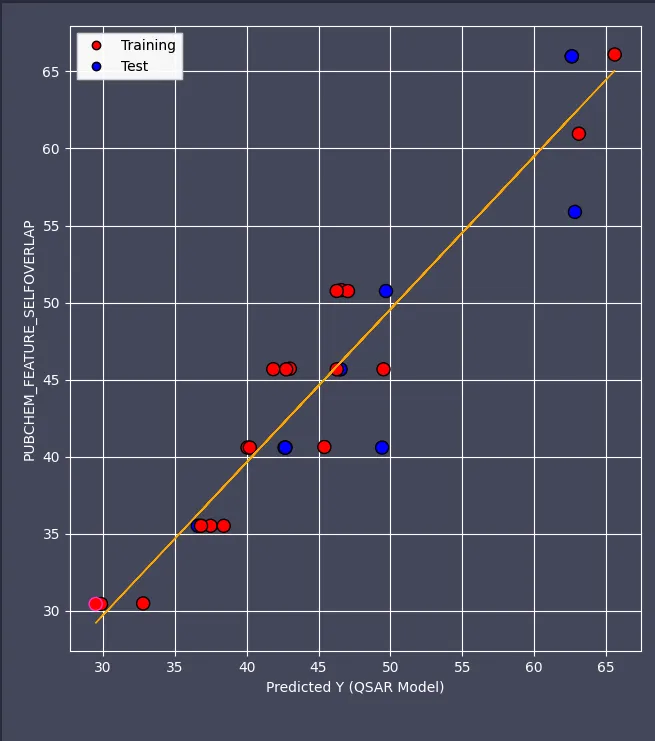
Figure 2: QSAR MODEL
The generated model demonstrated high internal and external predictive power
An R2 value exceeding 0.6 is considered the threshold for a reliable QSAR model; thus, our model’s 0.911 correlation indicates excellent fit [20]. The proximity of the test set R2 (0.836) to the training set suggests the model is robust and avoids overfitting.
The predictive equation is as follows:
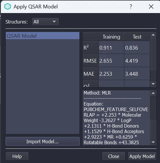
Figure 3: Regression Equation and Validation Parameters
Using CB-DOCK2, the 30 derivatives were docked into the lignd-binding domain (LBD) of the PPAR-Gamma receptor. The best performing compounds occupied the hydrophobic ‘Y-Shaped’ cavity, establishing critical hydrogen bonds with His323 and Tyr473. Stabilizing the Tyr473 residue is essential for the activation of the AF-2 surface, which facilitates the recruitment of co-activators necessary for the anti-diabetic response [22,23].
To address the historical safety concerns associated with glitazones (e.g., hepatotoxicity, cardiotoxicity, etc.) an in-depth ADMET profile was established for the series.
Table 8: In Silico ADMET and Safety profiling of the selected TZD derivatives
|
Sl. No. (Compounds) |
Clinical Toxicity |
Carcinogenicity |
Mutagenicity |
Drug Induced Liver Injury (DILI) |
Acute Toxicity (LD50) |
hERG Blocking |
Skin Reaction |
PPAR-Gamma |
|
1 |
0.3602 |
0.3483 |
0.3102 |
0.7783 |
2.2038 |
0.2481 |
0.6459 |
0.3669 |
|
2 |
0.2303 |
0.0415 |
0.2324 |
0.4114 |
2.7917 |
0.3286 |
0.3641 |
0.1182 |
|
3 |
0.2191 |
0.2759 |
0.0983 |
0.804 |
2.6666 |
0.4128 |
0.5003 |
0.1926 |
|
4 |
0.4804 |
0.2666 |
0.1951 |
0.9755 |
1.8492 |
0.017 |
0.3826 |
0.1666 |
|
5 |
0.3761 |
0.2201 |
0.1422 |
0.9201 |
2.5432 |
0.1907 |
0.3224 |
0.2189 |
|
6 |
0.3364 |
0.1706 |
0.1148 |
0.9259 |
2.2668 |
0.1221 |
0.2035 |
0.1278 |
|
7 |
0.2732 |
0.2509 |
0.2764 |
0.9637 |
2.5813 |
0.618 |
0.2604 |
0.3226 |
|
8 |
0.382 |
0.283 |
0.133 |
0.6539 |
2.8561 |
0.2004 |
0.304 |
0.0175 |
|
9 |
0.3718 |
0.4553 |
0.4237 |
0.9779 |
2.4361 |
0.2528 |
0.6017 |
0.4661 |
|
10 |
0.1066 |
0.1515 |
0.0618 |
0.4011 |
2.1961 |
0.4703 |
0.3803 |
0.0095 |
|
11 |
0.3227 |
0.2293 |
0.2783 |
0.9674 |
2.6744 |
0.6014 |
0.2216 |
0.3013 |
|
12 |
0.299 |
0.3205 |
0.1925 |
0.9319 |
2.1428 |
0.1511 |
0.2965 |
0.1759 |
|
13 |
0.3339 |
0.1275 |
0.1249 |
0.9175 |
2.2398 |
0.0928 |
0.2287 |
0.0898 |
|
14 |
0.3141 |
0.3854 |
0.2309 |
0.9777 |
2.1374 |
0.0816 |
0.3641 |
0.3811 |
|
15 |
0.2788 |
0.2509 |
0.1172 |
0.9009 |
2.2652 |
0.3723 |
0.4464 |
0.1416 |
|
16 |
0.2666 |
0.2631 |
0.1297 |
0.879 |
2.2352 |
0.4659 |
0.4333 |
0.161 |
|
17 |
0.6434 |
0.1437 |
0.1581 |
0.9644 |
2.2432 |
0.0503 |
0.2002 |
0.1968 |
|
18 |
0.4957 |
0.5151 |
0.1779 |
0.9468 |
1.8498 |
0.3547 |
0.6579 |
0.239 |
|
19 |
0.3997 |
0.4046 |
0.1306 |
0.9588 |
2.0091 |
0.2391 |
0.5005 |
0.26 |
|
20 |
0.3685 |
0.3163 |
0.0786 |
0.9416 |
1.899 |
0.3416 |
0.6208 |
0.3194 |
|
21 |
0.6518 |
0.3184 |
0.4049 |
0.9768 |
1.7452 |
0.1622 |
0.3797 |
0.1615 |
|
22 |
0.6435 |
0.3174 |
0.3559 |
0.9684 |
1.5125 |
0.2307 |
0.4744 |
0.1439 |
|
23 |
0.2217 |
0.2995 |
0.1452 |
0.806 |
2.352 |
0.2001 |
0.5274 |
0.0186 |
|
24 |
0.4408 |
0.5075 |
0.2015 |
0.8979 |
2.1776 |
0.2124 |
0.3913 |
0.0403 |
|
25 |
0.3046 |
0.2222 |
0.188 |
0.8373 |
2.3273 |
0.5481 |
0.2503 |
0.0815 |
|
26 |
0.3587 |
0.6066 |
0.2478 |
0.3537 |
2.3771 |
0.3812 |
0.1904 |
0.0066 |
|
27 |
0.5043 |
0.6925 |
0.2849 |
0.79 |
1.8785 |
0.1386 |
0.8405 |
0.0788 |
|
28 |
0.3851 |
0.6236 |
0.2786 |
0.9868 |
2.362 |
0.2786 |
0.6355 |
0.2454 |
|
29 |
0.4168 |
0.5913 |
0.2589 |
0.959 |
2.5032 |
0.2299 |
0.4898 |
0.0676 |
|
30 |
0.4979 |
0.5378 |
0.2027 |
0.9618 |
2.2775 |
0.2474 |
0.4668 |
0.1879 |
Based on the convergence of docking scores, QSAR reliability and safety parameters, five lead compounds were identified as candidates for further synthesis:
Table 9: Top 5 Least Toxic Compounds
|
Compound Serial Number |
Justification |
|
26 |
Lowest (favourable) DILI Good LD50 |
|
23 |
Balanced profile Moderate toxicity in all parameters |
|
3 |
Very Low Carcinogenicity Good LD50 but high DILI |
|
5 |
High Liver Toxicity Overall good |
|
18 |
Stable profile, Acceptable toxicity but not best |
SELECTION AND JUSTIFICATION OF LEAD CANDIDATE:
Among the 30 derivatives, Compound 26 was prioritized as the primary lead candidate. This section is justified by its balanced performance across all computational filters:
CONCLUSION
The study successfully integrated molecular docking, 2D-QSAR modelling and ADMET profiling to evaluate 30 TZD derivatives against the PPAR-Gamma (1KNU). The developed QSAR model proved highly reliable (R2=0.911), identifying molecular bulk and hydrogen bonding as key contributors to activity. Compound 26 emerged as the most promising lead candidate due to its excellent binding affinity and significantly reduced hepatotoxicity profile compared to traditional agents.
ACKNOWLEDGEMENT
The authors are thankful to Global College of Pharmaceutical Technology for giving us the opportunities to perform this research work.
CONFLICT OF INTEREST: None
REFERENCES



Swastika Mukherjee, Sayoni Roy, Soumallya Chakraborty, Somenath Bhattacharya, In Silico Design and Identification of Novel Thiazolidinedione Derivatives as PPAR-Gamma Receptor Agonists for the Treatment of Type 2 Diabetes Mellitus, Int. J. of Pharm. Sci., 2026, Vol 4, Issue 5, 7931-7951, https://doi.org/10.5281/zenodo.20444746
 10.5281/zenodo.20444746