

We use cookies to ensure our website works properly and to personalise your experience. Cookies policy
Delonix society baramati college of pharmacy barhanpur baramati. Dr. Babasaheb Ambedkar technology university lonere.
being employed. Because of its high surface to volume ratio, nanomedicine's special ability to bind, absorb, and transport tiny biomolecules like DNA, RNA, medications, proteins, etc increases the effectiveness of medicinal medicines by delivering additional molecules to the targeted region. Objective: This article's goal is to give readers an understanding of a number of aspects of nanotechnology in cancer treatment, including its function in cancer therapy, drug release techniques, and different nanomaterials as drug vehicles.Methods: In order to find research publications on nanotechnology and cancer treatments, we conducted a thorough search of bibliographic databases. We then combined the relevant material from these articles to create the current article.Results: Through the use of nanoparticles and quantum dots, cancer nanotechnology offers a novel approach to cancer prevention, early diagnosis, and customized treatment. One significant factor in the identification of cancer biomarkers is nanobiotechnology. Gold nanoparticles, carbon nanotubes, magnetic nanoparticles, quantum dots, and Biomolecules that can identify cancer biomarkers have been designed to be carried by nanowires and other similar materials. Biomolecules such as proteins, antibody fragments, DNA fragments, and RNA fragments serve as the foundation for cancer biomarkers in nanoparticle-assisted cancer diagnosis and monitoring.Conclusion: This review focuses on many ways that cancer nanotechnology is being used to improve cancer treatment.
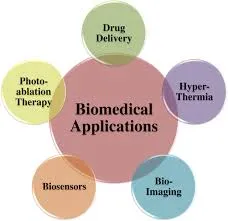
Fig no. 1 Therapeutic applications of nanotechnology in different biomedical field.
Numerous methods of nanomedicine are being thoroughly studied, including those employing lipid and micelle-based nanoparticles, dendrimers, quantum specks, carbon nanotubes, and polymeric and non-polymeric nanoparticles. Furthermore, the potential of nanomedicine multivalent ligand targeting and the ability to transport a large payload are essential components of cancer treatment. This circumvents defense mechanisms and provides precision for tissue targeting. The main challenges facing these upcoming medications are the possible toxicity of nanoparticles, which necessitates a thorough evaluation before nanomedicines may be employed as cancer (malignancy) treatments [3]. Creating a biocompatible nanosystem—such as nanocrystals, strong lipid nanoparticles, nanostructured lipid carriers, lipid drug conjugates, nanoliposomes, dendrimers, nanoshells, emulsions, nanotubes, quantum dots, etc.—is essential to delivering nanomaterial conjugated medicine to the intended tumor site. Delivery of drug complexes and nanomaterials involves both passive and active targeting.
Techniques that can also be applied to the delivery of nanodrugs. The Enhanced Permeability and Maintenance (EPR) effect of the vasculature surrounding tumors is necessary for passive targeting. For medication delivery, active targeting employs ligand-coordinated binding of nanoparticles to tumor cell receptors. Temperature and pH variations in the body can control how much medication is released from nanoparticles. Different biodistribution profiles and the anticancer efficacy of nano-drugs in vivo are caused by a variety of nanomaterial characteristics, including their size, surface charge, PEGylation, and other biophysical characteristics [4]. A research team concentrated on creating biocompatible nanoparticles that might target particular cancer indicators and offer therapeutic effects in order to identify and treat cancer. Imagery as well as medicinal substances. More sophisticated nanoparticle drug complexes that can release different nanomedicines for improved treatment efficacy have been created in recent study [5]. By conjugating tumor-specific cell surface receptors, active targeting of nanoparticles increases the viability of nanoparticle drug delivery devices while effectively decreasing poisoning. Multifunctional nanoparticulate devices for simultaneous drug delivery and tumor mass imaging are among the most interesting developNanomaterials are referred to as nanovehicles because they serve as a vehicle for conjugated drug delivery. When it comes to cancer treatment, novel drug delivery systems based on nanoparticles target the tumor cells and the milieu that supports cancer cells. Therapeutic drug combinations carried by nanovehicles for the targeted and targeted removal of tumor cells, targeted drug delivery to tumor cells, cancer stem/tumor-initiating cells, and/or the supportive cancer cell microenvironment [7] ments in nanomedicine [6]. . Quantum dots, a frequently utilized nanomaterial in cancer cell imaging, have special photochemical and photophysical characteristics because they are orders of magnitude brighter than Conventional fluorophores have extremely narrow emission spectra that can be adjusted by changing the dot size. A novel type of fluorescent markers with enhanced brightness and resistance to photo bleaching is called quantum dots. These characteristics have the potential to increase biological imaging and detection sensitivity by at least 10–100 times. [8]. Other nanomaterials, such as dendrimers and carbon nanotubes (CNTs), offer intriguing qualities that can be used for thermal ablation, diagnostics, and delivery of medications for cancer. Because of their axial symmetry and nanoscale widths, carbon nanotubes (CNTs) are tubular materials with intriguing characteristics that may be used to diagnose and treat cancer. Similarly, CNTs may be able to carry medications straight to the cells and tissues that need them. Clarifying the toxicity of nanoparticles is also crucial given the quick advancements in the creation of materials based on nanotechnology. Furthermore, by balancing the hydrophobicity and hydrophilicity of the micelle framing block copolymers, polymeric micelles may be produced with improved drug stacking capabilities. They can also be efficiently cancer-focused by altering their surface in response to tumor-homing ligands.
Nonetheless, it might still be difficult to maintain the self-assembly scaffolding in circulation and disassemble it for drug release at the site of action [9]. Recent developments have led to the enhancement of Guided nanoparticle attached medications for the treatment of tumors, bioaffinity assays based on nanoparticles for atomic and cell imaging, and integrated nanodevices for disease detection and early screening. These developments create exciting opportunities for personalized oncology, where cancer is analyzed and treated based on the subatomic profiles of individual individuals using genetic and protein biomarkers. This review article provides an overview of the use of different nanotechnology-based methods for cancer diagnosis and treatment.
2 NANOTECHNOLOGIES IN CANCER DIAGNOSIS:
A mutation in a few certain genes within the cells is the root cause of cancer. A mass of mutant cells develops in a given tissue or organ as a result of this mutation, which changes the synthesis of certain macromolecules and ultimately causes unchecked cell division. Known as a tumor. Tumor cells are referred to as benign when they are contained, but as malignant when they spread to the surrounding tissues. The majority of cancer diagnostic and treatment approaches were created to stop cancer cells from proliferating and dividing. The most crucial aspect of cancer treatment is early and precise diagnosis, which is typically accomplished by ultrasonography, Positron Emission Tomography (PET), Magnetic Resonance Imaging (MRI), Computed Tomography (CT), etc. [10] Successful treatment and patient outcomes have become extremely challenging due to the new imaging and investigative techniques' inability to give comprehensive clinical information about different tumor types and stages [3, 4]. The majority of anticancer medications on the market now do not distinguish between healthy and malignant cells, resulting in unfavorable consequences including systemic damage. Moreover, a significant issue with cancer is that it is often diagnosed too late, after the disease has spread.
2.1 Nanotechnology Assisted Tumor Imaging:
Over the last few decades, there has been an increase in interest in the use of nanoparticles for molecular imaging and for the diagnosis and monitoring of cancer. The fundamental idea behind imaging based on nanomaterials is particle size, which gives nanoparticles their unique characteristics. For example, semiconductor nanoparticles utilized in cancer The optical, magnetic, and structural characteristics of iron oxide nanocrystals and quantum dots are uncommon in bulk materials an. Nanoparticles can be employed with a variety of anticancer medicines, such as medications and biomolecules, such as various peptides, antibodies, or other compounds, to designate tumors with high emOnly for personal use; do not distribute. Pathy and specificity, and as shown in Fig. (2), this compound is helpful in the early identification and screening of cancer cells. d Because of its vast surface area and tiny diameter, the nanoparticle can readily bind to functional groups of various optical, radio isotopic, or magnetic diagnostic and therapeutic agents, making the more effective and persuasive cancer diagnosis. A significant breakthrough in the diagnosis and treatment of cancer was made possible by these developments in nanotechnology [11]. molecules.
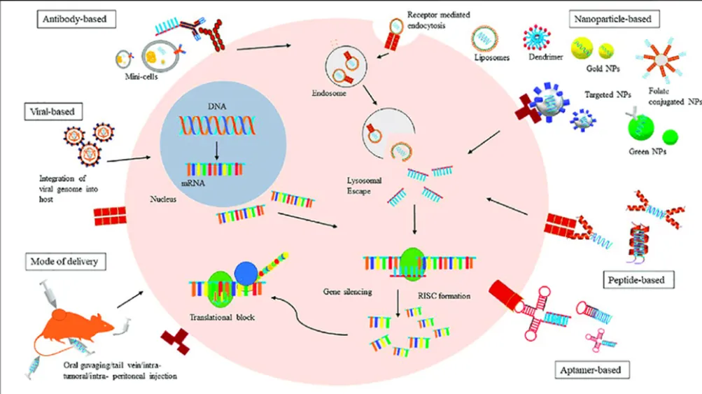
Fig. (2). Schematic mechanism of SERS mediated bio-imaging and anticancer drug delivery by using AuNGO
It's shocking to learn that researchers are working on a detecting device that may detect cancer inside the body by being worn on the wrist. This significant advancement in medical technology is because to this field's application of nanomaterials. Certain active magnetic molecules in the device above sliced through the wrist's blood vessels [12]. These magnetic molecules use a wrist device to show the findings of their detection of changes in the smart nanoparticles circulating in the blood. Nanomaterials' remarkable behavior and adaptability are the reason for this advancement in cancer diagnostics. These days, nanotechnology has confirmed imaging of cancer at the tissue, cell, and molecular level. One example is lanthanide-based up conversion nanoparticles, which use autofluorescence to detect deep tissue by upconverting low-energy photons to high-energy ones [13]. In addition, nanotechnology investigated the For instance, fibroblast activation protein-a on the membrane of tumor-associated fibroblasts can be detected by a pH-responsive fluorescent nanoprobe, which can be used to target and image cancerous tumors [14]. Here, we've covered a few high temporal and spatial nanotechnology-based approaches that can be useful for precisely tracking living cells and tracking dynamic biological activities within tumors.
The primary issue with visible spectrum imaging is its poor detection and penetration. Scientists created quantum dots that fluoresce in the near-infrared spectrum as a solution to this issue, i.e. NIR quantum dots are more suited for in vivo imaging of cancer in tissues such as the colon, liver, pancreas, and lymphatic tissue because of their wavelength of 700–1000 nm [15–17]. Live animals with multicolor quantum dot (QD) imaging capabilities. On a host animal, 1–2 million of each color were subcutaneously injected in three nearby places. Excitation from tungsten or mercury lamps produced the images. Gao X, Cui Y, Levinson RM, Chung LW, Nie S. In vivo cancer targeting and imaging with semiconductor quantum dots, reprinted with permission from Macmillan Publishers Ltd. 22:969-976; Nat Biotechnology, 2004.
2.1.2 Nano shells:
Nano shells are dielectric cores that range in size from 10 to 300 nm. They are typically composed of silica and have a thin metal shell, usually constructed of gold [18, 19]. These nano shells change electrical signals mediated by Plasmon. With an emission/absorption array spanning from the ultraviolet to the infrared, they can also be optically tuned to convert energy into light [20]. Because heavy metal toxicity does not affect their imaging, nano shells are appealing. The size of the nano shell is one of its issues.
2.1.3 Colloidal Gold Nanoparticles:
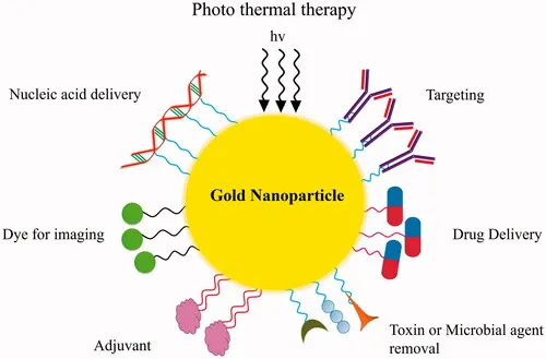
One of the most appealing types of agents for cancer diagnostics is gold nanoparticles. This is because gold has been approved for use in the treatment of human illness [21] and is simple to synthesize [22].
By dispersing visible light in in vitro samples, these gold nanoparticles serve as contrast agents. Additionally, gold nanoparticles can be conjugated with antibodies for biopsies for the detection of pancreatic and cervical malignancies. In addition, photoacoustic tomography can be performed using gold nanoparticles. Gold nanoparticles are therefore a priority-based detection tool for several malignancies [23].
3 Nanotechnology in Cancer Therapy:
3.1 Tools of Nanotechnology for Cancer Therapy:
Recent developments in the creation of different vehicles for effective drug administration are driving research effort in the field of nanotechnology. a variety of vehicles, including nanocarriers such carbon nanotubes, liposomes, micelles, dendrimers, and quantum dots. have been created thus far, as illustrated in Fig. (3).
3.1.1 Liposomes:
Liposomes are phospholipid-based vesicles that are at least 400 nm in size, with a bilayer membrane made of cholesterol and a hydrophilic head and hydrophobic tail [24]. Liposomes' special ability to solubilize the water-insoluble organic material makes them a vehicle for medication targeting. compounds, making them appropriate for the treatment of cancer and other illnesses. Drugs included in the membrane of liposomes have a number of benefits, including efficient distribution to the intended location, low nonspecific toxicity, and protection against degradation [25, 26]. Because endothelial cells' tight connections prevent particles from leaking out of vessels, liposomes are kept in the bloodstream in normal, healthy tissues. However, tumor vasculature are more prone to leakage than blood channels in healthy tissues, which allowsthe specific tumor site by allowing the nanosized liposome to escape from the circulation.
Liposomes have the ability to target cancer cells and are biodegradable, biocompatible, and more stable in colloidal fluids [27–29]. After comparing the toxicity and drug delivery efficacy of various liposomal compositions and free drug delivery with anticancer medicines, researchers came to the conclusion that liposomes are less harmful to tumor sites than free medications [30, 31].
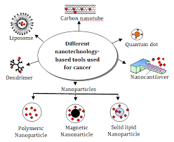
Fig. (3). Tools of nanotechnology. Schematic representation of various nanotechnology-based tools used for cancer therapeutics.
There are numerous anti-cancer medications on the market and in clinical trials [30]. Doxil (Liposomal Doxorubicin), the first FDA-approved anticancer nanodrug, is made Kaposi's sarcoma in AIDS patients is often treated with a PEGylated liposomal formulation [32, 33]. Johnson & Johnson sells PEGylated liposomes carrying the anti-cancer medication doxorubicin under the trade names Doxil (in the USA) and Caelyx (outside the USA). A manufacturing plant was shut down in 2011 as a result of problems with Doxil's quality control that led to an imbalance between the drug's supply and demand [33, 34]. Additionally, the FDA approved LipoDox as a substitute medication to address the Doxil shortage in the United States. LipoDox and Doxil have the same chemical makeup. Sun Pharma Company manufactures and sells Doxil in India; in 2013, the FDA approved the first generic version of the medication [34, 35]. Several studies demonstrate that Doxil can prevent ovarian cancer, and the FDA has also approved it to treat ovarian cancer that has returned [36, 37]. Doxil has been approved in the United States for the treatment of breast cancer [37], and in Europe and Canada, it is used in conjunction with Valadec to treat multiple myeloma [37, 38]. Celator Pharmaceuticals Inc. created CPX351, a liposomal mixture of daunorubicin and cytarabine. In the phase III clinical trial for individuals with acute myeloid leukemia (AML), CPX-351 demonstrated encouraging outcomes [39]. MBP-426 targets the transferrin receptor. Mebiopharm created the liposomal version of oxaliplatin, which is being tested in a phase II clinical trial to treat patients with stomach cancer [40]. Merrimack Pharma created MM-398, a liposomal sphere that encapsulates the medication irinotecan. that can cure malignancies that are resistant to chemotherapy, including gliomas, lung, pancreatic, and colorectal cancers [41–43].
3.1.2 Carbon Nanotubes:
Carbon nanotubes (CNTs) can be classified into two classes based on their diameter and structure: single-walled CNTs (SWNTs) and multiwalled CNTs (MWNTs). CNTs with a single wall are made up of a multiwalled carbon nanotubes, which are made out of many concentric graphene sheets, and a single sheet of cylindrical graphene [44]. The structure, surface area, mechanical strength, metallic behavior, electrical and thermal conductivity, and ultra-lightweight of carbon nanotubes are all related to their physical and chemical characteristics. Because of their many unique physical and chemical characteristics, CNTs are a good option for a wide range of biomedical applications [45]. CNTs have been utilized to target cancer cells because of their ability to absorb near-infrared (NIR) light, which causes the nanotubes to heat up [46–48]. This phenomenon is known as the thermal effect. Folic acid (FA) receptors are overexpressed in cancer cells, and Numerous research teams have created synthetic nanocarriers using conjugated biomaterials of FA derivatives. Furthermore, compared to spherical nanocarriers, it has been shown that CNTs are kept in the lymph nodes for longer periods of time [49]. Another study found that gemcitabine, an anticancer drug, had strong activity against lymph nodes when put onto magnetic MWNTs and administered subcutaneously to mice [50]. When drug delivery systems interact, CNTs can recognize the surface receptors that cause receptor-mediated endocytosis of CNTs. with cancer cells (NDDs) [51]. CNT-based drug delivery improves medicines' blood circulation and biodistribution, resulting in lower dosages and greater pharmacological efficacy. Liu and colleagues created DOX-loaded branching PEGfunctionalized SWNTs and administered the SWNT-DOX complex to the tumor location in mice, taking into account the extended blood circulation caused by CNTs. They discovered that SWNTs can be removed from the systemic blood circulation via renal excretion, and that DOX can be introduced into tumors. A chemotherapeutic medication called paclitaxel (PTX) is used to treat a variety of malignancies, but its poor solubility in aqueous solution makes it challenging to physically load PTX at the intended tumor site [52]. In order to address this issue, Lay and colleagues created PEG-graft SWCNTs and PEG-graft MWCNTs, which improve loading capacity and enable persistent PTX administration for up to 40 days in vitro [53]. To increase the effectiveness of anticancer medication delivery, researchers have also altered SWNTs to function as the Epidermal Growth Factor (EGF) mediated SWNT carrier [54]. As a carbon-based nanomaterial, MWNTs can be utilized for thermal ablation, which kills cancer cells by causing hyperthermia. In order for the medications to better limit the spread of cancer cells, the cancer cells absorb the CNTs complex and release the chemotherapeutic chemicals into the intracellular space. Therefore, the drug delivery method based on carbon nanotubes (CNTs) offers a number of benefits, including fewer side effects and less cytotoxicity [54]. Due to their high specific surface area, SWNTs demonstrated a greater capacity for drug loading than both dendrimer drug carriers and conventional liposomes [55].
3.1.3 Polymeric Micelles:
Micelles are typically utilized in targeted drug delivery to deliver less soluble or water-insoluble medications to tumor locations. Micelles are composed of both hydrophilic and amphiphilic co-polymers. and monomer units that are hydrophobic. Polymeric micelles, which have a hydrophilic PEG shell and range in size from 10 to 100 nm, are an effective drug delivery mechanism for anticancer medications that are poorly water soluble. The polymeric micelles exhibit prolonged blood circulation and greater accumulation at the tumor location. Micelles can be classified into a number of forms based on their structure and bonding. One type is block copolymer micelles, which can also be classified into amphiphilic micelles that are hydrophobically formed, polyion-complex micelles, and micelles that result from metal complexation [56]. Typically, macromolecules with distinct hydrophobic and hydrophilic domains are seen in hydrophobic micelles.
Certain commonly used copolymer block segments are only for personal use. Not for Publication been covered by Sutton and colleagues. Water-insoluble medications can be weighted down into the hydrophobic cores of these polymers as the amphiphilic molecules spontaneously self-assemble into super molecular core/shell configurations upon entering the aquatic system. Lam and associates have recently created a novel kind of micelle composed of PEG-cholic acid conjugates [57]. A carrier combination of Cholic Acid and PEG (PRG5K-CA8) can carry a large amount of paclitaxel; in this case, eight Cholic Acid molecules join with one PEG5000 chain to produce a carrier core that ranges in size from 20 to 60 nm [57]. Paclitaxel-loaded PEG5K-CA8 micelles showed improved antitumor efficacy and tolerance in a phase I clinical trial, as shown in Table 1 [58]. Furthermore, it was shown that a micellar carrier PEG2K-CA4 had a larger doxorubicin loading capacity and a more prolonged drug release profile than PEG5K-CA8 micelles. Lipid makes up the hydrophobic core of another kind of polymeric micelle.
For instance, the hydrophobic regions of some PEG diacyl lipid conjugates, such as phosphatidylethanolamine (PEG-PE), are lipids of different acyl chains that were produced by Torchilin's group [59]. In order to create stable micelles with an extremely low CMC value (~10-5 M), PEG-PE conjugates were formed as a result of strong hydrophobic interactions between the double acyl chains [60, 61]. Numerous weakly water-soluble medications, such as paclitaxel, tamoxifen, porphyrin, camptothecin, and vitamin K3 [62, 63], can be dissolved by these micelles. Micelles' extremely small size makes them more effective at carrying drugs than other drug carriers, especially for solid tumors, especially those with low vascularization [64]. Using PEG-PLA micelles and the weakly water-soluble chemical β-lapachone (βlap), Blanco et al. created an anticancer medication [65]. They discovered that the combination of PLA micellar core and β-lap was extremely stable and remained inside the micelles in the blood. circulation for an extended length of time, which results in the accumulation of drugs at the tumor site. Additionally, he discovered that PEG-PLA-β-lap works better in lab settings for treating orthotopic Lewis lung cancer (LCC) and subcutaneous lung cancer (NSCLC) A549. In a similar vein [66], Mu et al. treated multidrug-resistant (MDR) oral epidermal carcinoma with docetaxel-loaded PEG-PLA (MPP) and found that micelles increased the circulation time of docetaxel in the blood of mice and inhibited the growth of tumor more efficiently than Taxotere® (the clinically approved formulation of docetaxel). It was surprising that the combination of MPP micelles and Pluronic 85 (P85) in a ratio 6:1 (w/w) inhibit the tumor progression by increasing the delivery of docetaxel without affecting the plasma drug levels of mice blood [67] . An approved medication for the treatment of cancer, paclitaxel has been shown to be more effective against cancer when combined with drug carrier PEG-PCL-PEG (PCEC) micelles [68]. In paclitaxel-resistant SKOV-3 s.c. xenograft mice, Wang et al. investigated the paclitaxel-loaded P105/PCL50 micelles and discovered that PCLmodified Pluronic P105 (P105/PCL50) micelles were more effective. More effective than Taxol® at slowing the growth of tumors [69].
3.1.4 Dendrimers:
Another type of nanocarrier is a dendrimer, which has a spherical polymeric core with branches spaced regularly. Dendritic macromolecules tend to take on a more globular shape as their diameter increases linearly [70]. Two techniques are typically used to synthesis dendrimers: the first is a divergent technique that allows dendrimers to develop outward from a central core [71, 72], and the The second approach, known as the convergent method, synthesizes the dendrimer from the margin inward, culminating in the core. The dendrimers are often made from a variety of molecules, such as poly (propylene imine), poly (glycerol-cosuccinic acid), poly (L-lysine), poly (glycerol), poly (2,2-bis(hydroxymethyl)propionic acid), and melamine dendrimers. These dendrimers exhibit various chemical structures and characteristics, such as charge, basicity, and hydrogen bonding ability, which can be adjusted either byincreasing dendrimer production or by altering the dendrimer surface groups. Antineoplastic agents are typically covalently attached to the peripheral groups of dendrimers to form dendrimer-drug conjugates. As a result, a number of pharmacological compounds can be affixed to every dendrimer molecule, and the type of connections helps regulate how these therapeutic chemicals are released. Dendrimers have emerged as a promising nanocarrier because of their clear characteristics, numerous linkage groups, polymer size, charge, and biologically associated characteristics such lipid bilayer interactions, cytotoxicity, internalization, blood plasma retention time, biodistribution, and filtration [73]. An asymmetric doxorubicin-functionalized bow-tie dendrimer was created by PEGylating one side of a 2,2-bis(hydroxymethyl) propionic acid dendrimer (G3) and connecting the drug to the other side via acyl hydrazone linkage (G4). Fréchet and Szoka's work serves as an example of an architecturally-optimized dendritic drug delivery system. 8–10% of the whole system is doxorubicin [74]. Then, following the entire medication and It was discovered that the absorption of the dendriticdoxorubicin complex is greater than that of doxorubicin alone when the dendritic system was injected into BALB/c mice with s.c. C-26 colon cancer. . It was unexpected that when doxorubicin-conjugated dendrimer was administered to malignant mice after 60 days, the tumor entirely disappeared and Every mouse made it out alive. On the other hand, mice given doxorubicin alone show no change in tumor size. Another study discovered that folic acid attached dendrimers can thwart cancer cells that express more folic acid receptors [75, 76, 77]. . The ability of the dendrimer to assemble with DNA in clusters, such as the DNA-Polyamidoamine cluster, or DNAPAMAM, provides an additional benefit. Cancer cells are effectively killed by this compound, which highly express folic acid receptors. Dendrimer-antibody conjugates attach to prostate-specific membrane antigen-positive (LNCaP.FGC) cells more efficiently than they do to normal cells, and tumor cells absorbed the conjugate far more readily than unconjugated dendrimer. Glycopeptide dendrimers conjugated to the anti-mitotic substance colchicine and dendrimers that include sugar moieties [78] in their structure [79] are examples of another kind of dendrimers, sometimes known as glycodendrimers. These conjugates were examined in healthy cells (non-transformed mouse embryonic fibroblasts, or MEFs) and cancer cell lines (HeLa) [63], and the findings demonstrated that Non-glycosylated dendrimers demonstrated ten times less selectivity for HeLa cells, whereas dendrimers reduced HeLa cell proliferation 20–100 times more effectively than MEFs [80].
3.1.5 Quantum Dots:
In 1980, Ekimov and associates created the first quantum dots (QDs), which are tiny particles or nanocrystals of semiconducting material that range in size from 2 to 10 nanometers [81]. The electrical characteristics of quantum dots are in between those of mass semiconductors and distinct atoms, which results from these particles' high surface-to-volume ratios [82]. Over time, a number of QD-based technologies have emerged, including modified QD conjugates and QD immunostaining. Along with the enhanced multiplexing capabilities, the improved QDs conjugates provide a significant time and cost-effectiveness improvement over singlecolor tests. Furthermore, compared to conventional immunochemistry experiments, QD immunostaining has been demonstrated to be more accurate, precise, and background-free at low protein expression levels. QD immunostaining could be used to detect different tumor biomarkers, such as cell proteins or other elements of diverse tumor samples, in the context of cancer diagnosis. QDs have the ability to group together in particular bodily areas and then transfer the cancer-causing medications that are attached to them. QDs have the potential to replace untargeted medication administration and so circumvent the negative effects of chemotherapy because of their ability to aggregate in a single human organ. Recent advancements in QD surface modification and in vivo coupling of QDs with biomolecules, such as peptides and antibodies, might allow for the targeting of cancers and
possible use in imaging for cancer. With the aid of proteins or peptides targeted against overexpressed surface receptors on cancer cells, such as the transferrin receptor, antigen claudin-4, and urokinase plasminogen activator receptor [84], QD-based imaging probes Personal Use Only Not For Distribution can detect pancreatic cancer at an extremely early stage [83]. When Gao and colleagues tagged human prostate cancer cells using QDs coupled with an antibody for Prostate-Specific Membrane Antigen (PSMA), they established the use of QDs in cancer detection. Bostick et al. used QD-based multiplexed imaging to identify five biomarkers on a single tissue slide; additional biomarkers may be assessed using multiple slides each. stained using each of the five biomarkers [85]. Additionally, they suggested creating a workflow for the quantitative analysis of every biomarker. The suggested system was so practical and effective that it could evaluate six biomarkers in just seven hours, which was beneficial for clinical use. When compared to traditional fluorescent immunolabelling, Ruan et al. demonstrated that QD-based immunolabelling had a more constant photo-intensity [86]. It has been shown that very sensitive QD-based probes may image cancer cells in vivo using multicolor fluorescence [87]. Additionally, QDs can identify the ovarian cancer marker CA125 in a variety of specimen types, including. Additionally, QDs can be utilized to identify the ovarian cancer marker CA125 in a variety of tissues, including tissue slices, fixed cells, and xenograft fragments.Furthermore, compared to traditional organic dye, QD signals exhibit more selective and brighter photostability [88]. Chen et al. used QD-based probes to successfully detect BC, proving that QD-Immunohistochemistry (IHC) could clearly detect lower HER2 expression than conventional IHC and achieve multiplexed QD-based detection at the same time [89]. This method was expanded by Yezhelyev et al. to specifically label MCF-7 and BT-474 BC cells for HER2, Progesterone, Estrogen Receptor (ER), and Epidermal Growth Factor Receptor (EGFR). QD-based nanotechnology is an effective way to provide multiplexed cancer biomarker imaging in situ on intact tumor tissue specimens for tumor pathology study at the histological and molecular levels simultaneously, according to research on receptor (PR) and mammalian target of rapamycin (m-TOR) by visible and NIR QDs [90]. In SKOV-3 human ovarian cancer cells, Liu et al. produced pH-dependent, pH-sensitive photoluminescent CdSe/ZnSe/ZnS QDs, indicating potential uses for intracellular pH sensors [91]. EGFR single molecules in human ovarian epidermal carcinoma cells (A431) were successfully addressed by Kawashima et al. [92].
3.2 Drug Targeting Approaches for Cancer Therapy:
3.2.1 Active Targeting:
The best targeting strategy for effectively delivering nanoparticles into malignant cells without producing any toxicity is active targeting of the medication. This particular targeting method often depends on ligand-receptor interaction, wherein nanoparticles have a ligand that binds selectively to the tumor's receptor. the cell surface as shown in Figure (4). By providing strong ligand-receptor binding to deliver the medication in peripheral tissues, active targeting reduces nonspecific contact. In one study, mice treated with both free Doxil and Doxil combined with antibody F5 conjugated PEG showed a quick and notable decrease in tumor volume in mice treated with Doxil conjugated F5 as opposed to mice treated with free Doxil [93]. More than 90% of patients with ovarian carcinoma and several other cancer forms (choriocarcinomas, uterine sarcomas, and osteosarcomas) have overexpressed the Folate Receptor (FR), a highly specific tumor marker. Because of their increased need for folate for DNA synthesis, cancer cells often overexpress folate receptors. A folate moiety's engagement with the tumor cells' folate receptor triggers an endocytic transport that causes cytosolic accumulation. Folate-coated liposomes have been shown to improve the cytotoxicity of chemotherapeutic drugs by increasing their accumulation in several tumor cell types [94], but they can also get beyond MDR of tumor cells [95]. Additionally, because KB (human epidermal carcinoma) and HeLa (cervical cancer) cells overexpress folate receptors, folate-coated liposomes have been employed as a delivery vehicle for DOX and improve its in vitro uptake in these cells [96]. To induce specific medication targeting, polymeric nanoparticles (NPs) with surface-conjugated folate and derivatives, ranging in size from 50 to 100 nm, have been produced [97]. Matrix metalloproteinases (MMPs), angiopoeitins and their receptors (tie1 and tie2), platelet-derived growth factor (PDGF), fibroblast growth factor (FGF), endothelial growth factor (EGF), and their receptors are examples of potential targets in vascular endothelium that are present in the subendothelial matrix and involved in angiogenesis. The study by Bibby and colleagues, in which they screened two antibodies, F5 and C1, to the human breast tumor cell line SK-BR3, which in turn binds to overexpressed growth factor ErbB2 in human breast cancer and several other adenocarcinomas, is a classic example of active targeting-based identification of the ideal ligand [98].
Another method of actively targeting the distribution of anti-cancer medications is the use of antibody conjugated liposomes, also known as immunoliposomes. Similar to liposomes, immunoliposomes contain anti-cancer medicines; but, because of the coupled tumor-specific antibody, they provide highly concentrated cancer cell targeting. When compared to naked PEGylated liposomes, DOX-loaded anti-HER-2 (anti-human epidermal growth factor receptor-2) immunoliposomes have been shown to exhibit enhanced therapeutic efficacy against several breast cancer xenograft models [99]. Additionally, transferrin receptors are among the other surface receptors that are overexpressed on a variety of tumor cells. The cellular absorption of transferrin-conjugated PLGA NPs loaded with paclitaxel was three times more than the unconjugated ones in human prostate cancer cells (PC3), according to in vitro research based on transferrin receptors. It's interesting to note that in a mouse model, PC3 cells treated with paclitaxeltransferrin-conjugated NPs (paclitaxel dose 24 mg/Kg) administered subcutaneously caused total tumor shrinkage. Due to a higher intracellular retention of paclitaxel, this NP conjugated-paclitaxel delivery system has also been tested with MCF-7 breast cancer cells and shown to be more effective in these cells [100]. Since integrins are essential for cell invasion and migration, a different method of actively targeting anticancer drugs has also been proposed: using nanoparticle coupled integrin ligand to deliver genes selectively to the angiogenic blood arteries in tumor-bearing mice. Avb3 ligand-carrying DNA-encapsulated cationic polymerized liposomes were created by Hood and a colleague's research group and utilized to target the integrins of M21-melanoma xenograft tumors. The delivery of a mutant Raf gene inhibited endothelial cell signaling and angiogenesis, resulting in a marked reduction in tumor size after a single injection, according to the results, which also showed a selective increase in gene expression in the tumor [101].
As seen in Fig. (4), passive targeting is the diffusion-mediated transport of pharmaceuticals that entails creating a drug carrier complex that can evade the body's defense mechanisms. The medication
The carrier complex travels through the bloodstream and is delivered to the intended receptor. Effective passive drug targeting depends on a number of drug carrier complex characteristics, including molecular weight, surface charge, surface hydrophobicity or hydrophilia, and size. For example, PEG-coated stealth liposomes go through the bloodstream, and the surface charge on PEG-containing liposomes plays a crucial role in how long they stay there. The most widely used strategy for medication delivery in cancerous cells is passive targeting.
The enhanced permeability and retention (EPR) impact is caused by both this vascular malformation and inadequate lymphatic waste, which aids in the penetration and upkeep of nanoparticles at the tumor site [102]. Indifferent Since they have a lengthy half-life in the bloodstream, nanocarriers with no net charge make the best passive targeting candidates. The body's immune system may eradicate the charged nanocarriers; opsonization removes positively charged nanocarriers, while Kupffer cells remove negatively charged nanocarriers. Because nanoparticles easily pass through the thin dividers of vessels and enter lymphatic vessels, passive targeting of drugs conjugated with nanoparticles is coordinated to lymphoid organs, lymphatic vessels, and the spleen.
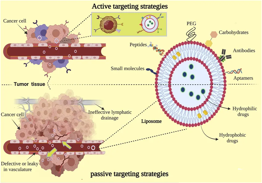
Fig no .4 Liposome mediated active and passive drug targeting. Active targeting involves conjugation of ligands to the liposome that bind to a specific target cell receptor. Passive targeting can be mediated by internalization or local high-concentration release of the drug.
because nanoparticles effortlessly cross through the thin dividers of vessels and thus infiltrate into
lymphatic vessels. Nanoparticles have a harder time penetrating the tight junctions of the arteries found in normal tissues. The tranquillizing fixations in powerful tumors of a few overlays can then increase with passive
distribution compared to those obtained with freeDoxorubicin (Doxil1/Caelyx1 and Myocet1) and daunorubicin (Daunosome1) are two examples of liposomal formulations that can deliver certain anticancer medications.
Diffusion of the drug from the carrier into the tumor site and subsequent absorption of the released drug by tumor cells comprise the drug release mechanism from liposomes. However, the precise drug release mechanism that hinders the development of passive targeting in in vivo applications is still poorly known. Additionally, the FDA approved Abraxane, a formulation of albumin-bound paclitaxel nanoparticles, to treat breast cancer [104
Recent advances in nanotechnology have expanded its application in conventional cancer therapies i.e. photothermal and gene therapy.
As shown in Fig. (5), photothermal therapy is a controlled and successful cancer treatment that uses a photothermal agent to selectively heat the target malignant area, causing thermal damage to the tumor. Metal nanoparticles, natural chromophores, or light-absorbing dyes like indocyanine green, naphthalocyanine, porphyrin coupled with transition metal, etc. are examples of these photothermal agents. Electromagnetic energy like microwaves and radiowaves injure cells during thermal therapy of malignancies by denaturing proteins and membranes, which ultimately leads to cell death. Because tumor cells are heat-sensitive and do not harm healthy cells, photothermal therapy targets the tumor cells directly [105]. Drug-carrying photothermal agents, such as nanoparticles, absorb light and transform it into heat [106]. Carbon nanotubes and gold nanoparticles In the NIR range, (CNT) and nanorods exhibit high absorption between 650 and 900 nm [107]. Nanoparticles between 10 and 100 nm in size have the ability to transform light into heat, which provides the lower energy needed to destroy tumor cells. In addition to heat-mediated cellular death, heating metal nanoparticles, such as gold nanoparticles, results in cavitation and bubble formation around the particle, which frequently produces mechanical stress that eventually damages cells [108]. Since the targeted tumor cells in nanoparticle-mediated photothermal therapy are optically sensitive, lower threshold lasers are needed to increase the heat surrounding the infected cells, which is sufficient to cause cellular harm. Researchers have investigated the use of gold nanoparticles conjugated with antiEGFR antibodies in photothermal therapy for cancer and discovered that the resulting cell death was induced by thermal abrasion at about 70° to 80°C [109Furthermore, because iron oxide nanoparticles have a high particle density in water and a huge surface area, they are another widely utilized photothermal agent with amazing power absorption potential. It has been demonstrated that when water-suspended iron oxide nanoparticles are injected straight into tumors when an external oscillating magnetic field is present, heat is produced [110].
By altering the expression of tumor genes, transferring genes that produce therapeutic proteins, or turning a non-toxic compound into a lethal drug, gene therapy offers a potent tool for cancer treatment and may be able to eliminate the decreased efficacy and off-target toxicity of chemotherapy. Because of this, several strategies for cancer gene therapy have been developed, including gene silencing using siRNA/shRNA, miRNA-mediated gene therapy, and suicide gene therapy using a transgene that inhibits tumor growth after being inserted into tumor cells [111]. By employing tiny interfering RNA (siRNA) and short hairpin RNA (shRNA) to decrease tumor-specific oncogenes and mutant tumor suppressor genes, systemic toxicity can be prevented by precisely targeting tumor cells. The short siRNA, which is 20–25 nucleotides long, is created when ribonuclease breaks down double-stranded RNA. As shown in Fig. (5) [112], siRNA binds to the multifunctional protein Argonaute to create the RNA Induced Silencing Complex (RISC), which then breaks down the passenger RNA strand upon contacting the targeted complementary mRNA. Furthermore, tumor-associated miRNA, which are essential for tumor development, progression, and metastasis, are suppressed and induced by miRNA-based cancer therapy [113]. Moreover, a number of suicide genes have been thoroughly studied in different tumor cells, including truncated Bid, carboxyl esterase/Irinotecan, human tumor necrosis factor α-related apoptosis-inducing ligand (TRAIL), herpes simplex virus type-1 thymidine kinase (HSV-tk), cytosine deaminase (CDK), BCL-2 like protein (BAX2), and others [111]. The delivery of genes or siRNA/miRNA in specific tumor cells is one of the largest challenges in the cancer gene therapy strategy, despite the fact that gene therapy has many benefits. Continuing, unchanged. The size of siRNA and the presence of serum nucleases make it difficult for them to cross the cell membrane, whereas RNAi targeting via miRNA results in electrostatic repulsion because of the identical anionic charge on the cell membrane. Recent developments in nanoparticles offer a strict method for effectively delivering tiny RNAs and genes. Because of their tiny size and great surface area, NPs can easily pass through cell membranes and carry siRNA [114]. It is possible to conjugate genes and short RNAs onto the surface of NPs or bind them to them via electrostatic contact. Numerous nanocarriers for cancer genes have been developed.
therapy, inorganic and polymeric nanoparticles have been widely used in numerous cancer treatment investigations. Small size, limited distribution, the capacity to contain a wide range of gene therapies, resistance to enzymatic degradation, and exceptional stability are only a few benefits of polymer-based nanoparticles [115]. Polysaccharides (chitosan, alginate, and hyaluronic acid, or HA), synthetic polymers (polyethyleneimine, or PEI), poly (lactic-co-glycolic acid, or PLGA), and polylysine are among the extensively researched polymeric nanoparticles for gene delivery at specific tumor sites. Su and colleagues' study team used PLGA-PEI nanoparticles to deliver the stat3 siRNA and anti-cancer medication PTX to the A549 lung cancer cell line concurrently. The findings demonstrated that stst3 silencing increased the cancer cells' sensitivity to PTX and thereby induced apoptosis in cancer cells [116]. Recently, Matheolabakis et al. created a hybrid polymer nanoparticle based on PEI that contained HA and PEG. They then combined it with survivin silencing siRNA to create a polyplex. Significant tumor growth inhibition was seen in lung cancer cells when polyplex and CCD-C8 were cotransfected [117]. Moreover, inorganic nanoparticles such as quantum dots, gold nanoparticles, carbon nanotubes, etc. have been applied to gene therapy for cancer. Oishi and colleagues' research team was the first to report incorporating siRNA into gold nanoparticles and delivering it to liver cancer.HuH7 cell line [118]. Furthermore, Davis and colleagues conducted the first proof-of-principle study in which siRNA was delivered to cancer cells in cyclodextrin-polymer-based nanoparticle form to inhibit the expression of the M2 subunit of ribonucleotide reductase (RRM2). The delivery system used human transferrin protein to target the cancer cells specifically, and PEG was used to increase the stability of the nanoparticles [119]. Therefore, it is evident that cancer gene therapy aided by nanotechnology has the potential to be a successful and efficient cancer treatment strategy.
A new idea in cancer nanotechnology, cancer theragnosis is very advantageous for patients and physicians alike. Targeted therapy and diagnostic testing are done concurrently in a single integrated system for cancer diagnosis [120]. The most important step in the procedure is the use of theragnostic agents, which are outlined in Fig. and serve both diagnostic and therapeutic purposes, allowing for simultaneous diagnosis, treatment, and therapeutic monitoring.
(6). Actually, cancer theragnosis relies on the identification of different cellular phenotypes identified at the targeted tumor site by a theragnostic agent. This allows for both monitoring, or imaging of theragnostic agents, and real-time cancer therapy. Nanotechnology has emerged as a promising technique in cancer theragnosis due to its wide range of uses. Numerous nanoparticles, such as silver, gold, and chitosan-based nanoparticles (CNPs), among others, have been transformed into multimodal therapeutic nanoparticles with imaging, targeting, and therapeutic capabilities. Both active and passive targeting are used to deliver theragnostic NPs to tumors, just like the nanoparticles. Through the EPR effect, cancer cells that are particular to a tumor's leakier blood artery gather around the tumor location. Non-invasive imaging techniques such as optical imaging (fluorescence or bioluminescence), magnetic resonance imaging, computed tomography (CT), and Positron Emission Tomography (PET) imaging are labeled to nanoparticles containing an anti-cancer medication to create cancer theragnostic NPs.
While theragnostic NPs are passively targeted, ligand-conjugated theragnostic NPs are actively targeted. When it comes to cancer diagnostics, non-invasive imaging methods help with early detection, targeted delivery, and cancer therapy monitoring. Chemotherapy, photothermal therapy, and siRNA/miRNA therapy are among the various cancer treatments for which theragnostic NPs are being thoroughly investigated. In the SCC7 mouse model, Ryu and colleagues' research team administered chitosan-based theragnostic nanoparticles (NPs) labeled with the NIR fluorescent dye Cy5.5 and encapsulating the medication PTX to the tumor location [121]. By attaching the ApoP1 peptide, which binds to an apoptotic biomarker, to PEG NPs loaded with doxorubicin (L-Dox), Wang et al. developed an apoptosis focused drug delivery of an anticancer medication that allows for simultaneous drug delivery and therapeutic response monitoring [122]. A pH-responsive DOX-loaded chitosan theragnostic NP tagged with Cy5 fluorescent dye was created by Chen et al. in order to comprehend intracellular drug release [123]. Kwon et al. recently created a theragnostic netic gold nanoparticle cluster (SPAuNCs) on the capsid of the hepatitis B virus that was designed to deliver an EGFR ligand that targets the tumor cells [124]. Furthermore, because theragnostic NP administration reduces enzymatic degradation of siRNA and increases its stability in the circulation following intravenous injection, it can be a highly helpful tool in cancer gene therapy [125]. Consistent with this fact, the research group of Huh and colleagues developed glycosyl
chitosan (GC) and polyethyleneimine (PEI) nanoparticle and PEI provides a site for Cy5.5 dye conjugated siRNA binding. In tumorbearing mouse model, a significant increase in NIR fluorescence of Cy5.5 indicated the accumulation of siRNA in targeted tumor site [126]. In addition, theragnostic NPs could contribute to the determination of patient-specific optimum anti-cancer drug dosage and monitor the tumor growth as well [114]. Due to a surge in research into the design of theragnosis NPs, the drug release behavior of the nanoparticle at the targeted tumor site is being monitored using the special fluorescence resonance energy transfer (FRET), one type of optical imaging [126]. Thus it is apparent that continuous effort in the advancement of cancer theragnostic NPs would open a new avenue in cancer diagnosis and therapy.
FUTURE PROSPECTIVE:
Numerous research groups have used nanotechnology extensively in cancer detection and treatment studies, and it may be the next big thing in the fight against cancer. Even though a great deal of study has been done in the field of cancer nanotechnology thus far, much more work has not yet been completed. Notwithstanding the inherent limitations, there is little question about the promise of DNA-based nanomaterials in cancer treatment, and additional development in this field would offer a significant approach to cancer diaFurthermore, it is important to highlight the synthesis of a successful multimodal nanoparticle that could combat cancer by providing both rapid diagnostic and efficient treatment. Therefore, it is evident that in the not-too-distant future, cancer nanotechnology will undoubtedly offer an effective, reliable, and secure cancer diagnosis and treatment approach.gnostics and treatment
CONCLUSION
Over the past few decades, the use of nanomaterials in various scientific, engineering, and technological domains has grown significantly. Nowadays, nanoparticles are widely employed in biomedical research as a therapeutic strategy or as a drug delivery mechanism. Accordingly, the application of nanotechnology in cancer treatment and diagnosis has created a new field of study called nanooncology. Recent developments in nanotechnology have fueled study in the field of nano-oncology over the years, establishing the field as a viable cancer treatment strategy. Despite this, scientists have created a plethora of novel anticancer medications and diagnostic compounds that can quickly identify and treat a variety of cancer forms. One major issue with cancer treatment is the accurate delivery of anticancer medications to the intended tumor site, which is mostly accomplished by creating various nanopolymers as drug carriers, such as dendrimers, micelles, and nanocantilevers. We can significantly improve the properties of nanopolymers and create new molecules that are more efficient and compatible with biological systems by modifying their structure and architecture. All things considered, we can say that nano-oncology has created countless opportunities for the search and development of medications and drug delivery systems for the treatment of cancer. Nano-oncology will soon become a well-known cancer treatment strategy because to extensive and ongoing research.
LIST OF ABBREVIATIONS:
AIDS = Acquired Immune Deficiency Syndrome
AML = Acute Myeloid Leukemia
CNTs = Carbon Nanotubes
CT = Computed Tomography
EGF = Endothelial Growth Factor
EGF = Epidermal Growth Factor
FA = Folic Acid
FDA = Food and Drug Administration
FGF = Fibroblast Growth Factor
FR = Folate Receptor
MCF-7 = Michigan Cancer Foundation
MMPs = Matrix Metalloproteinase
MRI = Magnetic Resonance Imaging
MWNTs = Multi-Walled Nanotubes
NIR = Near Infrared
NP = Nanoparticle
PDGF = Platelet Derived Growth Factor
PEG = Polyethylene glycol
PET = Positron Emission Tomography
PLGA = PolyLactic-co-Glycolic Acid
QDs = Quantum Dots
siRNA = Small Interfering RNA
REFERENCES
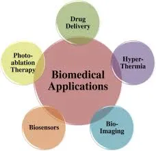
Fig no. 1 Therapeutic applications of nanotechnology in different biomedical field.
Numerous methods of nanomedicine are being thoroughly studied, including those employing lipid and micelle-based nanoparticles, dendrimers, quantum specks, carbon nanotubes, and polymeric and non-polymeric nanoparticles. Furthermore, the potential of nanomedicine multivalent ligand targeting and the ability to transport a large payload are essential components of cancer treatment. This circumvents defense mechanisms and provides precision for tissue targeting. The main challenges facing these upcoming medications are the possible toxicity of nanoparticles, which necessitates a thorough evaluation before nanomedicines may be employed as cancer (malignancy) treatments [3]. Creating a biocompatible nanosystem—such as nanocrystals, strong lipid nanoparticles, nanostructured lipid carriers, lipid drug conjugates, nanoliposomes, dendrimers, nanoshells, emulsions, nanotubes, quantum dots, etc.—is essential to delivering nanomaterial conjugated medicine to the intended tumor site. Delivery of drug complexes and nanomaterials involves both passive and active targeting.
Techniques that can also be applied to the delivery of nanodrugs. The Enhanced Permeability and Maintenance (EPR) effect of the vasculature surrounding tumors is necessary for passive targeting. For medication delivery, active targeting employs ligand-coordinated binding of nanoparticles to tumor cell receptors. Temperature and pH variations in the body can control how much medication is released from nanoparticles. Different biodistribution profiles and the anticancer efficacy of nano-drugs in vivo are caused by a variety of nanomaterial characteristics, including their size, surface charge, PEGylation, and other biophysical characteristics [4]. A research team concentrated on creating biocompatible nanoparticles that might target particular cancer indicators and offer therapeutic effects in order to identify and treat cancer. Imagery as well as medicinal substances. More sophisticated nanoparticle drug complexes that can release different nanomedicines for improved treatment efficacy have been created in recent study [5]. By conjugating tumor-specific cell surface receptors, active targeting of nanoparticles increases the viability of nanoparticle drug delivery devices while effectively decreasing poisoning. Multifunctional nanoparticulate devices for simultaneous drug delivery and tumor mass imaging are among the most interesting developNanomaterials are referred to as nanovehicles because they serve as a vehicle for conjugated drug delivery. When it comes to cancer treatment, novel drug delivery systems based on nanoparticles target the tumor cells and the milieu that supports cancer cells. Therapeutic drug combinations carried by nanovehicles for the targeted and targeted removal of tumor cells, targeted drug delivery to tumor cells, cancer stem/tumor-initiating cells, and/or the supportive cancer cell microenvironment [7] ments in nanomedicine [6]. . Quantum dots, a frequently utilized nanomaterial in cancer cell imaging, have special photochemical and photophysical characteristics because they are orders of magnitude brighter than Conventional fluorophores have extremely narrow emission spectra that can be adjusted by changing the dot size. A novel type of fluorescent markers with enhanced brightness and resistance to photo bleaching is called quantum dots. These characteristics have the potential to increase biological imaging and detection sensitivity by at least 10–100 times. [8]. Other nanomaterials, such as dendrimers and carbon nanotubes (CNTs), offer intriguing qualities that can be used for thermal ablation, diagnostics, and delivery of medications for cancer. Because of their axial symmetry and nanoscale widths, carbon nanotubes (CNTs) are tubular materials with intriguing characteristics that may be used to diagnose and treat cancer. Similarly, CNTs may be able to carry medications straight to the cells and tissues that need them. Clarifying the toxicity of nanoparticles is also crucial given the quick advancements in the creation of materials based on nanotechnology. Furthermore, by balancing the hydrophobicity and hydrophilicity of the micelle framing block copolymers, polymeric micelles may be produced with improved drug stacking capabilities. They can also be efficiently cancer-focused by altering their surface in response to tumor-homing ligands.
Nonetheless, it might still be difficult to maintain the self-assembly scaffolding in circulation and disassemble it for drug release at the site of action [9]. Recent developments have led to the enhancement of Guided nanoparticle attached medications for the treatment of tumors, bioaffinity assays based on nanoparticles for atomic and cell imaging, and integrated nanodevices for disease detection and early screening. These developments create exciting opportunities for personalized oncology, where cancer is analyzed and treated based on the subatomic profiles of individual individuals using genetic and protein biomarkers. This review article provides an overview of the use of different nanotechnology-based methods for cancer diagnosis and treatment.
2 NANOTECHNOLOGIES IN CANCER DIAGNOSIS:
A mutation in a few certain genes within the cells is the root cause of cancer. A mass of mutant cells develops in a given tissue or organ as a result of this mutation, which changes the synthesis of certain macromolecules and ultimately causes unchecked cell division. Known as a tumor. Tumor cells are referred to as benign when they are contained, but as malignant when they spread to the surrounding tissues. The majority of cancer diagnostic and treatment approaches were created to stop cancer cells from proliferating and dividing. The most crucial aspect of cancer treatment is early and precise diagnosis, which is typically accomplished by ultrasonography, Positron Emission Tomography (PET), Magnetic Resonance Imaging (MRI), Computed Tomography (CT), etc. [10] Successful treatment and patient outcomes have become extremely challenging due to the new imaging and investigative techniques' inability to give comprehensive clinical information about different tumor types and stages [3, 4]. The majority of anticancer medications on the market now do not distinguish between healthy and malignant cells, resulting in unfavorable consequences including systemic damage. Moreover, a significant issue with cancer is that it is often diagnosed too late, after the disease has spread.
2.1 Nanotechnology Assisted Tumor Imaging:
Over the last few decades, there has been an increase in interest in the use of nanoparticles for molecular imaging and for the diagnosis and monitoring of cancer. The fundamental idea behind imaging based on nanomaterials is particle size, which gives nanoparticles their unique characteristics. For example, semiconductor nanoparticles utilized in cancer The optical, magnetic, and structural characteristics of iron oxide nanocrystals and quantum dots are uncommon in bulk materials an. Nanoparticles can be employed with a variety of anticancer medicines, such as medications and biomolecules, such as various peptides, antibodies, or other compounds, to designate tumors with high emOnly for personal use; do not distribute. Pathy and specificity, and as shown in Fig. (2), this compound is helpful in the early identification and screening of cancer cells. d Because of its vast surface area and tiny diameter, the nanoparticle can readily bind to functional groups of various optical, radio isotopic, or magnetic diagnostic and therapeutic agents, making the more effective and persuasive cancer diagnosis. A significant breakthrough in the diagnosis and treatment of cancer was made possible by these developments in nanotechnology [11]. molecules.
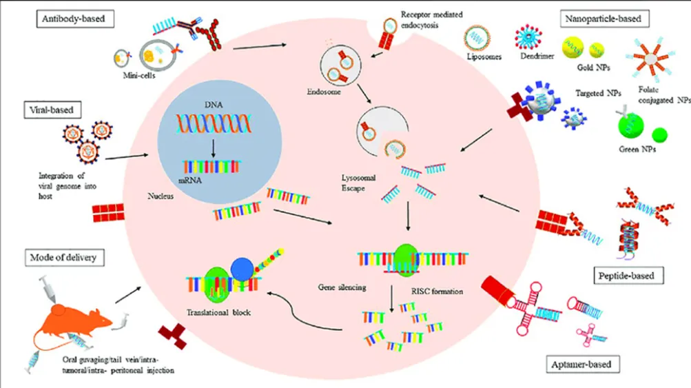
Fig. (2). Schematic mechanism of SERS mediated bio-imaging and anticancer drug delivery by using AuNGO
It's shocking to learn that researchers are working on a detecting device that may detect cancer inside the body by being worn on the wrist. This significant advancement in medical technology is because to this field's application of nanomaterials. Certain active magnetic molecules in the device above sliced through the wrist's blood vessels [12]. These magnetic molecules use a wrist device to show the findings of their detection of changes in the smart nanoparticles circulating in the blood. Nanomaterials' remarkable behavior and adaptability are the reason for this advancement in cancer diagnostics. These days, nanotechnology has confirmed imaging of cancer at the tissue, cell, and molecular level. One example is lanthanide-based up conversion nanoparticles, which use autofluorescence to detect deep tissue by upconverting low-energy photons to high-energy ones [13]. In addition, nanotechnology investigated the For instance, fibroblast activation protein-a on the membrane of tumor-associated fibroblasts can be detected by a pH-responsive fluorescent nanoprobe, which can be used to target and image cancerous tumors [14]. Here, we've covered a few high temporal and spatial nanotechnology-based approaches that can be useful for precisely tracking living cells and tracking dynamic biological activities within tumors.
The primary issue with visible spectrum imaging is its poor detection and penetration. Scientists created quantum dots that fluoresce in the near-infrared spectrum as a solution to this issue, i.e. NIR quantum dots are more suited for in vivo imaging of cancer in tissues such as the colon, liver, pancreas, and lymphatic tissue because of their wavelength of 700–1000 nm [15–17]. Live animals with multicolor quantum dot (QD) imaging capabilities. On a host animal, 1–2 million of each color were subcutaneously injected in three nearby places. Excitation from tungsten or mercury lamps produced the images. Gao X, Cui Y, Levinson RM, Chung LW, Nie S. In vivo cancer targeting and imaging with semiconductor quantum dots, reprinted with permission from Macmillan Publishers Ltd. 22:969-976; Nat Biotechnology, 2004.
2.1.2 Nano shells:
Nano shells are dielectric cores that range in size from 10 to 300 nm. They are typically composed of silica and have a thin metal shell, usually constructed of gold [18, 19]. These nano shells change electrical signals mediated by Plasmon. With an emission/absorption array spanning from the ultraviolet to the infrared, they can also be optically tuned to convert energy into light [20]. Because heavy metal toxicity does not affect their imaging, nano shells are appealing. The size of the nano shell is one of its issues.
2.1.3 Colloidal Gold Nanoparticles:
One of the most appealing types of agents for cancer diagnostics is gold nanoparticles. This is because gold has been approved for use in the treatment of human illness [21] and is simple to synthesize [22].
By dispersing visible light in in vitro samples, these gold nanoparticles serve as contrast agents. Additionally, gold nanoparticles can be conjugated with antibodies for biopsies for the detection of pancreatic and cervical malignancies. In addition, photoacoustic tomography can be performed using gold nanoparticles. Gold nanoparticles are therefore a priority-based detection tool for several malignancies [23].
3 Nanotechnology in Cancer Therapy:
3.1 Tools of Nanotechnology for Cancer Therapy:
Recent developments in the creation of different vehicles for effective drug administration are driving research effort in the field of nanotechnology. a variety of vehicles, including nanocarriers such carbon nanotubes, liposomes, micelles, dendrimers, and quantum dots. have been created thus far, as illustrated in Fig. (3).
3.1.1 Liposomes:
Liposomes are phospholipid-based vesicles that are at least 400 nm in size, with a bilayer membrane made of cholesterol and a hydrophilic head and hydrophobic tail [24]. Liposomes' special ability to solubilize the water-insoluble organic material makes them a vehicle for medication targeting. compounds, making them appropriate for the treatment of cancer and other illnesses. Drugs included in the membrane of liposomes have a number of benefits, including efficient distribution to the intended location, low nonspecific toxicity, and protection against degradation [25, 26]. Because endothelial cells' tight connections prevent particles from leaking out of vessels, liposomes are kept in the bloodstream in normal, healthy tissues. However, tumor vasculature are more prone to leakage than blood channels in healthy tissues, which allowsthe specific tumor site by allowing the nanosized liposome to escape from the circulation.
Liposomes have the ability to target cancer cells and are biodegradable, biocompatible, and more stable in colloidal fluids [27–29]. After comparing the toxicity and drug delivery efficacy of various liposomal compositions and free drug delivery with anticancer medicines, researchers came to the conclusion that liposomes are less harmful to tumor sites than free medications [30, 31].
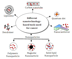
Fig. (3). Tools of nanotechnology. Schematic representation of various nanotechnology-based tools used for cancer therapeutics.
There are numerous anti-cancer medications on the market and in clinical trials [30]. Doxil (Liposomal Doxorubicin), the first FDA-approved anticancer nanodrug, is made Kaposi's sarcoma in AIDS patients is often treated with a PEGylated liposomal formulation [32, 33]. Johnson & Johnson sells PEGylated liposomes carrying the anti-cancer medication doxorubicin under the trade names Doxil (in the USA) and Caelyx (outside the USA). A manufacturing plant was shut down in 2011 as a result of problems with Doxil's quality control that led to an imbalance between the drug's supply and demand [33, 34]. Additionally, the FDA approved LipoDox as a substitute medication to address the Doxil shortage in the United States. LipoDox and Doxil have the same chemical makeup. Sun Pharma Company manufactures and sells Doxil in India; in 2013, the FDA approved the first generic version of the medication [34, 35]. Several studies demonstrate that Doxil can prevent ovarian cancer, and the FDA has also approved it to treat ovarian cancer that has returned [36, 37]. Doxil has been approved in the United States for the treatment of breast cancer [37], and in Europe and Canada, it is used in conjunction with Valadec to treat multiple myeloma [37, 38]. Celator Pharmaceuticals Inc. created CPX351, a liposomal mixture of daunorubicin and cytarabine. In the phase III clinical trial for individuals with acute myeloid leukemia (AML), CPX-351 demonstrated encouraging outcomes [39]. MBP-426 targets the transferrin receptor. Mebiopharm created the liposomal version of oxaliplatin, which is being tested in a phase II clinical trial to treat patients with stomach cancer [40]. Merrimack Pharma created MM-398, a liposomal sphere that encapsulates the medication irinotecan. that can cure malignancies that are resistant to chemotherapy, including gliomas, lung, pancreatic, and colorectal cancers [41–43].
3.1.2 Carbon Nanotubes:
Carbon nanotubes (CNTs) can be classified into two classes based on their diameter and structure: single-walled CNTs (SWNTs) and multiwalled CNTs (MWNTs). CNTs with a single wall are made up of a multiwalled carbon nanotubes, which are made out of many concentric graphene sheets, and a single sheet of cylindrical graphene [44]. The structure, surface area, mechanical strength, metallic behavior, electrical and thermal conductivity, and ultra-lightweight of carbon nanotubes are all related to their physical and chemical characteristics. Because of their many unique physical and chemical characteristics, CNTs are a good option for a wide range of biomedical applications [45]. CNTs have been utilized to target cancer cells because of their ability to absorb near-infrared (NIR) light, which causes the nanotubes to heat up [46–48]. This phenomenon is known as the thermal effect. Folic acid (FA) receptors are overexpressed in cancer cells, and Numerous research teams have created synthetic nanocarriers using conjugated biomaterials of FA derivatives. Furthermore, compared to spherical nanocarriers, it has been shown that CNTs are kept in the lymph nodes for longer periods of time [49]. Another study found that gemcitabine, an anticancer drug, had strong activity against lymph nodes when put onto magnetic MWNTs and administered subcutaneously to mice [50]. When drug delivery systems interact, CNTs can recognize the surface receptors that cause receptor-mediated endocytosis of CNTs. with cancer cells (NDDs) [51]. CNT-based drug delivery improves medicines' blood circulation and biodistribution, resulting in lower dosages and greater pharmacological efficacy. Liu and colleagues created DOX-loaded branching PEGfunctionalized SWNTs and administered the SWNT-DOX complex to the tumor location in mice, taking into account the extended blood circulation caused by CNTs. They discovered that SWNTs can be removed from the systemic blood circulation via renal excretion, and that DOX can be introduced into tumors. A chemotherapeutic medication called paclitaxel (PTX) is used to treat a variety of malignancies, but its poor solubility in aqueous solution makes it challenging to physically load PTX at the intended tumor site [52]. In order to address this issue, Lay and colleagues created PEG-graft SWCNTs and PEG-graft MWCNTs, which improve loading capacity and enable persistent PTX administration for up to 40 days in vitro [53]. To increase the effectiveness of anticancer medication delivery, researchers have also altered SWNTs to function as the Epidermal Growth Factor (EGF) mediated SWNT carrier [54]. As a carbon-based nanomaterial, MWNTs can be utilized for thermal ablation, which kills cancer cells by causing hyperthermia. In order for the medications to better limit the spread of cancer cells, the cancer cells absorb the CNTs complex and release the chemotherapeutic chemicals into the intracellular space. Therefore, the drug delivery method based on carbon nanotubes (CNTs) offers a number of benefits, including fewer side effects and less cytotoxicity [54]. Due to their high specific surface area, SWNTs demonstrated a greater capacity for drug loading than both dendrimer drug carriers and conventional liposomes [55].
3.1.3 Polymeric Micelles:
Micelles are typically utilized in targeted drug delivery to deliver less soluble or water-insoluble medications to tumor locations. Micelles are composed of both hydrophilic and amphiphilic co-polymers. and monomer units that are hydrophobic. Polymeric micelles, which have a hydrophilic PEG shell and range in size from 10 to 100 nm, are an effective drug delivery mechanism for anticancer medications that are poorly water soluble. The polymeric micelles exhibit prolonged blood circulation and greater accumulation at the tumor location. Micelles can be classified into a number of forms based on their structure and bonding. One type is block copolymer micelles, which can also be classified into amphiphilic micelles that are hydrophobically formed, polyion-complex micelles, and micelles that result from metal complexation [56]. Typically, macromolecules with distinct hydrophobic and hydrophilic domains are seen in hydrophobic micelles.
Certain commonly used copolymer block segments are only for personal use. Not for Publication been covered by Sutton and colleagues. Water-insoluble medications can be weighted down into the hydrophobic cores of these polymers as the amphiphilic molecules spontaneously self-assemble into super molecular core/shell configurations upon entering the aquatic system. Lam and associates have recently created a novel kind of micelle composed of PEG-cholic acid conjugates [57]. A carrier combination of Cholic Acid and PEG (PRG5K-CA8) can carry a large amount of paclitaxel; in this case, eight Cholic Acid molecules join with one PEG5000 chain to produce a carrier core that ranges in size from 20 to 60 nm [57]. Paclitaxel-loaded PEG5K-CA8 micelles showed improved antitumor efficacy and tolerance in a phase I clinical trial, as shown in Table 1 [58]. Furthermore, it was shown that a micellar carrier PEG2K-CA4 had a larger doxorubicin loading capacity and a more prolonged drug release profile than PEG5K-CA8 micelles. Lipid makes up the hydrophobic core of another kind of polymeric micelle.
For instance, the hydrophobic regions of some PEG diacyl lipid conjugates, such as phosphatidylethanolamine (PEG-PE), are lipids of different acyl chains that were produced by Torchilin's group [59]. In order to create stable micelles with an extremely low CMC value (~10-5 M), PEG-PE conjugates were formed as a result of strong hydrophobic interactions between the double acyl chains [60, 61]. Numerous weakly water-soluble medications, such as paclitaxel, tamoxifen, porphyrin, camptothecin, and vitamin K3 [62, 63], can be dissolved by these micelles. Micelles' extremely small size makes them more effective at carrying drugs than other drug carriers, especially for solid tumors, especially those with low vascularization [64]. Using PEG-PLA micelles and the weakly water-soluble chemical β-lapachone (βlap), Blanco et al. created an anticancer medication [65]. They discovered that the combination of PLA micellar core and β-lap was extremely stable and remained inside the micelles in the blood. circulation for an extended length of time, which results in the accumulation of drugs at the tumor site. Additionally, he discovered that PEG-PLA-β-lap works better in lab settings for treating orthotopic Lewis lung cancer (LCC) and subcutaneous lung cancer (NSCLC) A549. In a similar vein [66], Mu et al. treated multidrug-resistant (MDR) oral epidermal carcinoma with docetaxel-loaded PEG-PLA (MPP) and found that micelles increased the circulation time of docetaxel in the blood of mice and inhibited the growth of tumor more efficiently than Taxotere® (the clinically approved formulation of docetaxel). It was surprising that the combination of MPP micelles and Pluronic 85 (P85) in a ratio 6:1 (w/w) inhibit the tumor progression by increasing the delivery of docetaxel without affecting the plasma drug levels of mice blood [67] . An approved medication for the treatment of cancer, paclitaxel has been shown to be more effective against cancer when combined with drug carrier PEG-PCL-PEG (PCEC) micelles [68]. In paclitaxel-resistant SKOV-3 s.c. xenograft mice, Wang et al. investigated the paclitaxel-loaded P105/PCL50 micelles and discovered that PCLmodified Pluronic P105 (P105/PCL50) micelles were more effective. More effective than Taxol® at slowing the growth of tumors [69].
3.1.4 Dendrimers:
Another type of nanocarrier is a dendrimer, which has a spherical polymeric core with branches spaced regularly. Dendritic macromolecules tend to take on a more globular shape as their diameter increases linearly [70]. Two techniques are typically used to synthesis dendrimers: the first is a divergent technique that allows dendrimers to develop outward from a central core [71, 72], and the The second approach, known as the convergent method, synthesizes the dendrimer from the margin inward, culminating in the core. The dendrimers are often made from a variety of molecules, such as poly (propylene imine), poly (glycerol-cosuccinic acid), poly (L-lysine), poly (glycerol), poly (2,2-bis(hydroxymethyl)propionic acid), and melamine dendrimers. These dendrimers exhibit various chemical structures and characteristics, such as charge, basicity, and hydrogen bonding ability, which can be adjusted either byincreasing dendrimer production or by altering the dendrimer surface groups. Antineoplastic agents are typically covalently attached to the peripheral groups of dendrimers to form dendrimer-drug conjugates. As a result, a number of pharmacological compounds can be affixed to every dendrimer molecule, and the type of connections helps regulate how these therapeutic chemicals are released. Dendrimers have emerged as a promising nanocarrier because of their clear characteristics, numerous linkage groups, polymer size, charge, and biologically associated characteristics such lipid bilayer interactions, cytotoxicity, internalization, blood plasma retention time, biodistribution, and filtration [73]. An asymmetric doxorubicin-functionalized bow-tie dendrimer was created by PEGylating one side of a 2,2-bis(hydroxymethyl) propionic acid dendrimer (G3) and connecting the drug to the other side via acyl hydrazone linkage (G4). Fréchet and Szoka's work serves as an example of an architecturally-optimized dendritic drug delivery system. 8–10% of the whole system is doxorubicin [74]. Then, following the entire medication and It was discovered that the absorption of the dendriticdoxorubicin complex is greater than that of doxorubicin alone when the dendritic system was injected into BALB/c mice with s.c. C-26 colon cancer. . It was unexpected that when doxorubicin-conjugated dendrimer was administered to malignant mice after 60 days, the tumor entirely disappeared and Every mouse made it out alive. On the other hand, mice given doxorubicin alone show no change in tumor size. Another study discovered that folic acid attached dendrimers can thwart cancer cells that express more folic acid receptors [75, 76, 77]. . The ability of the dendrimer to assemble with DNA in clusters, such as the DNA-Polyamidoamine cluster, or DNAPAMAM, provides an additional benefit. Cancer cells are effectively killed by this compound, which highly express folic acid receptors. Dendrimer-antibody conjugates attach to prostate-specific membrane antigen-positive (LNCaP.FGC) cells more efficiently than they do to normal cells, and tumor cells absorbed the conjugate far more readily than unconjugated dendrimer. Glycopeptide dendrimers conjugated to the anti-mitotic substance colchicine and dendrimers that include sugar moieties [78] in their structure [79] are examples of another kind of dendrimers, sometimes known as glycodendrimers. These conjugates were examined in healthy cells (non-transformed mouse embryonic fibroblasts, or MEFs) and cancer cell lines (HeLa) [63], and the findings demonstrated that Non-glycosylated dendrimers demonstrated ten times less selectivity for HeLa cells, whereas dendrimers reduced HeLa cell proliferation 20–100 times more effectively than MEFs [80].
3.1.5 Quantum Dots:
In 1980, Ekimov and associates created the first quantum dots (QDs), which are tiny particles or nanocrystals of semiconducting material that range in size from 2 to 10 nanometers [81]. The electrical characteristics of quantum dots are in between those of mass semiconductors and distinct atoms, which results from these particles' high surface-to-volume ratios [82]. Over time, a number of QD-based technologies have emerged, including modified QD conjugates and QD immunostaining. Along with the enhanced multiplexing capabilities, the improved QDs conjugates provide a significant time and cost-effectiveness improvement over singlecolor tests. Furthermore, compared to conventional immunochemistry experiments, QD immunostaining has been demonstrated to be more accurate, precise, and background-free at low protein expression levels. QD immunostaining could be used to detect different tumor biomarkers, such as cell proteins or other elements of diverse tumor samples, in the context of cancer diagnosis. QDs have the ability to group together in particular bodily areas and then transfer the cancer-causing medications that are attached to them. QDs have the potential to replace untargeted medication administration and so circumvent the negative effects of chemotherapy because of their ability to aggregate in a single human organ. Recent advancements in QD surface modification and in vivo coupling of QDs with biomolecules, such as peptides and antibodies, might allow for the targeting of cancers and
possible use in imaging for cancer. With the aid of proteins or peptides targeted against overexpressed surface receptors on cancer cells, such as the transferrin receptor, antigen claudin-4, and urokinase plasminogen activator receptor [84], QD-based imaging probes Personal Use Only Not For Distribution can detect pancreatic cancer at an extremely early stage [83]. When Gao and colleagues tagged human prostate cancer cells using QDs coupled with an antibody for Prostate-Specific Membrane Antigen (PSMA), they established the use of QDs in cancer detection. Bostick et al. used QD-based multiplexed imaging to identify five biomarkers on a single tissue slide; additional biomarkers may be assessed using multiple slides each. stained using each of the five biomarkers [85]. Additionally, they suggested creating a workflow for the quantitative analysis of every biomarker. The suggested system was so practical and effective that it could evaluate six biomarkers in just seven hours, which was beneficial for clinical use. When compared to traditional fluorescent immunolabelling, Ruan et al. demonstrated that QD-based immunolabelling had a more constant photo-intensity [86]. It has been shown that very sensitive QD-based probes may image cancer cells in vivo using multicolor fluorescence [87]. Additionally, QDs can identify the ovarian cancer marker CA125 in a variety of specimen types, including. Additionally, QDs can be utilized to identify the ovarian cancer marker CA125 in a variety of tissues, including tissue slices, fixed cells, and xenograft fragments.Furthermore, compared to traditional organic dye, QD signals exhibit more selective and brighter photostability [88]. Chen et al. used QD-based probes to successfully detect BC, proving that QD-Immunohistochemistry (IHC) could clearly detect lower HER2 expression than conventional IHC and achieve multiplexed QD-based detection at the same time [89]. This method was expanded by Yezhelyev et al. to specifically label MCF-7 and BT-474 BC cells for HER2, Progesterone, Estrogen Receptor (ER), and Epidermal Growth Factor Receptor (EGFR). QD-based nanotechnology is an effective way to provide multiplexed cancer biomarker imaging in situ on intact tumor tissue specimens for tumor pathology study at the histological and molecular levels simultaneously, according to research on receptor (PR) and mammalian target of rapamycin (m-TOR) by visible and NIR QDs [90]. In SKOV-3 human ovarian cancer cells, Liu et al. produced pH-dependent, pH-sensitive photoluminescent CdSe/ZnSe/ZnS QDs, indicating potential uses for intracellular pH sensors [91]. EGFR single molecules in human ovarian epidermal carcinoma cells (A431) were successfully addressed by Kawashima et al. [92].
3.2 Drug Targeting Approaches for Cancer Therapy:
3.2.1 Active Targeting:
The best targeting strategy for effectively delivering nanoparticles into malignant cells without producing any toxicity is active targeting of the medication. This particular targeting method often depends on ligand-receptor interaction, wherein nanoparticles have a ligand that binds selectively to the tumor's receptor. the cell surface as shown in Figure (4). By providing strong ligand-receptor binding to deliver the medication in peripheral tissues, active targeting reduces nonspecific contact. In one study, mice treated with both free Doxil and Doxil combined with antibody F5 conjugated PEG showed a quick and notable decrease in tumor volume in mice treated with Doxil conjugated F5 as opposed to mice treated with free Doxil [93]. More than 90% of patients with ovarian carcinoma and several other cancer forms (choriocarcinomas, uterine sarcomas, and osteosarcomas) have overexpressed the Folate Receptor (FR), a highly specific tumor marker. Because of their increased need for folate for DNA synthesis, cancer cells often overexpress folate receptors. A folate moiety's engagement with the tumor cells' folate receptor triggers an endocytic transport that causes cytosolic accumulation. Folate-coated liposomes have been shown to improve the cytotoxicity of chemotherapeutic drugs by increasing their accumulation in several tumor cell types [94], but they can also get beyond MDR of tumor cells [95]. Additionally, because KB (human epidermal carcinoma) and HeLa (cervical cancer) cells overexpress folate receptors, folate-coated liposomes have been employed as a delivery vehicle for DOX and improve its in vitro uptake in these cells [96]. To induce specific medication targeting, polymeric nanoparticles (NPs) with surface-conjugated folate and derivatives, ranging in size from 50 to 100 nm, have been produced [97]. Matrix metalloproteinases (MMPs), angiopoeitins and their receptors (tie1 and tie2), platelet-derived growth factor (PDGF), fibroblast growth factor (FGF), endothelial growth factor (EGF), and their receptors are examples of potential targets in vascular endothelium that are present in the subendothelial matrix and involved in angiogenesis. The study by Bibby and colleagues, in which they screened two antibodies, F5 and C1, to the human breast tumor cell line SK-BR3, which in turn binds to overexpressed growth factor ErbB2 in human breast cancer and several other adenocarcinomas, is a classic example of active targeting-based identification of the ideal ligand [98].
Another method of actively targeting the distribution of anti-cancer medications is the use of antibody conjugated liposomes, also known as immunoliposomes. Similar to liposomes, immunoliposomes contain anti-cancer medicines; but, because of the coupled tumor-specific antibody, they provide highly concentrated cancer cell targeting. When compared to naked PEGylated liposomes, DOX-loaded anti-HER-2 (anti-human epidermal growth factor receptor-2) immunoliposomes have been shown to exhibit enhanced therapeutic efficacy against several breast cancer xenograft models [99]. Additionally, transferrin receptors are among the other surface receptors that are overexpressed on a variety of tumor cells. The cellular absorption of transferrin-conjugated PLGA NPs loaded with paclitaxel was three times more than the unconjugated ones in human prostate cancer cells (PC3), according to in vitro research based on transferrin receptors. It's interesting to note that in a mouse model, PC3 cells treated with paclitaxeltransferrin-conjugated NPs (paclitaxel dose 24 mg/Kg) administered subcutaneously caused total tumor shrinkage. Due to a higher intracellular retention of paclitaxel, this NP conjugated-paclitaxel delivery system has also been tested with MCF-7 breast cancer cells and shown to be more effective in these cells [100]. Since integrins are essential for cell invasion and migration, a different method of actively targeting anticancer drugs has also been proposed: using nanoparticle coupled integrin ligand to deliver genes selectively to the angiogenic blood arteries in tumor-bearing mice. Avb3 ligand-carrying DNA-encapsulated cationic polymerized liposomes were created by Hood and a colleague's research group and utilized to target the integrins of M21-melanoma xenograft tumors. The delivery of a mutant Raf gene inhibited endothelial cell signaling and angiogenesis, resulting in a marked reduction in tumor size after a single injection, according to the results, which also showed a selective increase in gene expression in the tumor [101].
As seen in Fig. (4), passive targeting is the diffusion-mediated transport of pharmaceuticals that entails creating a drug carrier complex that can evade the body's defense mechanisms. The medication
The carrier complex travels through the bloodstream and is delivered to the intended receptor. Effective passive drug targeting depends on a number of drug carrier complex characteristics, including molecular weight, surface charge, surface hydrophobicity or hydrophilia, and size. For example, PEG-coated stealth liposomes go through the bloodstream, and the surface charge on PEG-containing liposomes plays a crucial role in how long they stay there. The most widely used strategy for medication delivery in cancerous cells is passive targeting.
The enhanced permeability and retention (EPR) impact is caused by both this vascular malformation and inadequate lymphatic waste, which aids in the penetration and upkeep of nanoparticles at the tumor site [102]. Indifferent Since they have a lengthy half-life in the bloodstream, nanocarriers with no net charge make the best passive targeting candidates. The body's immune system may eradicate the charged nanocarriers; opsonization removes positively charged nanocarriers, while Kupffer cells remove negatively charged nanocarriers. Because nanoparticles easily pass through the thin dividers of vessels and enter lymphatic vessels, passive targeting of drugs conjugated with nanoparticles is coordinated to lymphoid organs, lymphatic vessels, and the spleen.
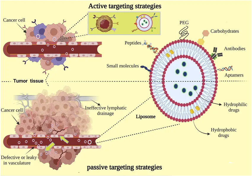
Fig no .4 Liposome mediated active and passive drug targeting. Active targeting involves conjugation of ligands to the liposome that bind to a specific target cell receptor. Passive targeting can be mediated by internalization or local high-concentration release of the drug.
because nanoparticles effortlessly cross through the thin dividers of vessels and thus infiltrate into
lymphatic vessels. Nanoparticles have a harder time penetrating the tight junctions of the arteries found in normal tissues. The tranquillizing fixations in powerful tumors of a few overlays can then increase with passive
distribution compared to those obtained with freeDoxorubicin (Doxil1/Caelyx1 and Myocet1) and daunorubicin (Daunosome1) are two examples of liposomal formulations that can deliver certain anticancer medications.
Diffusion of the drug from the carrier into the tumor site and subsequent absorption of the released drug by tumor cells comprise the drug release mechanism from liposomes. However, the precise drug release mechanism that hinders the development of passive targeting in in vivo applications is still poorly known. Additionally, the FDA approved Abraxane, a formulation of albumin-bound paclitaxel nanoparticles, to treat breast cancer [104
Recent advances in nanotechnology have expanded its application in conventional cancer therapies i.e. photothermal and gene therapy.
As shown in Fig. (5), photothermal therapy is a controlled and successful cancer treatment that uses a photothermal agent to selectively heat the target malignant area, causing thermal damage to the tumor. Metal nanoparticles, natural chromophores, or light-absorbing dyes like indocyanine green, naphthalocyanine, porphyrin coupled with transition metal, etc. are examples of these photothermal agents. Electromagnetic energy like microwaves and radiowaves injure cells during thermal therapy of malignancies by denaturing proteins and membranes, which ultimately leads to cell death. Because tumor cells are heat-sensitive and do not harm healthy cells, photothermal therapy targets the tumor cells directly [105]. Drug-carrying photothermal agents, such as nanoparticles, absorb light and transform it into heat [106]. Carbon nanotubes and gold nanoparticles In the NIR range, (CNT) and nanorods exhibit high absorption between 650 and 900 nm [107]. Nanoparticles between 10 and 100 nm in size have the ability to transform light into heat, which provides the lower energy needed to destroy tumor cells. In addition to heat-mediated cellular death, heating metal nanoparticles, such as gold nanoparticles, results in cavitation and bubble formation around the particle, which frequently produces mechanical stress that eventually damages cells [108]. Since the targeted tumor cells in nanoparticle-mediated photothermal therapy are optically sensitive, lower threshold lasers are needed to increase the heat surrounding the infected cells, which is sufficient to cause cellular harm. Researchers have investigated the use of gold nanoparticles conjugated with antiEGFR antibodies in photothermal therapy for cancer and discovered that the resulting cell death was induced by thermal abrasion at about 70° to 80°C [109Furthermore, because iron oxide nanoparticles have a high particle density in water and a huge surface area, they are another widely utilized photothermal agent with amazing power absorption potential. It has been demonstrated that when water-suspended iron oxide nanoparticles are injected straight into tumors when an external oscillating magnetic field is present, heat is produced [110].
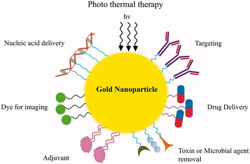
By altering the expression of tumor genes, transferring genes that produce therapeutic proteins, or turning a non-toxic compound into a lethal drug, gene therapy offers a potent tool for cancer treatment and may be able to eliminate the decreased efficacy and off-target toxicity of chemotherapy. Because of this, several strategies for cancer gene therapy have been developed, including gene silencing using siRNA/shRNA, miRNA-mediated gene therapy, and suicide gene therapy using a transgene that inhibits tumor growth after being inserted into tumor cells [111]. By employing tiny interfering RNA (siRNA) and short hairpin RNA (shRNA) to decrease tumor-specific oncogenes and mutant tumor suppressor genes, systemic toxicity can be prevented by precisely targeting tumor cells. The short siRNA, which is 20–25 nucleotides long, is created when ribonuclease breaks down double-stranded RNA. As shown in Fig. (5) [112], siRNA binds to the multifunctional protein Argonaute to create the RNA Induced Silencing Complex (RISC), which then breaks down the passenger RNA strand upon contacting the targeted complementary mRNA. Furthermore, tumor-associated miRNA, which are essential for tumor development, progression, and metastasis, are suppressed and induced by miRNA-based cancer therapy [113]. Moreover, a number of suicide genes have been thoroughly studied in different tumor cells, including truncated Bid, carboxyl esterase/Irinotecan, human tumor necrosis factor α-related apoptosis-inducing ligand (TRAIL), herpes simplex virus type-1 thymidine kinase (HSV-tk), cytosine deaminase (CDK), BCL-2 like protein (BAX2), and others [111]. The delivery of genes or siRNA/miRNA in specific tumor cells is one of the largest challenges in the cancer gene therapy strategy, despite the fact that gene therapy has many benefits. Continuing, unchanged. The size of siRNA and the presence of serum nucleases make it difficult for them to cross the cell membrane, whereas RNAi targeting via miRNA results in electrostatic repulsion because of the identical anionic charge on the cell membrane. Recent developments in nanoparticles offer a strict method for effectively delivering tiny RNAs and genes. Because of their tiny size and great surface area, NPs can easily pass through cell membranes and carry siRNA [114]. It is possible to conjugate genes and short RNAs onto the surface of NPs or bind them to them via electrostatic contact. Numerous nanocarriers for cancer genes have been developed.
therapy, inorganic and polymeric nanoparticles have been widely used in numerous cancer treatment investigations. Small size, limited distribution, the capacity to contain a wide range of gene therapies, resistance to enzymatic degradation, and exceptional stability are only a few benefits of polymer-based nanoparticles [115]. Polysaccharides (chitosan, alginate, and hyaluronic acid, or HA), synthetic polymers (polyethyleneimine, or PEI), poly (lactic-co-glycolic acid, or PLGA), and polylysine are among the extensively researched polymeric nanoparticles for gene delivery at specific tumor sites. Su and colleagues' study team used PLGA-PEI nanoparticles to deliver the stat3 siRNA and anti-cancer medication PTX to the A549 lung cancer cell line concurrently. The findings demonstrated that stst3 silencing increased the cancer cells' sensitivity to PTX and thereby induced apoptosis in cancer cells [116]. Recently, Matheolabakis et al. created a hybrid polymer nanoparticle based on PEI that contained HA and PEG. They then combined it with survivin silencing siRNA to create a polyplex. Significant tumor growth inhibition was seen in lung cancer cells when polyplex and CCD-C8 were cotransfected [117]. Moreover, inorganic nanoparticles such as quantum dots, gold nanoparticles, carbon nanotubes, etc. have been applied to gene therapy for cancer. Oishi and colleagues' research team was the first to report incorporating siRNA into gold nanoparticles and delivering it to liver cancer.HuH7 cell line [118]. Furthermore, Davis and colleagues conducted the first proof-of-principle study in which siRNA was delivered to cancer cells in cyclodextrin-polymer-based nanoparticle form to inhibit the expression of the M2 subunit of ribonucleotide reductase (RRM2). The delivery system used human transferrin protein to target the cancer cells specifically, and PEG was used to increase the stability of the nanoparticles [119]. Therefore, it is evident that cancer gene therapy aided by nanotechnology has the potential to be a successful and efficient cancer treatment strategy.
A new idea in cancer nanotechnology, cancer theragnosis is very advantageous for patients and physicians alike. Targeted therapy and diagnostic testing are done concurrently in a single integrated system for cancer diagnosis [120]. The most important step in the procedure is the use of theragnostic agents, which are outlined in Fig. and serve both diagnostic and therapeutic purposes, allowing for simultaneous diagnosis, treatment, and therapeutic monitoring.
(6). Actually, cancer theragnosis relies on the identification of different cellular phenotypes identified at the targeted tumor site by a theragnostic agent. This allows for both monitoring, or imaging of theragnostic agents, and real-time cancer therapy. Nanotechnology has emerged as a promising technique in cancer theragnosis due to its wide range of uses. Numerous nanoparticles, such as silver, gold, and chitosan-based nanoparticles (CNPs), among others, have been transformed into multimodal therapeutic nanoparticles with imaging, targeting, and therapeutic capabilities. Both active and passive targeting are used to deliver theragnostic NPs to tumors, just like the nanoparticles. Through the EPR effect, cancer cells that are particular to a tumor's leakier blood artery gather around the tumor location. Non-invasive imaging techniques such as optical imaging (fluorescence or bioluminescence), magnetic resonance imaging, computed tomography (CT), and Positron Emission Tomography (PET) imaging are labeled to nanoparticles containing an anti-cancer medication to create cancer theragnostic NPs.
While theragnostic NPs are passively targeted, ligand-conjugated theragnostic NPs are actively targeted. When it comes to cancer diagnostics, non-invasive imaging methods help with early detection, targeted delivery, and cancer therapy monitoring. Chemotherapy, photothermal therapy, and siRNA/miRNA therapy are among the various cancer treatments for which theragnostic NPs are being thoroughly investigated. In the SCC7 mouse model, Ryu and colleagues' research team administered chitosan-based theragnostic nanoparticles (NPs) labeled with the NIR fluorescent dye Cy5.5 and encapsulating the medication PTX to the tumor location [121]. By attaching the ApoP1 peptide, which binds to an apoptotic biomarker, to PEG NPs loaded with doxorubicin (L-Dox), Wang et al. developed an apoptosis focused drug delivery of an anticancer medication that allows for simultaneous drug delivery and therapeutic response monitoring [122]. A pH-responsive DOX-loaded chitosan theragnostic NP tagged with Cy5 fluorescent dye was created by Chen et al. in order to comprehend intracellular drug release [123]. Kwon et al. recently created a theragnostic netic gold nanoparticle cluster (SPAuNCs) on the capsid of the hepatitis B virus that was designed to deliver an EGFR ligand that targets the tumor cells [124]. Furthermore, because theragnostic NP administration reduces enzymatic degradation of siRNA and increases its stability in the circulation following intravenous injection, it can be a highly helpful tool in cancer gene therapy [125]. Consistent with this fact, the research group of Huh and colleagues developed glycosyl
chitosan (GC) and polyethyleneimine (PEI) nanoparticle and PEI provides a site for Cy5.5 dye conjugated siRNA binding. In tumorbearing mouse model, a significant increase in NIR fluorescence of Cy5.5 indicated the accumulation of siRNA in targeted tumor site [126]. In addition, theragnostic NPs could contribute to the determination of patient-specific optimum anti-cancer drug dosage and monitor the tumor growth as well [114]. Due to a surge in research into the design of theragnosis NPs, the drug release behavior of the nanoparticle at the targeted tumor site is being monitored using the special fluorescence resonance energy transfer (FRET), one type of optical imaging [126]. Thus it is apparent that continuous effort in the advancement of cancer theragnostic NPs would open a new avenue in cancer diagnosis and therapy.
FUTURE PROSPECTIVE:
Numerous research groups have used nanotechnology extensively in cancer detection and treatment studies, and it may be the next big thing in the fight against cancer. Even though a great deal of study has been done in the field of cancer nanotechnology thus far, much more work has not yet been completed. Notwithstanding the inherent limitations, there is little question about the promise of DNA-based nanomaterials in cancer treatment, and additional development in this field would offer a significant approach to cancer diaFurthermore, it is important to highlight the synthesis of a successful multimodal nanoparticle that could combat cancer by providing both rapid diagnostic and efficient treatment. Therefore, it is evident that in the not-too-distant future, cancer nanotechnology will undoubtedly offer an effective, reliable, and secure cancer diagnosis and treatment approach.gnostics and treatment
CONCLUSION
Over the past few decades, the use of nanomaterials in various scientific, engineering, and technological domains has grown significantly. Nowadays, nanoparticles are widely employed in biomedical research as a therapeutic strategy or as a drug delivery mechanism. Accordingly, the application of nanotechnology in cancer treatment and diagnosis has created a new field of study called nanooncology. Recent developments in nanotechnology have fueled study in the field of nano-oncology over the years, establishing the field as a viable cancer treatment strategy. Despite this, scientists have created a plethora of novel anticancer medications and diagnostic compounds that can quickly identify and treat a variety of cancer forms. One major issue with cancer treatment is the accurate delivery of anticancer medications to the intended tumor site, which is mostly accomplished by creating various nanopolymers as drug carriers, such as dendrimers, micelles, and nanocantilevers. We can significantly improve the properties of nanopolymers and create new molecules that are more efficient and compatible with biological systems by modifying their structure and architecture. All things considered, we can say that nano-oncology has created countless opportunities for the search and development of medications and drug delivery systems for the treatment of cancer. Nano-oncology will soon become a well-known cancer treatment strategy because to extensive and ongoing research.
LIST OF ABBREVIATIONS:
AIDS = Acquired Immune Deficiency Syndrome
AML = Acute Myeloid Leukemia
CNTs = Carbon Nanotubes
CT = Computed Tomography
EGF = Endothelial Growth Factor
EGF = Epidermal Growth Factor
FA = Folic Acid
FDA = Food and Drug Administration
FGF = Fibroblast Growth Factor
FR = Folate Receptor
MCF-7 = Michigan Cancer Foundation
MMPs = Matrix Metalloproteinase
MRI = Magnetic Resonance Imaging
MWNTs = Multi-Walled Nanotubes
NIR = Near Infrared
NP = Nanoparticle
PDGF = Platelet Derived Growth Factor
PEG = Polyethylene glycol
PET = Positron Emission Tomography
PLGA = PolyLactic-co-Glycolic Acid
QDs = Quantum Dots
siRNA = Small Interfering RNA
REFERENCES



Ritesh Nannaware, Sharad Mali, Dhananjay Ghodke, Shrikrishna Baokar, A New Revolution for Cancer Diagnosis and Therapy, Int. J. of Pharm. Sci., 2026, Vol 4, Issue 7, 4024-4051, https://doi.org/10.5281/zenodo.21394139
 10.5281/zenodo.21394139