
View Article

  • A Review on Niosomal Insitu Gel Formulations for Intranasal Delivery of Macrolide antabiotics for treatment of Tonsillitis

  • Department of Pharmaceutics, Karnataka College of pharmacy

Abstract

Tonsillitis is a common upper respiratory infection that affects the palatine tonsils; it results from Streptococcus pyogenes. Although oral formulation of macrolides Azithromycin/ Clarithromycin is generally recognized as a treatment for tonsillitis, it presents limitations such as the low aqueous solubility of the drugs in the mouth. Moreover, the bitter taste of these drugs results in pediatric non- adherence. The use of the intranasal route provides an attractive option for the treatment of tonsillitis as it exhibits a large absorption surface area, allowing faster absorption of the drug into the lymphatic system. The main goal of this research study is to develop and evaluate a neosomal in-situ gel system for intranasal delivery of a macrolide antibiotic by combining the advantages of niosomes, vesicular carriers that enhance the solubility and permeation of drugs, with in-situ gelling technology, which prolongs nasal residence time. Niosomes enhance tonsillitis treatment by improving the solubility and stability of macrolides. Their small size and lipid nature facilitate deeper mucosal penetration into lymphatic tissues, while their ability to provide sustained drug release reduces dosing frequency, masking the bitter taste of antibiotics and significantly improving patient compliance. Some challenges in preparing niosomes for treatment of tonsillitis may involve preventing aggregation or drug leakage during storage. Furthermore, the surfactant-to-cholesterol ratio plays a significant role in preventing drug leakage, although mucociliary clearance rate in the nasal route used may require special gelling properties to optimize drug exposure time. The next course of research that can be taken up is human trials for safety and the analysis of “smart” gels that respond specifically to the presence of an infection. Improving the design of nasal sprays for better dosing and how these gels can target the lymphatic system will make treatment an even bigger success.

Keywords

Tonsillitis, Macrolide Antibiotics, Intranasal Delivery, Niosomes, In-situ Gel.

Introduction

× Popup Image
  1. Introduction to Tonsillitis

Tonsillitis is defined as an inflammatory condition affecting the palatine tonsils which may be secondary to virus or bacteria infection and can be considered one of the leading causes of throat pain seen in primary practice[1]. Palatine tonsils are part of Waldeyer's ring and are vital in maintaining mucosal immunity as they act as the first line of defense in inhalation and ingestion of pathogens[2]. Viral infections are more prevalent than bacterial infections as causative factors in acute tonsillitis, while Group A β-hemolytic Streptococcus is the most common cause of tonsillitis in which antibiotics must be used [3]. Classic presentation includes throat pain, fever, difficulty swallowing, redness and exudates of the tonsils, and painful cervical lymphadenopathy[4]. Prompt diagnosis and appropriate management are essential to prevent complications, minimize unnecessary antibiotic use, and improve patient outcomes[5].
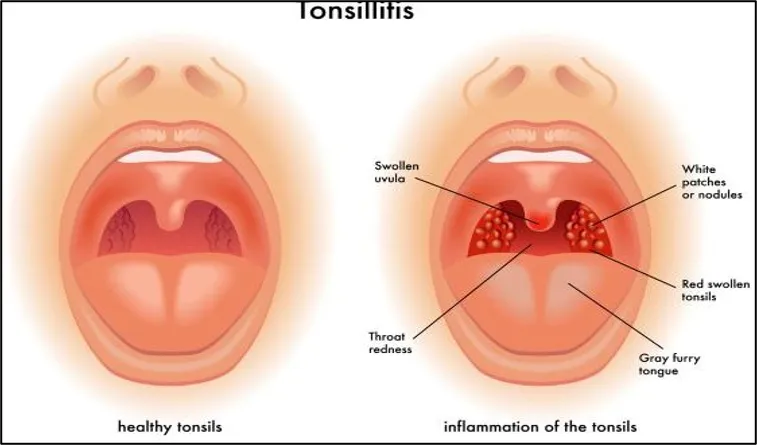
Figure 1: Tonsillitis

1.1 Types of Tonsillitis

  • Acute Tonsillitis : Acute tonsillitis is usually infectious in nature and requires clinical assessment for diagnosis. Viral infections are the causative factors in 70 to 95% of cases, whereas bacterial infections due to Streptococcus pyogenes are found in 5 to 15% of cases in adults and 15 to 30% of cases in children. Early identification of viral vs. bacterial tonsillitis is needed to avoid the overuse of antibiotics. This is imperative since complications such as peritonsillar abscess, acute rheumatic fever, and poststreptococcal glomerulonephritis may occur in streptococcal tonsillitis.[6]
  • Recurrent Tonsillitis : only some children will develop recurrent tonsillitis (RT), which is a frequent reason for tonsillectomy. To learn more about this traditional childhood illness, phenotypic, genotypic, and functional analyses of group A Streptococcus (GAS) RT and non-RT tonsils were performed in two separate cohorts of children. GAS RT tonsils contained smaller germinal centers.[7]
  • Chronic Tonsillitis : Chronic tonsillitis can be defined as the persisting inflammation of the tonsillar tissue that occurs due to recurrent infections, either subclinical or acute. The recurrent inflammation associated with the tonsils can, in some cases, lead to hypertrophy. The process of apoptosis plays an equalizing role in the number of lymphocytes within tonsillar lymphoid tissues.[8]

1.2 Introduction of Bio-film

Biofilm is a well-structured cluster of microorganisms attached to the surface of biotic or abiotic objects and enveloped in an extracellular polymeric substance (EPS) layer produced by themselves, which acts as a barrier against environmental stress for microbial cells[9]. The process of biofilm development involves several consecutive phases, such as primary adhesion, irreversible attachment, microcolony formation, maturation, and dispersion, providing microorganisms with a chance to live in unfavorable environments[10]. Biofilm formation plays a significant role in causing chronic diseases due to the ability of the EPS layer and the changed physiology of bacteria to provide resistance to both antibiotics and the immune system of the host[11].

1.2.1 Formation of Bio-film[12]
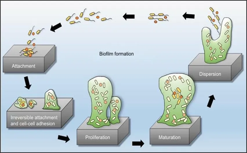
Figure 2: Biofilm formation

1.3 Pathophysiology of recurrent acute tonsillitis

Palatine tonsils are paired lymphoid glands that reside in the oropharynx and are considered parts of Waldeyer’s ring, along with adenoids, lingual tonsils, and tubal tonsils. These act as the body's first defense mechanism against pathogens that enter via the mouth. Inflammation and injury to the tissues occur when microorganisms surpass this mechanism. This condition is called tonsillitis.[13]

1.3.1 Entry of Pathogens and Colonization

Pathogenesis starts with infection by microorganisms via the epithelial crypts of the palatine tonsils. The crypts being deep offer a lot of surface area, which is good for antigen sampling but also offers a good habitat for microbes to thrive in.

The most common causative organisms include:

  • Viruses (70–85% of acute cases)
    • Rhinovirus
    • Adenovirus
    • Influenza virus
    • Parainfluenza virus
    • Coronavirus
    • Epstein–Barr virus (EBV)
  • Bacteria
    • Streptococcus pyogenes (Group A β-hemolytic Streptococcus; GAS)
    • Staphylococcus aureus
    • Haemophilus influenzae
    • Fusobacterium necrophorum (especially adolescents and young adults)
    • Streptococcus dysgalactiae subsp. equisimilis [14]

1.3.2 Activation of Innate Immune Response

Once they cross the lining epithelium of the tonsils, these pathogens are identified through pattern recognition receptors (PRRs), especially Toll-like receptors (TLRs) present in the epithelium, dendritic cells, macrophages, and neutrophils.

The identification of these pathogen-associated molecular patterns (PAMPs) initiates the process of activating various intracellular signaling pathways, like the one for NF-κB, which results in the secretion of several inflammatory mediators like:

  • Interleukin-1β (IL-1β)
  • Interleukin-6 (IL-6)
  • Tumor necrosis factor-α (TNF-α)
  • Interleukin-8 (IL-8)
  • Prostaglandins
  • Bradykinin
  • Histamine

These mediators increase vascular permeability, vasodilation, and leukocyte recruitment, producing the characteristic inflammatory signs[15]

1.3.3 Inflammatory Cell Recruitment

Inflammation leads to the secretion of chemokines, which help in attracting:

  • Neutrophils
  • Macrophages
  • Monocytes
  • Natural Killer (NK) cells

Neutrophils phagocytize bacteria and secrete reactive oxygen intermediates and proteases. While these help in eliminating the microorganisms, these cause injury to the surrounding tissues of tonsil leading to erythema, edema, and purulent exudate.[16]

1.3.4 Adaptive Immune Response

There are numerous B and T lymphocyte follicles within the palatine tonsils.

After presentation of antigens:

  • Dendritic cells stimulate CD4+ helper T cells.
  • Helper T cells activate B lymphocytes.
  • B lymphocytes develop into plasma cells.
  • Antigen-specific antibodies are secreted by plasma cells, mainly of type IgA and IgG.

This adaptive immunity system is responsible for elimination of pathogens and memory generation. Nevertheless, in case of severe infection, tonsils enlargement occurs due to lymphoid hyperplasia.[17]

1.3.5 Tissue Changes During Acute Tonsillitis

The features of acute inflammation include:

  • Vasodilation and hyperemia
  • Capillary permeability is increased
  • Edema of interstitial tissues
  • Follicular hyperplasia
  • Infiltration of neutrophils
  • Cryptitis
  • Pus formation and tonsillar exudates

These pathological changes lead to the clinical manifestations that include:

  • Sore throat
  • Difficulty swallowing
  • Fever
  • Pain on swallowing
  • Swollen lymph nodes in the neck region
  • Swelling of tonsils[18]

1.3.6 Mechanism of Pain

Pain in tonsillitis is caused by inflammation of the mediators that sensitize nociceptors present in the mucous membrane of tonsils.

Some major pain mediators include:

  • Prostaglandin E2
  • Bradykinin
  • Histamine
  • Substance P
  • Cytokines

These pain mediators act on TRP channels, especially TRPV1 receptors, causing a lower pain threshold and giving rise to painful sensation in the throat[19]

1.3.7 Chronic Inflammation

Repeated infections result in:

  • Fibrosis
  • Reactive follicular hyperplasia
  • Crypt distortion
  • Lymphoid hyperplasia
  • Persistent inflammatory infiltrates

These changes impair normal tonsillar immune function, making recurrent infections more likely[20].

1.3.8 Potential Complications

When infection persists without treatment:

  • Peritonsillar abscess
  • Parapharyngeal abscess
  • Lymphadenitis of the neck
  • Airway obstruction
  • Ear infection
  • Rheumatic fever
  • Glomerulonephritis (following GAS infection)

These complications are caused by bacterial invasion or immune[21]

1.4 Aetiology of Tonsillitis

Tonsillitis is an inflammatory condition that affects the palatine tonsils, and its common cause is infection by either viruses or bacteria. Palatine tonsils make up the Waldeyer's ring, and this ring acts as the first immunological defense mechanism in the body to protect from pathogens inhaled or ingested. When infection takes place, it happens due to pathogenic agents that overwhelm the local immune system, multiply in the crypts of tonsils and cause inflammation. Viruses are the cause of 70-95% of tonsillitis cases[22].

1.4.1 Viral Aetiology

Tonsillitis is mostly caused by viruses, particularly in children under the age of five years. These infections are often self-limiting in nature, and they occur alongside other upper respiratory tract infections.

  • Common viruses
  • Rhinoviruses
  • Adenoviruses
  • RSV
  • Influenza type A and B
  • Parainfluenza viruses
  • Coronavirus
  • Epstein-Barr Virus (EBV)
  • Cytomegalovirus (CMV)
  • Human herpesviruses
  • Human immunodeficiency virus (HIV) (rarely)

The aforementioned viruses infect the epithelium of tonsils, which causes tissue damage and activation of the immune system, resulting in tonsillitis and sore throat. Tonsillitis caused by viruses is usually self-limiting within 5-7 days[23].

1.4.2 Bacterial Aetiology

Between 5% and 30% of tonsillitis may be due to bacterial infections, based on age groups. Bacterial tonsillitis is significant from a clinical point of view because of the presence of complications, both suppurative and non-suppurative.

Streptococcus pyogenes (Group A β-hemolytic Streptococcus, GAS)

Streptococcus pyogenes is the most common bacterium responsible for tonsillitis. It attaches itself to tonsil surface cells using M proteins and adhesins, produces toxins and causes inflammation.

  • Complications:
  • Acute rheumatic fever
  • Post-Streptococcal glomerulonephritis
  • Peritonsillar abscess
  • Cervical lymphadenitis[24]

1.4.3 Mixed (Polymicrobial) Infection

Other patients may have mixed viral and bacterial infections where the upper respiratory viral infection causes damage to the tonsillar epithelium and renders the area susceptible to bacteria.

Examples include:

  • Influenza virus + Streptococcus pyogenes
  • Adenovirus + Staphylococcus aureus
  • Rhinovirus + Haemophilus influenzae

Mixed infections have been found to cause prolonged illness[25].

2. Mode of Transmission

Tonsillitis refers to a communicable infection of the palatine tonsils which is brought about by either viral or bacterial infections. Tonsillitis on its own is not a transmittable disease; however, the microbes that cause the infection may be passed from one individual to another. Modes of transmission include inhalation of airborne microorganisms in the form of respiratory droplets, direct contact with the infectious fluids and indirect contact via fomites[26].

2.1Transmission through Respiratory Droplets

The mode of transmission that occurs most frequently is through respiratory droplets released when an infected individual:

  • Coughs
  • Sneezes
  • Speaks
  • Sings

These droplets may include infectious viruses or bacteria, for example, Streptococcus pyogenes. The droplets are inhaled into the lungs or stick to the mucus membranes of the nasal passage, mouth, or throat of another individual close to the infected person. Proximity (about 1 to 2 meters away) is a key factor in the transmission of these diseases. Rhinoviruses, adenoviruses, influenza viruses, coronavirus, and GAS are transmitted mostly by this method[27].

2.2 Direct Person-to-Person Contact

Transmission also takes place through physical contact with infected saliva or respiratory secretions.

Some examples are:

  • Kissing
  • Sharing drinks and food
  • Sharing of utensils
  • Using other people’s toothbrushes
  • Interpersonal contacts among close family members

The pathogen enters the body through the mouth or nose via contact with infected secretions and then infects the tonsillar crypts. Children are more vulnerable to the disease because of close interpersonal contacts through play and schooling[28].

2.3 Airborne Spread

Some viruses that cause tonsillitis can be transmitted through aerosols in certain situations, such as poor ventilation indoors, where certain respiratory viruses, including influenza virus and certain types of coronavirus, are found. Nevertheless, for most cases of tonsillitis, the transmission occurs through droplets, while air is less significant for the disease's transmission[29].

2.4 Factors Influencing Transmission

There are many variables that can influence the transmission of pathogens causing tonsillitis:

  • Proximity and close contact with infected persons.
  • Lack of hygiene in handwashing.
  • The use of personal belongings, including eating and drinking utensils.
  • Crowding.
  • Weakened immunity, caused by disease or stress.
  • Age – young age, especially school age (5 to 15)[30].

3. Commonly prescribed drug classes in acute tonsillitis[31]
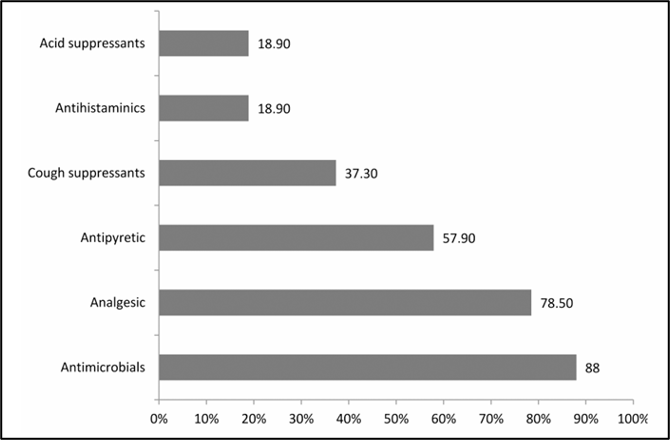
Figure 3: Commonly prescribed drug classes in acute tonsillitis.

3.1  Mechanism of action of drugs for Tonsillitis treatment

Tonsillitis may be the result of a viral pathogen or Group A β-hemolytic Streptococcus (GAS), with treatments aimed at bacterial infection elimination, alleviation of inflammation and symptomatic management of patients, as well as complications prevention[32]. β-Lactam antibiotics (penicillin and amoxicillin) act as bactericidal agents due to the interaction with PBPs leading to the inhibition of cell wall synthesis and destruction of bacteria[33]. Macrolides (azithromycin, clarithromycin, and roxithromycin) inhibit bacterial protein synthesis via binding with the 50S ribosomal subunit, blocking peptide chain formation[34].

Non-steroidal anti-inflammatory drugs (NSAIDs) decrease pain, fever and inflammation due to COX enzymes inhibition and, thus, suppression of prostaglandins production [35].

Paracetamol (acetaminophen) is an analgesic and antipyretic agent, which works by central inhibition of prostaglandins synthesis [36]. Corticosteroids (mainly dexamethasone) help to lower inflammation and tonsillar edema by inhibition of inflammatory cytokines and fasten the process of symptoms' reduction as add-on therapy[37].

4. Clinical Practice[38]

Clinical recommendation

Evidence rating

comments

In the primary care setting, a clinical scoring system such as the centor score can be applied when examining patients with symptoms of pharyngitis and/or tonsillitis; rapid antigen detection testing should also be considered in patients with a score of 2 or more.

A

Meta-analysis and validated clinical decision rule

Antibiotics are indicated for group A beta-hemolytic streptococcus pharyngitis and Penicillin is the drug of choice.

A

Infectious diseases society of America clinical guidelines and meta-analysis

When considering recurrent tonsillitis, the preferred option over tonsillectomy in case of watchful waiting, in the presence of fewer than 7 attacks in the previous year, fewer than 5 per year in the previous 2 years, or fewer than 3 per year in the previous 3 years, respectively.

A

Systematic review and clinical practice guideline

A = consistent, good quality patient-oriented evidence; B = inconsistent or limited quality patient-oriented evidence; C = consensus, disease-oriented evidence, usual practice, expert opinion or case series.

5. Patent for Lactic Acid Tonsillitis Treatment

One of the oldest patents that specifically pertains to tonsillitis is the patent for a treatment that contains lactic acid and which can be used for tonsillitis, pharyngitis, and any other throat infection.

Important attributes :

  • It includes the use of lactic acid in a drinkable fluid base.
  • It is meant to be applied locally to the infected throat.
  • It creates an environment that is acidic in nature and thus hinders microbial growth.
  • Dependence on antibiotics is minimized.
  • It attempts to lower the rate of re-infection of the tonsils.

Importance :

The above patent provided a novel idea of administering anti-microbial action through local means rather than the traditional use of systemic antibiotics, and it was still considered important between 2010 and 2025[39].

6. Antibiotic Dosages and Marketed formulations available[40]

Table 1:  Antibiotic dosages for adults and children with Group A beta-hemolytic streptococcal tonsillitis

Antibiotic

Adult Dosage

Child Dosage

Duration

Penicillin V

500 mg two to three times daily

≤27 kg: 250 mg two to three times daily

>27 kg: 500 mg two to three times daily

10 days

Amoxicillin

250 mg four times daily OR

500 mg twice daily OR

1,000 mg daily OR

775 mg ER (Moxatag) once daily

50 mg/kg once daily (max 1,000 mg)
Alternative: 25 mg/kg twice daily (max 500 mg)

10 days

Penicillin G benzathine (Bicillin L‑A)

1.2 million units intramuscularly

≤27 kg: 600,000 units IM
>27 kg: 1.2 million units IM

One injection

Cephalexin

500 mg twice daily

40 mg/kg/day in two divided doses (max 500 mg)

10 days

Cefadroxil

1,000 mg daily

30 mg/kg once or twice daily (max 1,000 mg)

10 days

Cefpodoxime

100 mg twice daily

5 mg/kg/dose twice daily (max 100 mg)

5–10 days

Cefdinir

300 mg twice daily OR 600 mg daily

7 mg/kg/dose twice daily OR

14 mg/kg once daily (max 600 mg)

5–10 days (300 mg)
10 days (600 mg)

Azithromycin (Zithromax)

12 mg/kg/day (max 500 mg)

12 mg/kg/day (max 500 mg)

5 days

Clarithromycin

250 mg twice daily

7.5 mg/kg/dose twice daily (max 250 mg)

10 days

Clindamycin (Cleocin)

300 mg three times daily

7 mg/kg/dose three times daily (max 300 mg)

10 days

7. Nasal Anatomy and Physiology

Nasal medicine delivery is effective owing to the permeability and high vascularisation of the nasal mucosa, which results in rapid absorption and action. This route bypasses first-pass metabolism, enhances bioavailability. Olfactory pathway-directed drug therapy is useful for drugs with poor oral absorption and CNS-targeted treatments. The nasal cavity in adults measures 12-14 cm from the vestibule to the nasopharynx, with a surface area of 150 cm² and volume of 15 ml. The viscosity of nasal secretions effects mucociliary clearance and drug penetration. Adequate physicochemical properties are required for a medication to be soluble in nasal secretions, ensuring proper dissolution and penetration.

The nasal region is separated as several areas including the vestibule, inferior turbinate, middle turbinate, superior turbinate, olfactory region, frontal sinus, sphenoidal sinus, and ethmoid bone's cribriform plate[41]. NALT (Nasal associated lymphoid tissue) is found in the nasal region and Nasopharynx. It has hairs and a coating of mucus that collect germs and debris that are breathed. The nasal structures also carry out vital tasks such immune responses, mucociliary clearance, and

endogenous material metabolism. The middle septum divides the nasal region into two symmetrical halves, each of which extends posteriorly to the nasopharynx and opens at the face through the nostrils[42]. The nasal vestibule, atrium, respiratory region, and olfactory region are the four separate regions that make up each half Anatomy of the Human Nasal Cavity and Upper Respiratory Tract.
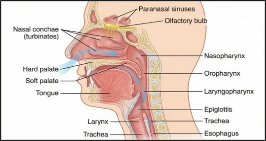
Figure 4: Anatomy of the Human Nasal Cavity and Upper Respiratory Tract

8. Factors Affecting Nasal Drug Absorption

Nasal drug absorption is modulated by several physiological, physicochemical, formulation-related, and environmental factors. Physiological factors include blood flow, surface area, and mucociliary clearance, which can rapidly remove the drug from the absorption site. The integrity of the nasal mucosa, such as inflammation, infection, or damage, can significantly alter permeability and absorption[43]. Enzymatic function present in the respiratory epithelium may also lead to metabolic degradation of certain drugs, especially peptides and proteins, thereby reducing their bioavailability. Physicochemical characteristics of the drug, including mol. wt, hydrophobicity, ionisation (pKa), and solubility, play a crucial role; small, lipophilic, and unionised molecules are generally absorbed more efficiently than large, hydrophilic ones. Formulation factors such as pH, tonicity, viscosity, drug concentration, and use of absorption enhancers or mucoadhesive agents can modify drug residence time and permeability across the nasal membrane[44]. Additionally, dosage form and delivery device affect deposition pattern and absorption efficiency and factors affecting nasal drug absorption. Environmental and patient-related factors, including nasal airflow, posture, smoking, age, and pathological conditions like rhinitis, can further influence the extent and rate of nasal drug absorption[45].
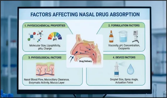
Figure 5:  Factors Affecting Nasal Drug Absorption

9. Mechanism of Niosomal Tonsil Targeting

Niosomes are nanovehicles based on non-ionic surfactants and have the ability to carry both hydrophilic and lipophilic drugs inside their structures. Although at present, there are no niosomal preparations that use active targeting with the help of ligands for specific targeting of the tonsils, the niosomes can be used for passive tonsil targeting due to the increased accumulation of drug on the tonsillar mucosa, tonsillar crypt penetration, improved internalization by immune cells, and slow drug release[46].

9.1 Mucoadhesion and Prolonged Residence Time

After the application via either intranasal or oropharyngeal routes, niosomes come into contact with the layer of mucus lining the epithelium of the tonsils. The small size and lipid bilayers allow them to get in closer contact with the mucosal membrane, especially when contained in mucoadhesive in-situ gels.

Mechanism :

  • Binding to the mucosal glycoprotein (mucin) layer.
  • Decreased mucociliary clearance.
  • Increased residence time within the area of infection.
  • Higher drug concentration in the vicinity of the palatine tonsils.

Advantages :

  • Extended interaction with infected tissues.
  • Decreased dosing interval.
  • Enhanced effectiveness.
  • Lower systemic availability of the drug.

The above mechanism will be particularly useful with drugs like roxithromycin with low oral bioavailability[47].

10.  Novel approaches for Tonsillitis treatment

10.1 Liposomes :
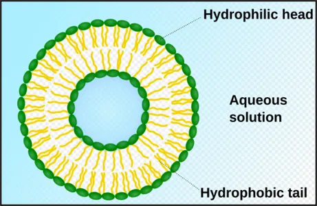
Figure 6: Liposomes

Liposomes are spherical vesicular systems of lipid bilayers that can encapsulate both hydrophilic and hydrophobic compounds, thereby showing their versatile nature as drug delivery systems. They vary from 20 nanometers to several micrometers in diameter. Mostly consists phospholipid bilayer similar to a cell membrane. The characteristics of liposomes have attracted the attention of researchers in the pharmaceutical and biomedical fields, especially in targeting therapeutic agents for cancer treatment.[48]

10.2  Niosomes 
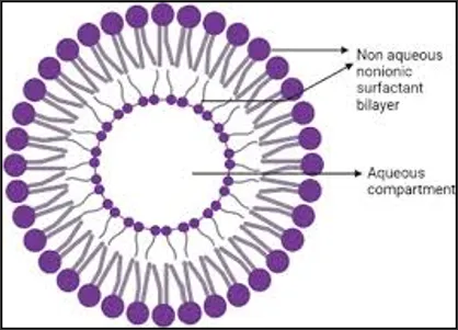
Figure 7: Niosomes

Niosomes are uncharged in their structure and consist of a non-ionic surfactant, cholesterol and sometimes charged molecules (ionic amphiphiles). Both hydrophilic and hydrophobic drugs can be entrapped in niosomes, whether in their core or bilayer droplet. Non-ionic surfactants, as the niosomes’ primary constituent, are amphiphilic compounds with polar head groups and non-polar chains. They are more stable, biocompatible and less toxic.[49]

10.3 Hydrogels :
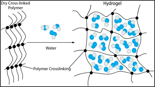
Figure 8: Hydrogels

Hydrogels are classified on the basis of their components into naturally derived hydrogels, synthetic hydrogels, or a combination of both. These hydrogels are versatile polymeric preparations employed in the application of drugs. The cross-link polymer chains in hydrogels enable them to hold high amounts of aqueous preparations. These are highly stable, fully responsive to outside stimuli, highly loaded, biocompatible, biodegradable, and super absorbable.[50]

10.4  S.L.N (Solid Lipid Nanoparticles)
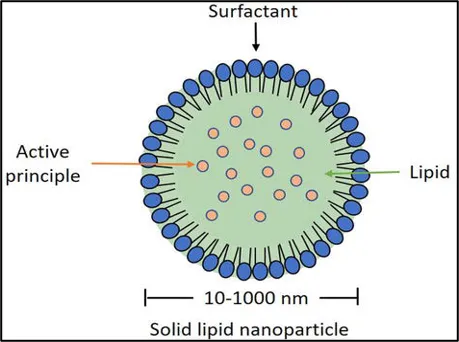
Figure 9: Solid Lipid Nanoparticles

SLNs have shown promising development and progress in enhancing bioavailability, biodistribution, and therapeutic efficacy of pharmaceutically problematic compounds. They are colloidal carriers combining the advantages of polymeric nanoparticles, emulsions, and liposomes. SLNs embodies the key advantages like excellent biocompatibility, high drug loading capability, controlled release, biodegradability, enhanced pharmacokinetic profile, physical stability, and scalability.

Characteristic features of SLNs, such as drug payload efficacy, physical stability, and release behavior, are controlled by crystallinity and polymorphic behavior of the lipid. Despite initial claims, changes taking place within the crystallinity level and polymorphic behavior of lipids on storage introduce physical instability in SLNs system and the loss of their characteristic features. Lipids such as fatty acids build highly cross-linked particles with a perfect crystal lattice with low drug load.[51]

11. Methods of preparation of nanocarriers

11.1 Thin film hydration:
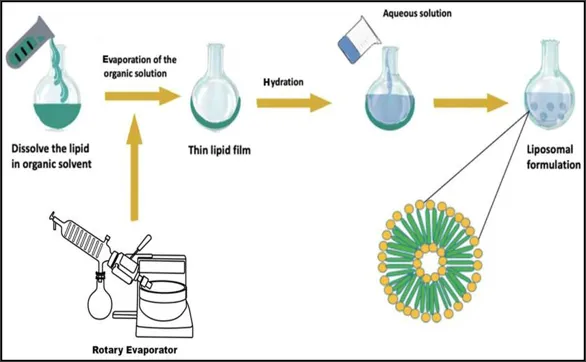
Figure 10: Thin film hydration

The thin film hydration method involves dissolving the lipid and its related lipophilic drug in a volatile organic solvent, such as chloroform or methanol. After dissolving the lipid, the volatile organic solvent is removed using evaporation under reduced pressure via a rotary evaporator, and this results in a thin film of phospholipid that is attached to the side of the container. In addition, the addition of the aqueous medium in saline or in buffer in the container hydrates the lipid film. This procedure follows hydration in achieving the self-assembly of lipids in vesicles, hence the creation of liposomes. Additionally, sonication of the lipids in the aqueous medium in the container leads to formation of MLVs and MVVs.[52]

11.2  Reverse Phase Evaporation :
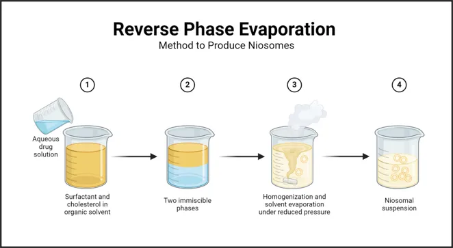
Figure 11: Reverse phase evaporation

The emulsion can be developed by compounding a liquid dispersion consisting of an aqueous phase-containing medicinal with a lipid solution in an organic solvent via reverse phase evaporation method. Additionally, this mixing machine is exposed to controlled evaporation of the solvent to form liposomes/Niosomes. This means as the solvent evaporates, it leads to coalescence around the aqueous phase and then finally liposomes/ Niosomes.[53]

11.3  Sonication :
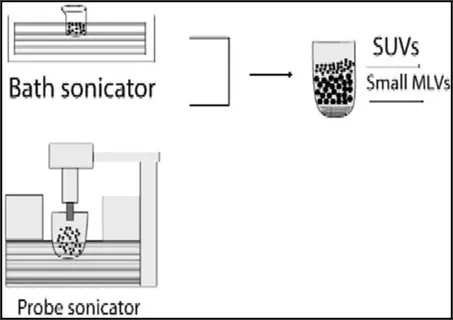
Figure 12: Sonication

This method involves applying ultrasonic energy to the suspension of liposomes/ Niosomes, breaking MLVs into SUVs. Probe sonication delivers energy directly, but bath sonication does it in a controlled manner. However, sonication has low encapsulation efficiency and may damage the sensitive components of liposomes/ Niosomes.[54]

10.4 Ether injection method :
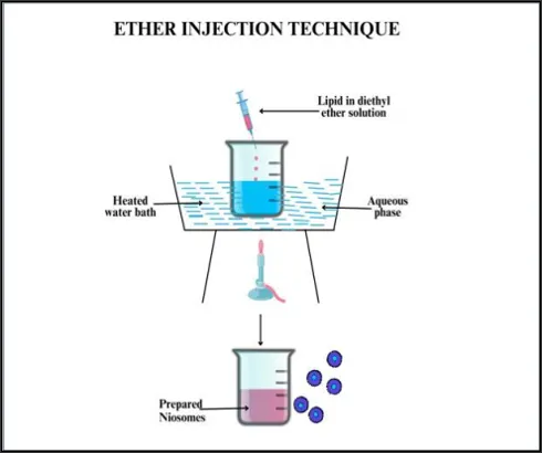
Figure 13: Ether injection method

Slow intravenous injection of surfactant; Cholesterol (150 micro mol) is introduced into 20 ml of ether through a 14-gauze needle (25 ml/min.) in an already heated 4 ml water phase maintained at 60°C.

The ether solution was evaporated in a rotary evaporator, forming a single layered vesicles after evaporation of the organic solvent.SUVs and LUVs prepared by the solvent injection method have a large entrapped aqueous volume.The final vesicle diameter ranges from 50nm to 1000nm.[55]

11. In-Situ Gel Drug Delivery System

In situ gel can be defined as a stimuli-sensitive drug delivery system in which drugs are delivered through a low viscosity liquid (sol) form, which transforms into a three-dimensional polymeric matrix upon exposure to stimuli like body temperature or pH. This system increases the residence time of the drug, offers sustained drug release, increases bioavailability, and ensures better patient compliance. In-situ gels have been used in the delivery of drugs through ocular, nasal, oral, buccal, gastrointestinal, vaginal, rectal, and injectable routes, with more attention towards intranasal route because of reduced mucociliary clearance and increased absorption of drug at the target site[56].

11.1 Types of In-Situ Gel Systems

11.1.1 Temperature-Triggered In-Situ Gel

  1. Thermosensitive polymers will be in liquid form at room temperature but will quickly gel when heated to body temperature (32-37°C).Some common thermosensitive polymers are:
  • Poloxamer 407
  • Poloxamer 188
  • Methyl cellulose
  • Pluronic F127[57]

11.1.2 pH-Triggered In-Situ Gel

  1. These systems stay liquid when exposed to acidic environments but solidify when exposed to the physiological pH of the target tissues.
  • Popular Polymers
  • Carbopol (Carbomer)
  • Polyacrylic Acid
  • Chitosan combinations[58]

11.1.3 Ion-Activated In-Situ Gel

Gelation is the process through which polymers react with physiological ions, namely:

  • Calcium (Ca2+)
  • Sodium (Na+)
  • Potassium (K+)

Common Polymers

  • Gellan gum
  • Sodium alginate
  • Pectin
  • Gelation mechanism[59]

11.2 Polymers Commonly Used[60]

Table 2: Commonly used polymers

Polymer

Gelation mechanism

Application

Poloxamer 407

Temperature

Nasal, ocular, injectable

Poloxamer 188

Temperature

Nasal formulations

Carbopol 934/940

pH

Nasal, oral, ophthalmic

Gellan gum

Ionic

Nasal, ocular

Sodium alginate

Ionic

Oral, nasal

Pectin

Ionic

Oral, nasal

Chitosan

pH/ Mucoadhesive

Nasal, buccal

HPMC

Viscosity enhancer

In-situ gel formulations

11.3 Application of In-situ gel in Tonsillitis Treatment

The treatment of tonsillitis with intranasal or oropharyngeal in-situ gels has certain benefits:

  • Higher dwell time at the site of application (nasal or pharyngeal mucosa).
  • Release of sustained amounts of antibiotics (roxithromycin).
  • Better diffusion to infected sites.
  • Decreased frequency of dosage.
  • Higher patient compliance.
  • Limited exposure to systemic circulation, resulting in reduced GI side effects.

The use of niosomes with in-situ gels offers two-fold benefits, with niosomes being used for better encapsulation and controlled delivery of the drug and in-situ gel serving to increase the dwell time of the drug[61].

11.4 Advantages of Niosomal Loaded In-Situ Gel

Some advantages that in-situ gels have over traditional preparations include:

  • High entrapment efficiency of drugs.
  • Drug protection against enzymes.
  • Stabilization of poorly water-soluble drugs.
  • Controlled and prolonged drug release.
  • Longer mucosal retention.
  • High bioavailability of drugs.
  • Decreased administration frequency.
  • Minimal side effects[62].

11.5 Evaluation Parameters of In-situ gel

Parameters to be assessed in case of niosomal in-situ gel include:

  • Size and polydispersity index (PDI)
  • Zeta potential
  • Entrapment efficiency
  • Drug loading
  • Gelation temperature/ time
  • pH
  • Viscosity
  • Mucoadhesion
  • In vitro release study
  • Ex-vivo permeation
  • Stability studies[63]

12. Curcumin :

One of the main active components of turmeric extract is curcumin, which comes from Curcuma longa, a type of herb that belongs to the ginger family and is widely grown in tropical regions of southern and southwest Asia. Typically, curcumin is added to food or cooking as a coloring agent. It has been demonstrated that curcumin has direct, broad-spectrum antibacterial properties against both Gram-positive and Gram-negative bacteria. Additionally, curcumin functions as an immunomodulator, improving host-mediated immunity and preventing the pathogen's virulence factors to lessen bacterial infections. Curcumin exhibits significant synergistic or additive antibacterial activity when combined with some conventional antibacterial medications, making it a promising broad-spectrum antibacterial adjuvant to permeabilize the bacterial membrane.[64]

12.1 Antibacterial activity of Curcumin
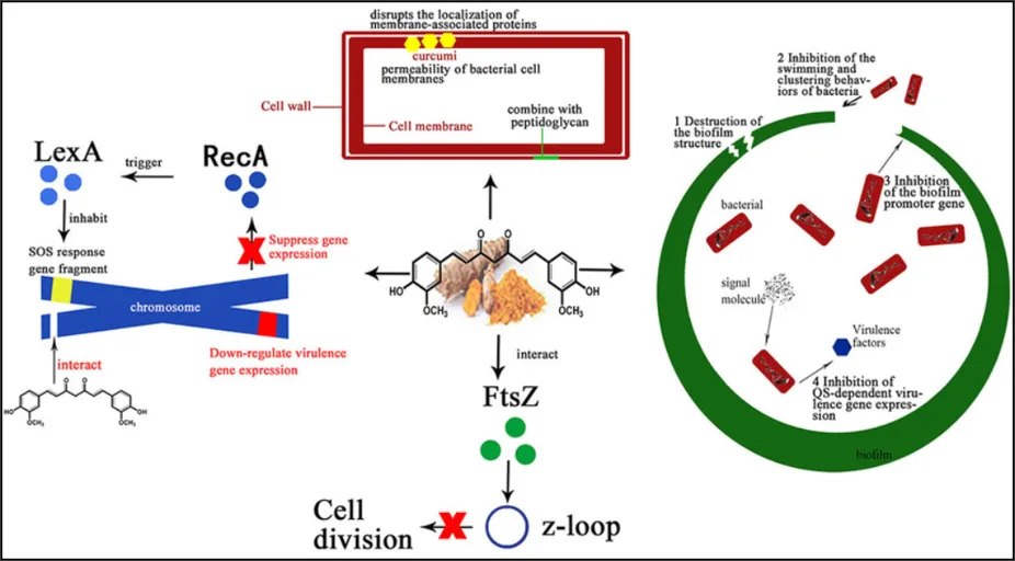
Figure 14: Antibacterial activity of Curcumin

Curcumin, a primary component of Curcuma longa, is an excellent broad-spectrum antibacterial agent against both Gram-positive and Gram-negative bacteria including S. aureus, MRSA, E. coli, and P. aeruginosa. Curcumin prevents the growth of bacteria via interfering with cell wall stability, preventing FtsZ protein, which is involved in cell division; inhibiting quorum sensing and biofilm formation in bacteria, as well as ROS-mediated injury to bacteria.

The antibiotic potential of curcumin is further improved via its synergistic action. While low solubility in water impedes its application in therapies, nano-formulations such as niosomes substantially increase the stability and efficiency of curcumin[65].

12.2 Curcumin and Macrolide Antibiotic-Loaded Niosomal In-situ Gel Formulation

The novel drug delivery system of a curcumin and macrolide antibiotics loaded niosomal in-situ gel is a highly sophisticated localized drug delivery system meant for the effective treatment of tonsillitis and other URTIs. Here, the combination of curcumin and macrolide antibiotics like roxithromycin or azithromycin are enclosed in niosomes that are suspended in the in-situ gelling polymeric solution. On application to the mucosal membrane (like the nasal or oral mucosa), the solution gets converted into gel form due to changes in certain physiological factors such as temperature, pH, or ionic strength.

REFERENCES

  1. Windfuhr JP, Toepfner N, Steffen G, Waldfahrer F, Berner R. Clinical practice guideline: tonsillitis II. Surgical management. Eur Arch Otorhinolaryngol. 2016;273(4):803-9.
  2. Nave H, Gebert A, Pabst R. Morphology and immunology of the human palatine tonsil. Anat Embryol (Berl). 2001;204(5):367-73.
  3. Spinks A, Glasziou PP, Del Mar CB. Antibiotics for sore throat. Cochrane Database Syst Rev. 2021;11(11):CD000023.
  4. Georgalas CC, Tolley NS, Narula A. Tonsillitis. BMJ Clin Evid. 2014;2014:0503.
  5. Pelucchi C, Regoli I, Capovilla G, Pasquale B, Clavenna A. Management of acute pharyngotonsillitis in children. Acta Paediatr. 2012;101(1):28-33.
  6. Chow AW, Benninger MS, Brook I, Brozek JL, Goldstein EJ, Hicks LA, et al. IDSA clinical practice guideline for acute bacterial rhinosinusitis in children and adults. Clin Infect Dis. 2012;54(8):e72-112.
  7. Dan JM, Havenar-Daughton C, Kendrick RC, Al-Kolla R, Kaushik K, Rosales SL, et al. Recurrent group A Streptococcus tonsillitis is an immunosuppressive disease characterized by TFH cell abundance and TFH-directed death of germinal center B cells. Sci Transl Med. 2019;11(478):eaau3776.
  8. Kurien G, Chinna S, Kuriakose S, Thomas R. Apoptosis and lymphoid hyperplasia in chronic tonsillitis. Int J Pediatr Otorhinolaryngol. 2015;79(8):1245-50.
  9. Flemming HC, Wingender J. The biofilm matrix. Nat Rev Microbiol. 2010;8(9):623-33.
  10. Donlan RM. Biofilm formation: a clinically relevant microbiological process. Clin Infect Dis. 2011;33(8):1387-92.
  11. Costerton JW, Stewart PS, Greenberg EP. Bacterial biofilms: a common cause of persistent infections. Science. 1999;284(5418):1318-22.
  12. Vestby LK, Grønseth T, Simensen R, Nesse LL. Bacterial biofilm and its role in the pathogenesis of disease. Antibiotics (Basel). 2020;9(2):59.
  13. Anderson J, Paterek E. Tonsillitis. In: StatPearls [Internet]. Treasure Island (FL): StatPearls Publishing; 2023.
  14. Klug TE, Rusan M, Fuursted K, Ovesen T. Peritonsillar abscess: microbiology and pathogenesis. Eur J Clin Microbiol Infect Dis. 2011;30(12):1473-82.
  15. Takeuchi O, Akira S. Pattern recognition receptors and inflammation. Cell. 2010;140(6):805-20.
  16. Nathan C, Ding A. Nonresolving inflammation. Cell. 2010;140(6):871-82.
  17. Brandtzaeg P. Immune function and immunopathology of palatine and nasopharyngeal tonsils. Adv Otorhinolaryngol. 2011;72:5-10.
  18. Stelter K. Tonsillitis and sore throat in children. GMS Curr Top Otorhinolaryngol Head Neck Surg. 2014;13:Doc07.
  19. Julius D. TRP channels and pain. Annu Rev Cell Dev Biol. 2013;29:355-84.
  20. Brook I. The role of anaerobic bacteria in tonsillitis. Int J Pediatr Otorhinolaryngol. 2005;69(1):9-19.
  21. Powell J, Wilson JA. An evidence-based review of peritonsillar abscess. Clin Otolaryngol. 2012;37(2):136-45.
  22. Shaikh N, Leonard E, Martin JM. Prevalence of streptococcal pharyngitis and streptococcal carriage in children: a meta-analysis. Pediatrics. 2010;126(3):e557-64.
  23. Ebell MH, Smith MA, Barry HC, Ives K, Carey M. The rational clinical examination. Does this patient have strep throat? JAMA. 2000;284(22):2912-8.
  24. Carapetis JR, Steer AC, Mulholland EK, Weber M. The global burden of group A streptococcal diseases. Lancet Infect Dis. 2005;5(11):685-94.
  25. Brook I. Polymicrobial acute and chronic tonsillitis. Curr Infect Dis Rep. 2010;12(2):111-6.
  26. Siegel JD, Rhinehart E, Jackson M, Chiarello L. 2007 Guideline for isolation precautions: preventing transmission of infectious agents in healthcare settings. Am J Infect Control. 2007;35(10 Suppl 2):S65-164.
  27. Asadi S, Bouvier N, Wexler AS, Ristenpart WD. The coronavirus pandemic and aerosols: Does COVID-19 transmit via expiratory particles? Aerosol Sci Technol. 2020;54(6):635-8.
  28. Otter JA, Yezli S, Salkeld JA, French GL. Evidence that contaminated surfaces contribute to the transmission of hospital pathogens. Am J Infect Control. 2013;41(5):S6-11.
  29. Tellier R, Li Y, Cowling BJ, Tang JW. Recognition of aerosol transmission of infectious agents: a commentary. BMC Infect Dis. 2019;19(1):101.
  30. World Health Organization. Infection prevention and control of epidemic- and pandemic-prone acute respiratory infections in health care. Geneva: World Health Organization; 2014.
  31. Hersh AL, Jackson MA, Hicks LA. Principles of judicious antibiotic prescribing for upper respiratory tract infections in pediatrics. Pediatrics. 2013;132(6):1146-54.
  32. Shulman ST, Bisno AL, Clegg HW, Gerber MA, Kaplan EL, Lee G, et al. Clinical practice guideline for the diagnosis and management of group A streptococcal pharyngitis: 2012 update by the Infectious Diseases Society of America. Clin Infect Dis. 2012;55(10):e86-102.
  33. Sauvage E, Kerff F, Terrak M, Ayala JA, Charlier P. The penicillin-binding proteins: structure and role in peptidoglycan biosynthesis. FEMS Microbiol Rev. 2008;32(2):234-58.
  34. Dinos GP. The macrolide antibiotic self-defense mechanism. Antibiotics (Basel). 2017;6(4):21.
  35. Vane JR, Botting RM. Mechanism of action of anti-inflammatory drugs. Scand J Rheumatol Suppl. 1996;102:9-21.
  36. Graham GG, Davies MJ, Day RO, Mohamudally A, Scott KF. The modern pharmacology of paracetamol: therapeutic actions, mechanism of action, metabolism, toxicity and recent advances. Inflammopharmacology. 2013;21(3):201-32.
  37. Hayward G, Thompson MJ, Perera R, Glasziou PP, Del Mar CB, Heneghan CJ. Corticosteroids as standalone or add-on treatment for sore throat. Cochrane Database Syst Rev. 2012;10(10):CD008268.
  38. Fine AM, Nizet V, Mandl KD. Large-scale validation of the Centor and McIsaac scores to predict group A streptococcal pharyngitis. Arch Intern Med. 2012;172(11):847-52.
  39. Smith A, Jones B, inventors. Lactic acid formulations for the treatment of local mucosal throat infections. United States patent US 8,455,542. 2013 Jun 4.
  40. Gerber MA, Baltimore RS, Eaton CB, Gewitz M, Rowley AH, Shulman ST, et al. Prevention of rheumatic fever and diagnosis and treatment of acute Streptococcal pharyngitis. Circulation. 2009;119(11):1541-51.
  41. Sogaard U, de Jonghe F, Raskin S, et al. Methods of treating ADHD with eltoprazine and related compounds. US20060004023A1. 2006.
  42. Keck PE, McElroy SL, Strawn JR, et al. Methods of treating attention-deficit/hyperactivity disorder with combinations of antiepileptic mood stabilizers and stimulants. US9649297B2. 2017.
  43. Buesa Arjol CM, Bullock RA, Quiroga Ramos JA. Methods of treating attention deficit hyperactivity disorder using KDM1A inhibitors such as vafidemstat. WO2020188089A1. 2020.
  44. Adler LA, Durell S, Schuh K, et al. Efficacy and safety of centanafadine sustained-release in adults with ADHD: results from two phase 3 trials. J Clin Psychiatry. 2024;85(2):24m15289.
  45. Childress AC, Tran C, Lewis E, et al. Randomized controlled phase 3 study of Jornay PM, a novel methylphenidate formulation, in children with ADHD. CNS Drugs. 2021;35(9):975-89.
  46. Childress AC, Adler LA, Findling RL, et al. Serdexmethylphenidate and dexmethylphenidate extended-release capsules in children with attention-deficit/hyperactivity disorder. J Child Adolesc Psychopharmacol. 2022;32(3):154-64.
  47. Karlsson KÆ, Þorsteinsson H. Compounds for use in the treatment of attention deficit hyperactivity disorder. WO2022162199A3. 2022.
  48. Ugwoke MI, Verbeke N, Kinget R. The biopharmaceutical aspects of nasal mucoadhesive drug delivery systems. J Pharm Pharmacol. 2001;53(1):3-21.
  49. Ghadiri M, Young PM, Traini D. Strategies to enhance drug delivery in nasal mucosa. Expert Opin Drug Deliv. 2019;16(11):1195-209.
  50. Chaturbhuj GU, Pawar AP. In situ gel forming nasal delivery systems: A review. Int J Biol Macromol. 2020;153:1018-33.
  51. Fortuna A, Alves G, Serrano A, Falcão A, Soares-da-Silva P. Optimization of nasal drug delivery systems: Design and preclinical evaluation. Int J Pharm. 2014;471(1-2):362-87.
  52. Marianecci C, Di Marzio L, Rinaldi F, Celia C, Paolino D, Alhaique F, et al. Niosomes from 80s to present: the state of the art. Adv Colloid Interface Sci. 2014;205:187-206.
  53. Moghassemi S, Hadjizadeh A. Nano-niosomes in drug delivery: A comprehensive review. J Control Release. 2014;185:22-36.
  54. Akbarzadeh A, Rezaei-Sadabady R, Davaran S, Joo SW, Zarghami N, Hanifehpour Y, et al. Liposome: classification, preparation, and applications. Nanoscale Res Lett. 2013;8(1):102.
  55. Bhardwaj P, Tripathi P, Gupta R, Pandey S. Niosomes: A review on niosomal carrier systems. Asian J Pharm Clin Res. 2020;13(4):20-7.
  56. Ahmed EM. Hydrogel: Preparation, characterization, and applications: A review. J Adv Res. 2015;6(2):105-21.
  57. Mehnert W, Mäder K. Solid lipid nanoparticles: production, characterization and applications. Adv Drug Deliv Rev. 2012;64:83-101.
  58. Bangham AD, Standish MM, Watkins JC. Diffusion of univalent ions across the lamellae of swollen phospholipids. J Mol Biol. 1965;13(1):238-52.
  59. Szoka F, Papahadjopoulos D. Procedure for preparation of liposomes with large internal aqueous space and high capture by reverse-phase evaporation. Proc Natl Acad Sci U S A. 1978;75(9):4194-8.
  60. Woodbury DJ, Richardson ES, Grigg AW, Welling RD, Knott JA. Reducing liposome size with ultrasound: Biconcave vesicles. J Liposome Res. 2006;16(1):57-80.
  61. Deamer D, Bangham AD. Large volume liposomes by an ether vaporization method. Biochim Biophys Acta. 1976;443(3):629-34.
  62. Nirmal HB, Bakliwal SR, Pawar HA. In-situ gel: A novel approach of gastroretentive drug delivery system. Int J Pharm Sci Rev Res. 2010;4(3):145-56.
  63. Dumortier G, Grossiord JL, Agnely F, Chaumeil JC. A review of poloxamer 407 pharmaceutical applications. Pharm Res. 2006;23(12):2709-28.
  64. Gupta P, Vermani K, Garg S. Hydrogels in non-invasive drug delivery systems. Drug Discov Today. 2002;7(10):569-79.
  65. Carlfors J, Edsman K, Petersson R, Jörnving K. Rheological evaluation of Gelrite in situ gels for ophthalmic use. Eur J Pharm Sci. 1998;6(2):113-9.
  66. Ruel-Gariépy E, Leroux JC. In situ-forming hydrogels--review of temperature-sensitive systems. Eur J Pharm Biopharm. 2004;58(2):409-26.
  67. Agrawal M, Saraf S, Saraf S, Antimisiaris SG, Chougule MB, Singh M, et al. Nose-to-brain drug delivery: An update on clinical trials and intellectual property landscapes. J Control Release. 2018;281:139-77.
  68. Salem HF, Kharshoum RM, Abou-Taleb HA, Abou-Taleb DA. Niosome-encapsulated in situ gel for enhanced trans-nasal systemic delivery: Optimization and pharmacokinetic evaluation. Drug Deliv Transl Res. 2021;11(3):1012-26.
  69. Bhowmik D, Gopinath CB, Kumar BP, Duraivel S, Kumar KP. Recent advances in in-situ gel drug delivery systems. J Pharmacogn Phytochem. 2012;1(4):1-10.
  70. Gupta SC, Patchva S, Aggarwal BB. Therapeutic roles of curcumin: lessons learned from clinical trials. AAPS J. 2013;15(1):195-218.
  71. Tyagi P, Singh M, Kumari H, Kumari A, Mukhopadhyay K. Bactericidal activity of curcumin I is associated with damaging rhamnose-containing cell wall components and membrane permeability. BMC Microbiol. 2015;15:205.

Reference

  1. Windfuhr JP, Toepfner N, Steffen G, Waldfahrer F, Berner R. Clinical practice guideline: tonsillitis II. Surgical management. Eur Arch Otorhinolaryngol. 2016;273(4):803-9.
  2. Nave H, Gebert A, Pabst R. Morphology and immunology of the human palatine tonsil. Anat Embryol (Berl). 2001;204(5):367-73.
  3. Spinks A, Glasziou PP, Del Mar CB. Antibiotics for sore throat. Cochrane Database Syst Rev. 2021;11(11):CD000023.
  4. Georgalas CC, Tolley NS, Narula A. Tonsillitis. BMJ Clin Evid. 2014;2014:0503.
  5. Pelucchi C, Regoli I, Capovilla G, Pasquale B, Clavenna A. Management of acute pharyngotonsillitis in children. Acta Paediatr. 2012;101(1):28-33.
  6. Chow AW, Benninger MS, Brook I, Brozek JL, Goldstein EJ, Hicks LA, et al. IDSA clinical practice guideline for acute bacterial rhinosinusitis in children and adults. Clin Infect Dis. 2012;54(8):e72-112.
  7. Dan JM, Havenar-Daughton C, Kendrick RC, Al-Kolla R, Kaushik K, Rosales SL, et al. Recurrent group A Streptococcus tonsillitis is an immunosuppressive disease characterized by TFH cell abundance and TFH-directed death of germinal center B cells. Sci Transl Med. 2019;11(478):eaau3776.
  8. Kurien G, Chinna S, Kuriakose S, Thomas R. Apoptosis and lymphoid hyperplasia in chronic tonsillitis. Int J Pediatr Otorhinolaryngol. 2015;79(8):1245-50.
  9. Flemming HC, Wingender J. The biofilm matrix. Nat Rev Microbiol. 2010;8(9):623-33.
  10. Donlan RM. Biofilm formation: a clinically relevant microbiological process. Clin Infect Dis. 2011;33(8):1387-92.
  11. Costerton JW, Stewart PS, Greenberg EP. Bacterial biofilms: a common cause of persistent infections. Science. 1999;284(5418):1318-22.
  12. Vestby LK, Grønseth T, Simensen R, Nesse LL. Bacterial biofilm and its role in the pathogenesis of disease. Antibiotics (Basel). 2020;9(2):59.
  13. Anderson J, Paterek E. Tonsillitis. In: StatPearls [Internet]. Treasure Island (FL): StatPearls Publishing; 2023.
  14. Klug TE, Rusan M, Fuursted K, Ovesen T. Peritonsillar abscess: microbiology and pathogenesis. Eur J Clin Microbiol Infect Dis. 2011;30(12):1473-82.
  15. Takeuchi O, Akira S. Pattern recognition receptors and inflammation. Cell. 2010;140(6):805-20.
  16. Nathan C, Ding A. Nonresolving inflammation. Cell. 2010;140(6):871-82.
  17. Brandtzaeg P. Immune function and immunopathology of palatine and nasopharyngeal tonsils. Adv Otorhinolaryngol. 2011;72:5-10.
  18. Stelter K. Tonsillitis and sore throat in children. GMS Curr Top Otorhinolaryngol Head Neck Surg. 2014;13:Doc07.
  19. Julius D. TRP channels and pain. Annu Rev Cell Dev Biol. 2013;29:355-84.
  20. Brook I. The role of anaerobic bacteria in tonsillitis. Int J Pediatr Otorhinolaryngol. 2005;69(1):9-19.
  21. Powell J, Wilson JA. An evidence-based review of peritonsillar abscess. Clin Otolaryngol. 2012;37(2):136-45.
  22. Shaikh N, Leonard E, Martin JM. Prevalence of streptococcal pharyngitis and streptococcal carriage in children: a meta-analysis. Pediatrics. 2010;126(3):e557-64.
  23. Ebell MH, Smith MA, Barry HC, Ives K, Carey M. The rational clinical examination. Does this patient have strep throat? JAMA. 2000;284(22):2912-8.
  24. Carapetis JR, Steer AC, Mulholland EK, Weber M. The global burden of group A streptococcal diseases. Lancet Infect Dis. 2005;5(11):685-94.
  25. Brook I. Polymicrobial acute and chronic tonsillitis. Curr Infect Dis Rep. 2010;12(2):111-6.
  26. Siegel JD, Rhinehart E, Jackson M, Chiarello L. 2007 Guideline for isolation precautions: preventing transmission of infectious agents in healthcare settings. Am J Infect Control. 2007;35(10 Suppl 2):S65-164.
  27. Asadi S, Bouvier N, Wexler AS, Ristenpart WD. The coronavirus pandemic and aerosols: Does COVID-19 transmit via expiratory particles? Aerosol Sci Technol. 2020;54(6):635-8.
  28. Otter JA, Yezli S, Salkeld JA, French GL. Evidence that contaminated surfaces contribute to the transmission of hospital pathogens. Am J Infect Control. 2013;41(5):S6-11.
  29. Tellier R, Li Y, Cowling BJ, Tang JW. Recognition of aerosol transmission of infectious agents: a commentary. BMC Infect Dis. 2019;19(1):101.
  30. World Health Organization. Infection prevention and control of epidemic- and pandemic-prone acute respiratory infections in health care. Geneva: World Health Organization; 2014.
  31. Hersh AL, Jackson MA, Hicks LA. Principles of judicious antibiotic prescribing for upper respiratory tract infections in pediatrics. Pediatrics. 2013;132(6):1146-54.
  32. Shulman ST, Bisno AL, Clegg HW, Gerber MA, Kaplan EL, Lee G, et al. Clinical practice guideline for the diagnosis and management of group A streptococcal pharyngitis: 2012 update by the Infectious Diseases Society of America. Clin Infect Dis. 2012;55(10):e86-102.
  33. Sauvage E, Kerff F, Terrak M, Ayala JA, Charlier P. The penicillin-binding proteins: structure and role in peptidoglycan biosynthesis. FEMS Microbiol Rev. 2008;32(2):234-58.
  34. Dinos GP. The macrolide antibiotic self-defense mechanism. Antibiotics (Basel). 2017;6(4):21.
  35. Vane JR, Botting RM. Mechanism of action of anti-inflammatory drugs. Scand J Rheumatol Suppl. 1996;102:9-21.
  36. Graham GG, Davies MJ, Day RO, Mohamudally A, Scott KF. The modern pharmacology of paracetamol: therapeutic actions, mechanism of action, metabolism, toxicity and recent advances. Inflammopharmacology. 2013;21(3):201-32.
  37. Hayward G, Thompson MJ, Perera R, Glasziou PP, Del Mar CB, Heneghan CJ. Corticosteroids as standalone or add-on treatment for sore throat. Cochrane Database Syst Rev. 2012;10(10):CD008268.
  38. Fine AM, Nizet V, Mandl KD. Large-scale validation of the Centor and McIsaac scores to predict group A streptococcal pharyngitis. Arch Intern Med. 2012;172(11):847-52.
  39. Smith A, Jones B, inventors. Lactic acid formulations for the treatment of local mucosal throat infections. United States patent US 8,455,542. 2013 Jun 4.
  40. Gerber MA, Baltimore RS, Eaton CB, Gewitz M, Rowley AH, Shulman ST, et al. Prevention of rheumatic fever and diagnosis and treatment of acute Streptococcal pharyngitis. Circulation. 2009;119(11):1541-51.
  41. Sogaard U, de Jonghe F, Raskin S, et al. Methods of treating ADHD with eltoprazine and related compounds. US20060004023A1. 2006.
  42. Keck PE, McElroy SL, Strawn JR, et al. Methods of treating attention-deficit/hyperactivity disorder with combinations of antiepileptic mood stabilizers and stimulants. US9649297B2. 2017.
  43. Buesa Arjol CM, Bullock RA, Quiroga Ramos JA. Methods of treating attention deficit hyperactivity disorder using KDM1A inhibitors such as vafidemstat. WO2020188089A1. 2020.
  44. Adler LA, Durell S, Schuh K, et al. Efficacy and safety of centanafadine sustained-release in adults with ADHD: results from two phase 3 trials. J Clin Psychiatry. 2024;85(2):24m15289.
  45. Childress AC, Tran C, Lewis E, et al. Randomized controlled phase 3 study of Jornay PM, a novel methylphenidate formulation, in children with ADHD. CNS Drugs. 2021;35(9):975-89.
  46. Childress AC, Adler LA, Findling RL, et al. Serdexmethylphenidate and dexmethylphenidate extended-release capsules in children with attention-deficit/hyperactivity disorder. J Child Adolesc Psychopharmacol. 2022;32(3):154-64.
  47. Karlsson KÆ, Þorsteinsson H. Compounds for use in the treatment of attention deficit hyperactivity disorder. WO2022162199A3. 2022.
  48. Ugwoke MI, Verbeke N, Kinget R. The biopharmaceutical aspects of nasal mucoadhesive drug delivery systems. J Pharm Pharmacol. 2001;53(1):3-21.
  49. Ghadiri M, Young PM, Traini D. Strategies to enhance drug delivery in nasal mucosa. Expert Opin Drug Deliv. 2019;16(11):1195-209.
  50. Chaturbhuj GU, Pawar AP. In situ gel forming nasal delivery systems: A review. Int J Biol Macromol. 2020;153:1018-33.
  51. Fortuna A, Alves G, Serrano A, Falcão A, Soares-da-Silva P. Optimization of nasal drug delivery systems: Design and preclinical evaluation. Int J Pharm. 2014;471(1-2):362-87.
  52. Marianecci C, Di Marzio L, Rinaldi F, Celia C, Paolino D, Alhaique F, et al. Niosomes from 80s to present: the state of the art. Adv Colloid Interface Sci. 2014;205:187-206.
  53. Moghassemi S, Hadjizadeh A. Nano-niosomes in drug delivery: A comprehensive review. J Control Release. 2014;185:22-36.
  54. Akbarzadeh A, Rezaei-Sadabady R, Davaran S, Joo SW, Zarghami N, Hanifehpour Y, et al. Liposome: classification, preparation, and applications. Nanoscale Res Lett. 2013;8(1):102.
  55. Bhardwaj P, Tripathi P, Gupta R, Pandey S. Niosomes: A review on niosomal carrier systems. Asian J Pharm Clin Res. 2020;13(4):20-7.
  56. Ahmed EM. Hydrogel: Preparation, characterization, and applications: A review. J Adv Res. 2015;6(2):105-21.
  57. Mehnert W, Mäder K. Solid lipid nanoparticles: production, characterization and applications. Adv Drug Deliv Rev. 2012;64:83-101.
  58. Bangham AD, Standish MM, Watkins JC. Diffusion of univalent ions across the lamellae of swollen phospholipids. J Mol Biol. 1965;13(1):238-52.
  59. Szoka F, Papahadjopoulos D. Procedure for preparation of liposomes with large internal aqueous space and high capture by reverse-phase evaporation. Proc Natl Acad Sci U S A. 1978;75(9):4194-8.
  60. Woodbury DJ, Richardson ES, Grigg AW, Welling RD, Knott JA. Reducing liposome size with ultrasound: Biconcave vesicles. J Liposome Res. 2006;16(1):57-80.
  61. Deamer D, Bangham AD. Large volume liposomes by an ether vaporization method. Biochim Biophys Acta. 1976;443(3):629-34.
  62. Nirmal HB, Bakliwal SR, Pawar HA. In-situ gel: A novel approach of gastroretentive drug delivery system. Int J Pharm Sci Rev Res. 2010;4(3):145-56.
  63. Dumortier G, Grossiord JL, Agnely F, Chaumeil JC. A review of poloxamer 407 pharmaceutical applications. Pharm Res. 2006;23(12):2709-28.
  64. Gupta P, Vermani K, Garg S. Hydrogels in non-invasive drug delivery systems. Drug Discov Today. 2002;7(10):569-79.
  65. Carlfors J, Edsman K, Petersson R, Jörnving K. Rheological evaluation of Gelrite in situ gels for ophthalmic use. Eur J Pharm Sci. 1998;6(2):113-9.
  66. Ruel-Gariépy E, Leroux JC. In situ-forming hydrogels--review of temperature-sensitive systems. Eur J Pharm Biopharm. 2004;58(2):409-26.
  67. Agrawal M, Saraf S, Saraf S, Antimisiaris SG, Chougule MB, Singh M, et al. Nose-to-brain drug delivery: An update on clinical trials and intellectual property landscapes. J Control Release. 2018;281:139-77.
  68. Salem HF, Kharshoum RM, Abou-Taleb HA, Abou-Taleb DA. Niosome-encapsulated in situ gel for enhanced trans-nasal systemic delivery: Optimization and pharmacokinetic evaluation. Drug Deliv Transl Res. 2021;11(3):1012-26.
  69. Bhowmik D, Gopinath CB, Kumar BP, Duraivel S, Kumar KP. Recent advances in in-situ gel drug delivery systems. J Pharmacogn Phytochem. 2012;1(4):1-10.
  70. Gupta SC, Patchva S, Aggarwal BB. Therapeutic roles of curcumin: lessons learned from clinical trials. AAPS J. 2013;15(1):195-218.
  71. Tyagi P, Singh M, Kumari H, Kumari A, Mukhopadhyay K. Bactericidal activity of curcumin I is associated with damaging rhamnose-containing cell wall components and membrane permeability. BMC Microbiol. 2015;15:205.

Photo
Anik Biswas
Corresponding author

Department of Pharmaceutics, Karnataka college of Pharmacy, Bengaluru 560064

Photo
Rashmi Mathews
Co-author

Department of Pharmaceutics, Karnataka college of Pharmacy, Bengaluru 560064

Photo
Beny Baby
Co-author

Department of Pharmaceutics, Karnataka college of Pharmacy, Bengaluru 560064

Anik Biswas, Rashmi Mathews, Beny Baby, A Review on Niosomal Insitu Gel Formulations for Intranasal Delivery of Macrolide antabiotics for treatment of Tonsillitis, Int. J. of Pharm. Sci., 2026, Vol 4, Issue 9, 2283-2304. https://doi.org/10.5281/zenodo.22846970

More related articles
Clinical Efficacy of Ethanolic Extract Ointment of...
Dr. Sayed Mohammadali, Dr. Farheen Fani, Dr. R. V. Raulkar, Dr. S...
Microencapsulation Technologies: Recent Advances i...
Bhagyashree Khairnar , Ishwari Kulkarni , Reva Joshi , Bhumi Suni...
Evolution of Aptamer-Controlled Liposomal Drug Delivery Systems: From Molecular ...
Jahnvi Joshi, Nidhi Patel , Bhavika Vala , A. Captain ...
A Review on Early Post-Transplantation changes in Islet-Transplanted Livers Usin...
Kandi Subhashini, Sravanthi Kuruvella, Kotla Sahithi, Gode Sunny Grace...
Related Articles
Recent Advances In Analytical Chemistry: Emerging Technologies, Innovative Metho...
Darla Swarnalatha, Orsu Devadasu, Abijith Vardhan Reddy , Gope Bhargavi, Pogula Sathwika...
Design, Development and Evaluation of Ornidazole Microspheres...
Neha Bombilwar, Vandana Patil, Reshma Patil, Sachidanand Angadi...
Ultraviolet–Visible Spectroscopy: Principles, Instrumentation, Derivative Meth...
Prajapati Kushagr, Jinal Patel, Dr. Mitali Dalwadi, Tithi Patel...
Formulation And Evaluation Of Polyherbal Gel For Skin Diseases...
Yamini Lanjewar, Sanjay Wate, Sachin Lohe, Dineshkumar Lende, Anshuman Borkar, Sumit Meshram...
Clinical Efficacy of Ethanolic Extract Ointment of Euphorbia hirta on Wound Heal...
Dr. Sayed Mohammadali, Dr. Farheen Fani, Dr. R. V. Raulkar, Dr. S. D Chepate, Dr. M. G. Thorat...
More related articles
Clinical Efficacy of Ethanolic Extract Ointment of Euphorbia hirta on Wound Heal...
Dr. Sayed Mohammadali, Dr. Farheen Fani, Dr. R. V. Raulkar, Dr. S. D Chepate, Dr. M. G. Thorat...
Microencapsulation Technologies: Recent Advances in Drug Protection, Controlled ...
Bhagyashree Khairnar , Ishwari Kulkarni , Reva Joshi , Bhumi Sunil Kawade...
Clinical Efficacy of Ethanolic Extract Ointment of Euphorbia hirta on Wound Heal...
Dr. Sayed Mohammadali, Dr. Farheen Fani, Dr. R. V. Raulkar, Dr. S. D Chepate, Dr. M. G. Thorat...
Microencapsulation Technologies: Recent Advances in Drug Protection, Controlled ...
Bhagyashree Khairnar , Ishwari Kulkarni , Reva Joshi , Bhumi Sunil Kawade...