

We use cookies to ensure our website works properly and to personalise your experience. Cookies policy
1 Assistant Professor, St. Ann's College for Women, Mehdipatnam, Hyderabad, Telangana, India.
2,3,4 Department of Pharmacy, University College of Technology, Osmania University, Hyderabad, Telangana, India
Pancreatic islet transplantations have been investigated in recent times as an appropriate therapeutic approach for type 1 Diabetes Mellitus with the aim of promoting the secretion of insulin from within the body instead of externally administering insulin. In spite of the many developments in the field of transplantation surgery, its long-term effectiveness has not been greatly achieved due to massive graft loss in the first 24 hours after engraftment. Loss of the ?-cells in the first few hours after transplantation can be traced to the instant blood mediated inflammatory response, among other causes. Recent developments in the field of metabolomics have opened up avenues to study these early biological events at a molecular level. High-resolution magic angle spinning nuclear magnetic resonance spectroscopy has been identified as an important technique for studying metabolic variations occurring in transplanted tissues without damaging the tissue architecture. The combination of studies using HRMAS NMR spectroscopy along with histology has shown that islet transplant leads to rapid formation of thrombin, granulocytes, and macrophages. Increased levels of glucose, alanine, glutamate, glutamine, aspartate, and glutathione observed during the early post-transplantation period indicate the presence of hypoxia-induced metabolic adaptation and oxidative stress responses. These findings highlight the complex interplay between inflammatory and metabolic pathways that contribute to graft dysfunction and loss. It is critical to understand such processes in order to design treatment modalities that would optimize islet transplantation, promote engraftment, and improve overall success in cases of T1DM patients. It should be noted that HRMAS NMR based metabolomics provides a great opportunity for the identification of new biomarkers associated with graft viability and transplantation results.
The transplantation of pancreatic islets within the liver is among the possible ways to treat individuals suffering from T1DM [1]. In this technique, islets are transferred into the liver through the portal vein with the aid of local anesthesia to help regain the production of insulin through replacing the lost β-cells [1]. It is worth mentioning that improvements have been witnessed in transplantation results with approximately 44% of the transplanted subjects becoming insulin-free for about 3 years after the procedure [3].
Even with these improvements, modern medical procedures still demand at least two donor pancreases for each patient.3 While 10-20% of the entire islet mass of the pancreas is enough to maintain euglycemia in normal conditions, a significant number of islets die immediately after being transferred [4]. More than half of the transplanted islets are killed during the first few hours after transplantation [5]. Such rejection is primarily due to the immediate blood-mediated inflammatory response (IBMIR), which is an innate immune response that is almost instantaneous once the islets are exposed to the blood [6]. This inflammation response is known to enhance coagulation, complement activation, and inflammation, leading to early graft rejection [7]. Moreover, islet transplantation results in hypoxia owing to damage to the existing blood vessels of the islets [8]. The importance of hypoxia as one of the factors affecting islet viability has long been established [9].
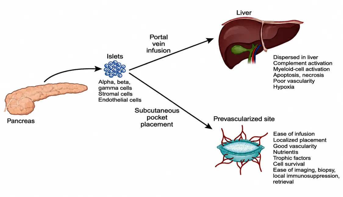
Figure:1. Pancreatic Islet Isolation and Implantation Schematic Diagram Showing the Process of Pancreatic Islets Isolated and Transplanted into the Liver by Infusion Through Portal Vein and Subcutaneous Pre-Vascularized Site, Discussing Strengths and Weaknesses of Each.
In order to make things clearer and also avoid these drawbacks, techniques of metabolomics such as HRMAS NMR spectroscopy have been used widely [10]. This method enables the analysis of intact tissue samples (<20 mg) and gives a complete metabolic profile which is physiologically accurate [15]. The techniques of metabolomics have been used in various phases of transplantation, such as assessing quality of donor organs, preserving the grafts and post-transplantation analysis of biofluids [11].
HRMAS NMR-based metabolomics has provided important insights into the early metabolic response of transplanted islets and the surrounding hepatic tissue. In particular, 1H HRMAS NMR spectroscopy combined with histological analysis has been used to investigate the metabolic and inflammatory events occurring during the first hours following intrahepatic islet transplantation [41]. This approach is advantageous because HRMAS NMR permits the analysis of intact or minimally processed tissue while preserving information about its metabolic state. The technique can simultaneously detect multiple endogenous metabolites, thereby providing a metabolic fingerprint of the tissue response to transplantation [41-43].
Donor pancreas preservation has been assessed using NMR-based metabolomics by measuring the levels of ATP, which serves as a marker for tissue oxygenation and viability [14]. In addition, several other metabolic biomarkers, such as high levels of lactate and lactate/alanine ratio, have been found as markers for hypoxia and low islet viability. Phosphocholine and mobile lipids have been noted as markers for cell death and injury [16]. A low ratio of phosphate diesters to phosphate monoesters has been associated with islet injury and necrosis. Nitric oxide metabolites have been suggested as markers of islet graft rejection [17].
A particularly important application of HRMAS NMR in pancreatic islet transplantation was reported by Vivot et al., who combined 1H HRMAS NMR spectroscopy with histological and immunohistochemical analysis to investigate early post-transplantation responses in islet-transplanted livers [41]. In this study, the investigators examined hepatic tissue during the first 24 h following intrahepatic islet transplantation. Histological analysis demonstrated thrombin formation around transplanted islets together with recruitment of granulocytes and macrophages, providing evidence of an early inflammatory response associated with IBMIR [41]. The combination of metabolic information obtained by HRMAS NMR with tissue-level histological observations therefore provides complementary information regarding the biochemical and morphological responses occurring during early graft injury [41].
The metabolic response observed during the early post-transplantation period was characterized by significant alterations in several metabolites. At approximately 12 h following transplantation, increased levels of glucose, alanine, aspartate, glutamate and glutathione were detected in transplanted liver tissue [41]. These metabolic changes were associated with hypoxia-induced metabolic adaptation, oxidative stress and tissue injury during the early engraftment period [41]. Increased glutathione levels may reflect activation of antioxidant mechanisms in response to oxidative stress, whereas alterations in glucose and amino-acid metabolism may indicate changes in cellular energy metabolism associated with hypoxic and inflammatory conditions [41,44].
While past research has mainly concentrated on transcriptomics, proteomics, and immunohistochemistry in order to study post-transplantation processes, the use of metabolomics presents an alternative and quick method of capturing biochemical alterations [18]. The HRMAS NMR technique allows for quick metabolic profiling and the direct analysis of tissue physiology [15]. When combined with other “omics” strategies, metabolomics adds a layer of knowledge regarding the biology involved in early islet engraftment processes [20].
An important advantage of combining HRMAS NMR metabolomics with histology is that biochemical and morphological information can be evaluated together. The detection of thrombin formation and inflammatory-cell infiltration by immunohistochemistry can be correlated with metabolic changes detected by 1H HRMAS NMR [41]. Such an integrated approach can provide information not only about which metabolites are altered but also about the cellular and inflammatory events occurring simultaneously within the transplanted tissue [41]. This is particularly relevant for understanding the relationship between IBMIR, inflammation, hypoxia, oxidative stress and metabolic adaptation during early graft engraftment.
The metabolic alterations observed following transplantation can provide potential information concerning graft viability and injury. Glucose, alanine, aspartate, glutamate and glutathione represent examples of metabolites that may reflect altered energy metabolism, hypoxic adaptation and antioxidant responses [41]. Other metabolite classes, including lactate, creatine, phospholipid-related metabolites and mobile lipids, may provide additional information concerning cellular energy status, membrane turnover and tissue injury [16,42]. However, individual metabolites should not be interpreted independently because metabolic profiles represent the combined outcome of multiple biochemical pathways. Therefore, multivariate analysis of metabolic patterns may provide a more comprehensive representation of the biological response than analysis of a single metabolite [42-44].
HRMAS NMR also offers several methodological advantages for transplantation research. The technique requires relatively limited sample preparation, permits simultaneous detection of multiple metabolites and can be applied to intact or minimally processed biological tissues [41-43]. The use of magic-angle spinning reduces line broadening arising from anisotropic interactions and improves the resolution of resonances in semi-solid biological samples [42,43]. These characteristics make HRMAS NMR particularly useful for investigating tissue metabolic changes associated with hypoxia, inflammation, oxidative stress and cellular injury.
Nevertheless, HRMAS NMR-based metabolomics has certain limitations. Metabolic profiles can be influenced by tissue collection time, ischemic delay, sample handling, temperature, storage conditions, pH, spinning conditions, water suppression and spectral-processing procedures [42,43]. Appropriate controls and standardized sample-processing protocols are therefore essential for obtaining reproducible results. Furthermore, NMR metabolomics generally identifies metabolic changes rather than providing direct evidence of the molecular mechanism responsible for each alteration. For this reason, integration with histology, immunohistochemistry, transcriptomics, proteomics and biochemical analyses can provide a more comprehensive understanding of transplantation-associated biological processes [20-43].
The integration of metabolomics with other “omics” approaches may therefore provide a more complete understanding of the complex biological processes involved in early islet engraftment [20]. HRMAS NMR can contribute an important metabolic dimension by revealing biochemical alterations associated with inflammation, hypoxia, oxidative stress and tissue injury. Identification of reproducible metabolic signatures may ultimately facilitate the discovery of biomarkers associated with graft viability, early graft injury and transplantation outcome [41-45]. Such biomarkers could be useful for evaluating strategies aimed at reducing early graft loss, improving islet survival and promoting successful revascularization and engraftment.
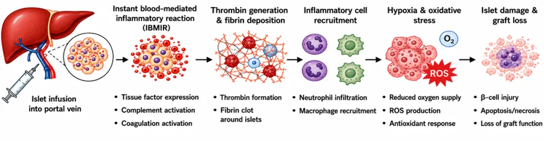
Figure: 2. Mechanism of early islet graft loss following intraportal transplantation.
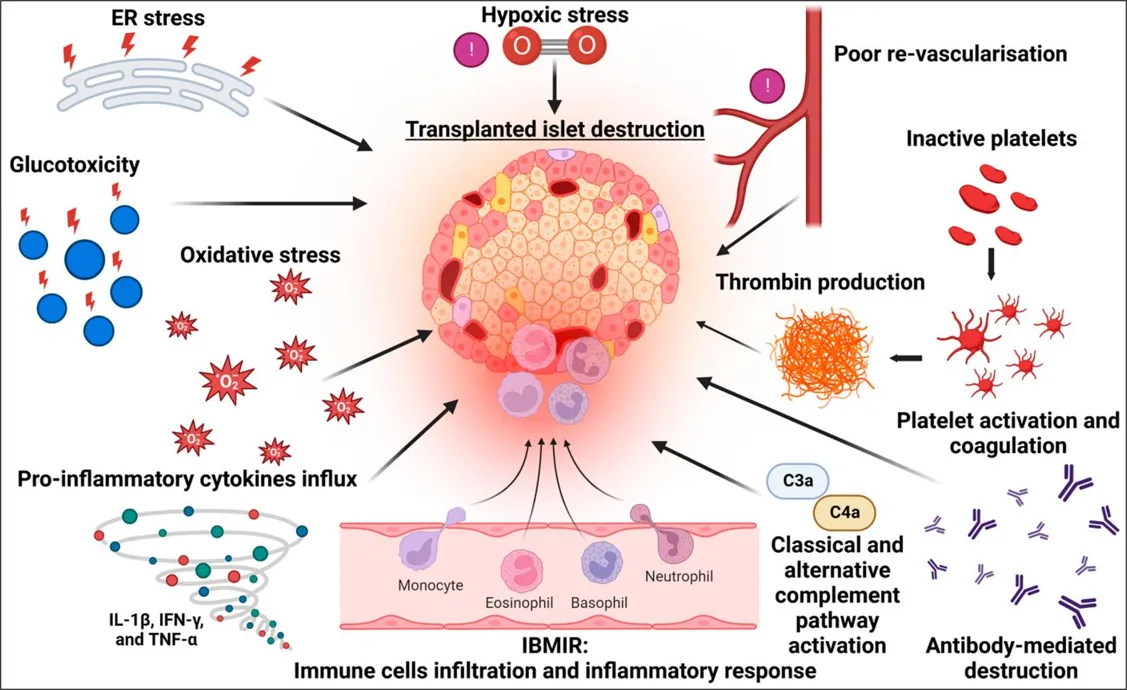
Figure: 3. Schematic Representation of Immune Cell Infiltration and Inflammatory Response Following Pancreatic Islet Transplantation, Showing the Recruitment of Inflammatory Cells and Their Contribution to Early Graft Injury and Islet Loss.
EXPERIMENTAL MODEL AND STUDY DESIGN
The experimental study summarized in this review was conducted in accordance with European Union recommendations for the care and use of laboratory animals (Directive 2007/526/CE) and was approved by the institutional Animal Care and Use Committee (CREMEAS; protocol no. AL/06/35/12/12). Male inbred Lewis rats weighing 225 ± 25 g were used to minimize allogenic immunological rejection and to investigate early islet graft injury following transplantation. Animals were maintained under controlled temperature and humidity conditions with a 12-h light/dark cycle and had free access to standard rodent diet and water. A total of 116 rats were allocated to experimental groups comprising non-diabetic (ND), diabetic control (D), bead-transplanted, and islet-transplanted groups, with an additional donor-islet group. Diabetes was induced by intraperitoneal administration of streptozotocin (75 mg/kg) dissolved in 0.1 M citrate buffer (pH 4.5). Diabetes was confirmed by blood glucose concentrations ≥3 g/L on two measurements using an Accu-Chek glucometer. NPH insulin (4 IU/day) was administered for one week before transplantation to reduce hyperglycemia-associated metabolic toxicity and maintain metabolic stability. Diabetic animals subsequently received either inert dextran beads as a procedural control or pancreatic islets as the transplantation intervention. Animals were sacrificed under isoflurane anesthesia by exsanguination at 0, 2, 4, 8, 12, and 24 h after transplantation, and portal venous blood was collected in heparinized tubes for biochemical analyses. Liver tissue was obtained from the caudate lobe, the principal region of islet engraftment following intraportal transplantation. Tissue specimens were prepared for histological/immunohistochemical and metabolomic analyses; samples intended for NMR analysis were preserved by freezing in liquid nitrogen and stored at −80 °C. These experimental procedures enabled evaluation of early inflammatory responses, IBMIR-associated changes, compromised oxygenation, and metabolic alterations associated with early islet graft injury.
PREPARATION OF SAMPLES FOR IMMUNOHISTOCHEMISTRY AND METABOLOMICS (NMR SPECIMENS)
Liver tissue samples were prepared for histological and metabolomics analyses. One set of samples was fixed in OCT compound, frozen in liquid nitrogen and kept at −80 °C for maintaining metabolism during NMR analysis. The immunohistochemistry test was done on cryosections to detect inflammation, while metabolic analysis of the graft and liver tissue was done by HRMAS NMR spectroscopy. Moreover, important physiopathological mechanisms related to early graft loss have been studied, such as IBMIR, hypoxia due to compromised vascularization, and inflammation caused by hyperglycemia, each one contributing largely to reduced islet viability and functionality.
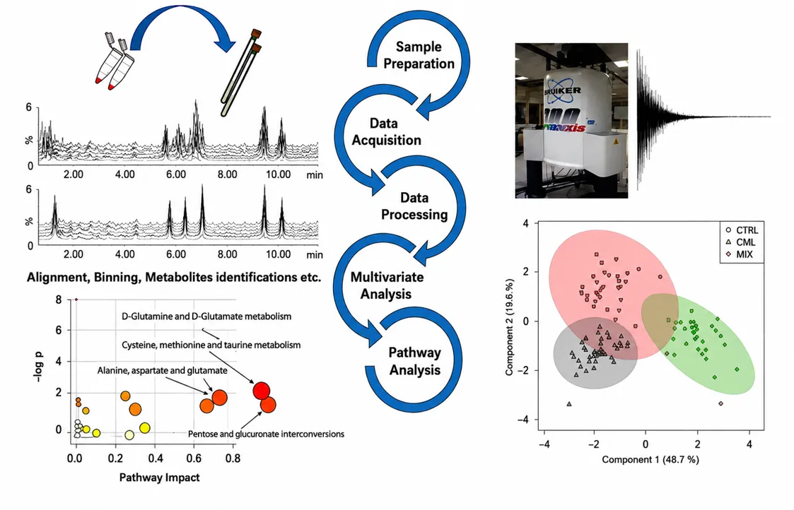
Figure: 4. Workflow of metabolomics in islet isolation, purification and culture.
ISLET ISOLATION, PURIFICATION AND CULTURE
Islets of pancreas were isolated from the donor rats' pancreas using the method of collagenase dissociation and purification. The technique allows successful separation of intact islets from exocrine tissues without damaging their function. Isolated islets were then cultured in M199 media supplemented with 10% FCS (fetal calf serum) and 1% ABAS (antibacterial-antimycotic solution) in a humidified incubator with 5% CO₂ at 37 °C for 24 hours. This period of time helps to eliminate non-viable islets and decrease the level of tissue factor expression to minimize inflammation response after transplantation [6].
INTRAPORTAL ISLET OR BEAD TRANSPORTATION
After seven days of induced diabetes, recipients received intraportal transplantation of islets or control beads. Intraportal injection of islets was conducted in anesthetized rats using inhalation anesthesia by isoflurane to gain access to the abdominal cavity and visualize the liver and the portal vein. Portal flow towards caudate lobes was achieved by temporarily occluding portal blood branches that supply right and left lobes by means of microvascular clamps. A total number of 500 islet equivalents or 500 nonfunctional dextran beads (about 150 µm in size which corresponds to an average pancreatic islet) were mixed in CMRL-1066 medium and injected into the portal vein.
Following the completion of the infusions, the clamps were then taken off, and manual pressure was applied at the point of infusion to reduce bleeding. Warming conditions were created to allow recovery through suturing of the abdominal incision.
SERUM ANALYSES
Systemic inflammation and functional analysis were performed by sampling blood drawn from the portal vein and analysis for markers. The plasma levels of IL-6, α₂-macroglobulin, and C-peptide were quantified using ELISA methodology. IL-6 acts as an indicator of acute inflammation, α₂-macroglobulin as an indicator of the systemic inflammation reaction, and C-peptide as an indicator of insulin release and graft function [26].
IMMUNOHISTOCHEMISTRY
Liver tissues were cut into sections of 7 µm thickness using cryostat and mounted on SuperFrost Plus slides. Sections were fixed with acetone kept at -20°C for 3 minutes to preserve cell structure and antigenicity. Prior to incubating with the primary antibodies, the sections were treated with blocking solutions, i.e., 5% normal goat serum and 0.5% Triton X-100 in phosphate buffer saline (PBS) solution for non-specific site binding.
For immunostaining, sections were treated overnight at 4°C with specific primary antibodies in humidifying conditions. Primary antibodies used were anti-insulin (from rabbit or mouse species as per co-labelling requirement), anti-thrombin, ED1 and HIS48 for detecting macrophages and granulocytes. Incubation of sections with primary antibodies was followed by washing with PBS for 10 minutes.
Sections were subsequently incubated with appropriate fluorescently labelled secondary antibodies. Insulin was detected with goat anti-rabbit or anti-mouse IgG conjugated with Cy3. Thrombin and immune cell markers were detected with Alexa Fluor 488-conjugated secondary antibodies. After final washing steps in PBS, sections were mounted in an aqueous fluorescence-preserving mounting medium to maintain signal integrity during imaging.
This immunohistochemical approach allows visualization and localization of transplanted islets, assessment of thrombotic events and evaluation of inflammatory cell recruitment in liver tissue after transplantation [27].
¹H AND ¹³C HRMAS NMR SPECTROSCOPY STUDIES
High resolution magic angle spinning (HRMAS) nuclear magnetic resonance (NMR) spectroscopy was performed on liver samples obtained from all experimental groups (diabetic (D), non-diabetic (ND), islet-transplanted and bead-transplanted animals). Spectra were collected on a Bruker Avance III 500 spectrometer. Tissue samples (approximately 15-20 mg) were taken from the lower half of the caudate lobe and prepared at -20 °C to preserve metabolic integrity [15].
One-dimensional (1D) 1H NMR spectra were recorded based on a one-pulse sequence followed by a Carr-Purcell-Meiboom-Gill (CPMG) pulse sequence with water presaturation. The CPMG sequence was used to suppress signals of macromolecules such as proteins and lipids with short transverse relaxation time (T2) in order to improve detection of low-molecular-weight metabolites [15].
The inter-pulse delay aimed for was 285 µs and was synchronized with sample spinning to achieve constancy of the acquired NMR data by eliminating signal loss caused by inhomogeneous magnetic fields. 128 scans were collected. Each scan had a spectral width of 14.2 ppm, a 2 s relaxation delay, and an acquisition time of 2.3 s. This resulted in an approximately 10 min acquisition time for each NMR spectrum.
NMR spectral data were processed by Fourier transformation, exponential line broadening function (0.3 Hz) and were loaded into the TOPSPIN software, where phase correction and baseline correction were applied. Chemical shift referencing was done using lactate at 1.33 ppm, with the (CH₃) resonance.
To confirm metabolic identifications, we implemented two-dimensional ¹H-¹³C HSQC NMR with phase-sensitive detection (echo/antiecho) for a set of samples. For the HSQC experiments, 73 ms of acquisition time with a 1.5 s relaxation delay and 116 repetitions per increment for 256 t₁ increments was performed. This created an approximate total HSQC acquisition time of 15 hours. Since tissue degradation could occur with long acquisition times, these 2D experiments were limited to resonance assignments and were not utilized for quantitative measurements.
Metabolic identifications were reported by using chemical shifts as reported in literature and supported by the Human Metabolome Database. A total of 30 metabolites were identified and confirmed using a combination of 1D and 2D spectral analyses. For metabolite quantifications, 1D CPMG spectra were used for all metabolites with the exception of fatty acids, which were analyzed with the one pulse sequence. For the 1D CPMG NMR spectra, the region from 0.5 to 4.7 ppm was divided into 0.01 ppm bins for quantitative analysis and was performed in AMIX, where the spectral area was also processed in MATLAB. The spectral data were also normalized by the sample weight.
Calculations for metabolites were completed from the integration of the spectra relative to a reference lactate solution from a calibrating lactate standard, and were expressed as nmol/mg tissue. Only metabolites with resolved spectral peaks that were free of spectral interference were included, while peaks of ethanol residue (1.23-1.14 ppm) from the surgical procedure were not included.
STATISTICAL ANALYSES
All stated statistics were computed by GraphPad Prism (version 4.03). Results were reported as mean ± standard error of the mean (SEM). This gives a good idea of the variability and precision of the results.
For comparisons between groups: non-diabetic (ND) versus diabetic (D), D versus bead group at time 0, D versus islet group at time 0, and bead versus islet groups at time 0, the non-parametric Mann-Whitney U test was applied. For comparisons of time effects after transplantation, within groups, the one-way analysis of variance (ANOVA I) was used, with the Newman-Keuls test, as a post hoc. Also, islet- and bead-transplanted groups (at the time points) were compared by two-way ANOVA (ANOVA II) with a Bonferroni post hoc test to determine the effect of multiple comparisons. A p value of < 0.05 was considered statistically significant.
For the analysis of the provided metabolomic data, the used one-dimensional HRMAS NMR spectrum was reduced to bins of 0.1 ppm for the segment of 0.5 to 4.7 ppm. The used software was AMIX (3.8). The spectral buckets were integrated and normalized to the total spectral area to limit the effects of concentration of samples. The shaped data were formatted as a matrix and imported to SIMCA for multivariate analysis.
The first step in this analysis was Principal Component Analysis (PCA). PCA was utilized to assess data quality as well as to identify groupings or clusters and outliers prior to any grouping of the data [20]. Following PCA, Partial Least Squares Discrimination Analysis (PLS-DA) helped create an unsupervised statistical model to maximize the difference between experimental groups (i.e., the groups of animals that underwent islet versus bead transplantation) [20]. This method was especially useful for advancing the understanding of the metabolic variation that is associated with the outcomes of different forms of transplantation.
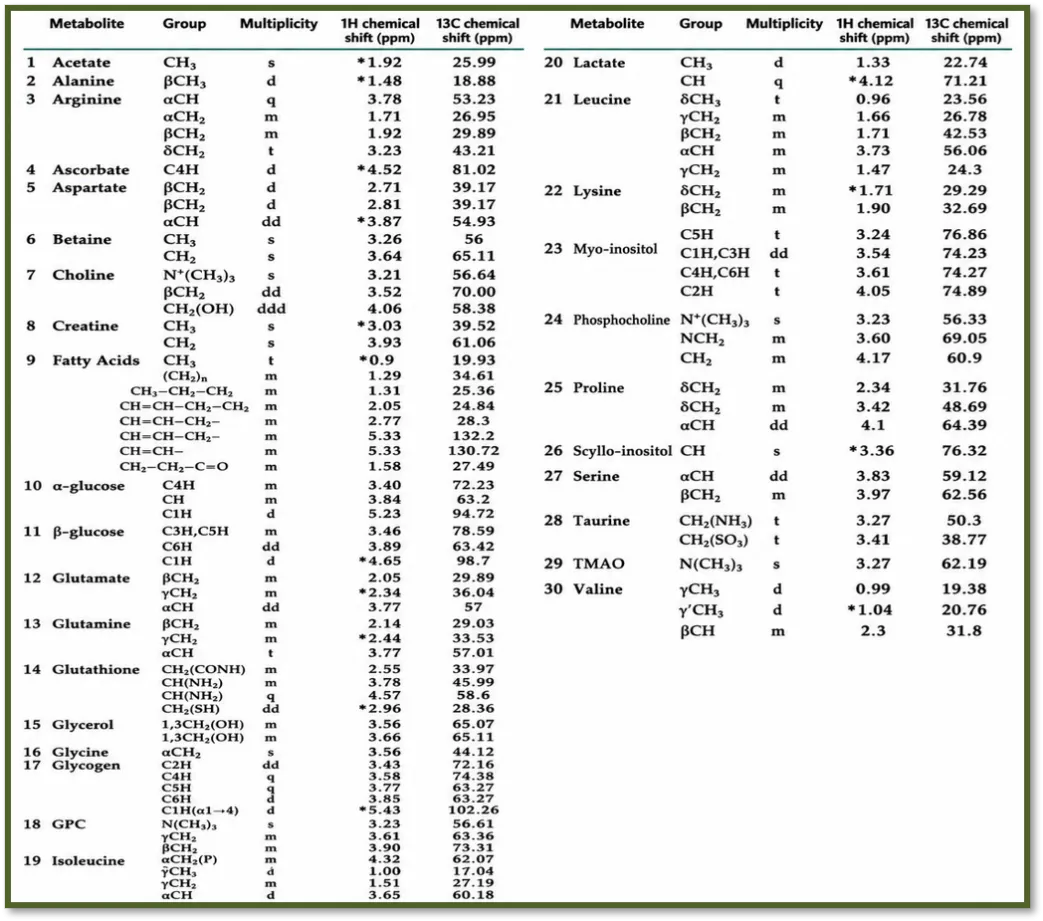
Table 1. Assignment of resonances for metabolites in rat liver tissue from high-resolution magic angle spinning (HRMAS) NMR spectra. Metabolites in bold were measured, and asterisks indicate the center of the integration window. The total choline concentration was quantified by integrating the signal between 3.203.25 ppm. Abbreviations like GPC (glycerophosphocholine), TMAO (trimethylamine-N-oxide), s (singlet), d (doublet), dd (doublet of doublets), dt (doublet of triplets), t (triplet), m (multiplet), ppm (ppm) [40].
ACUTE INFLAMMATORY PROCESSES AFTER ISLET TRANSPLANTATION (FIRST 24 HOURS)
Acute transplant inflammatory reactions in the graft leads to secretion of various pro-inflammatory cytokines (IL-6, which has crucial functions in neutrophils homing into the site of injury) [21]. Effects of IL-6 after islet transplantation are widely known. IL-6 is considered an active mediator for the regulation of the acute inflammatory process, the activation of various immune cell populations, and the inflammation related with islet graft. Furthermore, IL-6 drives the synthesis of acute- phase proteins, such as α2-macroglobulin, in the liver [25]. α2-macroglobulin was reported as a sensitive biomarker of inflammation in rats [23].
Serum levels IL-6 and macroglobulin was determined after infusion of islet graft or beads into rats in order to reflect local and systemic inflammation.
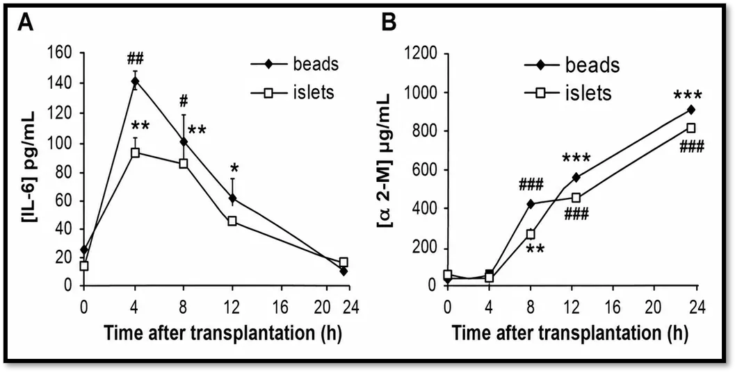
Figure 5. Time-dependent alterations of (A) interleukin-6 (IL-6) and (B) α2-macroglobulin (α2-M) levels after bead or islet cell transplantation into diabetic rats. IL-6 showed a transient increase after transplantation, peaking within the first few hours and thereafter decreasing. In contrast, α2-M levels rose during the whole observation period in both groups. Significant changes from baseline (0h) were analyzed using ANOVA I followed by the Newman-Keuls post hoc test. #P < 0.05, ##P < 0.01, ###P < 0.001 for the bead group; *P < 0.05, **P < 0.01, ***P < 0.001 for the islet group. No statistically significant differences were found when comparing the two groups at different times using ANOVA II [40].
Consistent with prior reports, IL-6 levels showed a transient and significant increase after transplantation in both groups, peaking at ~4 hrs (p<0.01 vs. baseline) and returning to near baseline levels by 24 hrs, reflecting a typical acute phase inflammatory response [24]. Importantly, there was no significant difference in IL-6 levels between bead and islet transplanted groups suggesting that this systemic cytokine elevation was largely procedure related versus islet grafting.
Similarly, α₂-macroglobulin levels increased significantly in both groups starting at 8 hrs post-transplantation (p<0.001), with values continuing to rise through 24 hrs. Again, as with IL-6, there was no significant difference seen between beads and islets over time
These results are consistent with previous studies showing an exaggerated systemic inflammatory response after intraportal infusions [22]. Nevertheless, the similarity across bead- versus islet-transplanted groups suggests this is a consequence of surgery, rather than unique to islet transplantation. Thus, systemic markers of inflammation such as IL-6 and α₂M may not consistently reflect the ‘immediate blood mediated inflammatory reaction’ (IBMIR), which is localized and transient. Previously, we have shown that α2-M can rise dramatically in response to surgical stress alone in rats [23].
C-PEPTIDE EVOLUTION AFTER ISLET TRANSPLANTATION
As C-peptide is well established as a marker of endogenous insulin secretion and is also released with β-cell injury, it is often used to assess islet integrity after transplantation [26]. In our study, plasma C-peptide levels were significantly raised in the islet-transplanted group versus diabetic non-transplanted controls (p<0.01) immediately post-transplant suggesting an immediate release following islet infusion. This initial peak was followed by a fall at 2 hours which then increased, reaching nearly double at 8 hours post-transplantation. Between 2 and 24 hours, C-peptide levels remained significantly raised in the islet-transplanted group compared with controls (p<0.05).
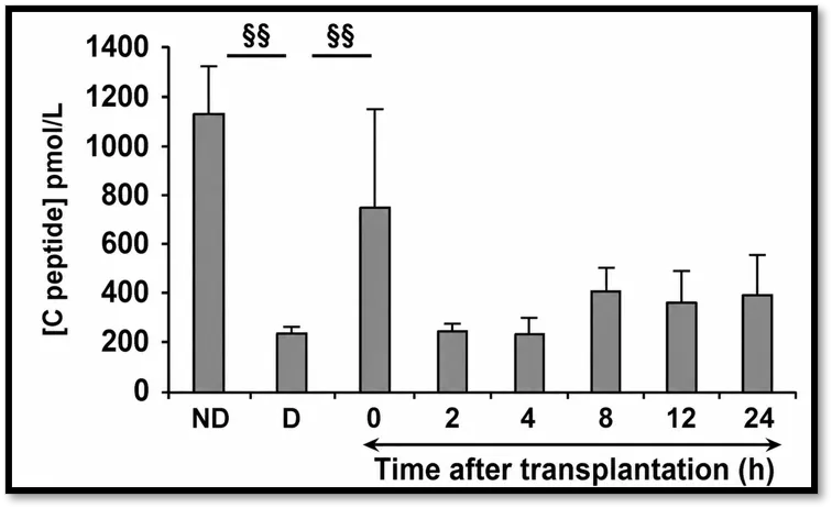
Figure 6. Plasma C-peptide concentrations (mean ± SEM) measured in non-diabetic (ND) rats, diabetic (D) rats, and diabetic rats following islet transplantation over a 24-hour period. C-peptide levels were markedly reduced in diabetic animals compared with non-diabetic controls, whereas transplantation of pancreatic islets significantly restored circulating C-peptide concentrations. Statistical comparisons between the ND and D groups, and between the D group and the 0-hours post-transplantation group were performed using the Mann-Whitney test. The symbol §§ denotes a statistically significant difference (P < 0.01). Temporal changes in C-peptide levels from 2 to 24 hrs after transplantation was assessed using ANOVA revealing a significant effect of time (P < 0.05) [40].
Our results demonstrate a biphasic pattern of C-peptide release after transplantation with an early peak at time 0 and a secondary increase at around 8 h. The quick and intense rise in C-peptide levels is concordant with previous reports and can be explained by mechanical and enzymatic stress-induced damage to islets during isolation and intraportal infusion processes [26]. The later increase at 8h could result from multiple processes such as early islet engraftment and functional insulin release, along with ongoing β-cell destruction due to inflammation and early graft loss.
In total, this biphasic picture highlights the delicate equilibrium between islet damage and repair during the initial post-transplant period and underlines the dynamic nature of graft adaptation within the liver milieu.
IN SITU CHARACTERIZATION OF INFLAMMATORY RESPONSES
To understand what happens specifically at the islet level, current study focuses on what happens in the liver, where intraportal islet engraftment occurs. Exposure of transplanted islets to blood triggers an innate immune response called the instant blood-mediated inflammatory reaction (IBMIR), which plays a crucial part in early graft loss [29]. This reaction involves coagulation activation, resulting in thrombin production, fibrin deposition, and consequently disrupted islet structure. Thrombin is a key player in all of this, acting as a chemoattractant and inducing activation/recruitment of neutrophils and monocytes, which directly kill engrafted islets [27].
To understand the kinetics of IBMIR in the first 24hours post-transplant, immunohistochemical analysis was performed on monitoring thrombin generation concurrently with granulocyte and macrophage recruitment in both islet- and bead-transplanted groups. In the bead group, thrombin could not be detected immediately post-transplant but appeared by 4hrs, accumulating around beads by 12hrs. Conversely, in the islet transplanted group, thrombin was readily detected from within minutes post-transplant and remained elevated throughout the 24hour observation period. Consistent with other studies showing a tight correlation between IBMIR and early thrombin generation [28].
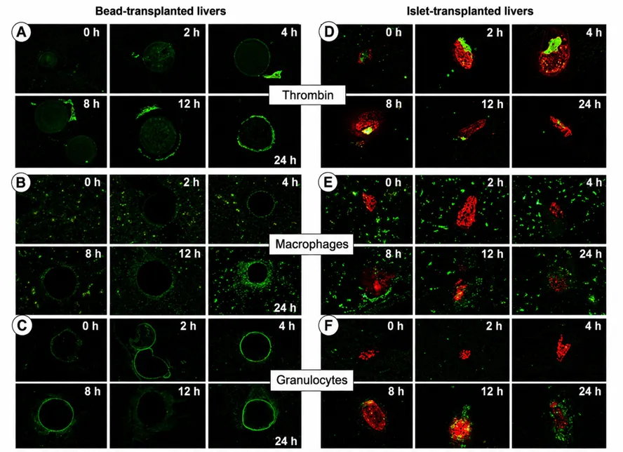
Figure 7. Immunofluorescence images
Figure 7. Immunofluorescence images show: After transplanting dextran beads or pancreatic islets into the liver, researchers tracked how thrombin formed and how immune cells gathered over time. Insulin-positive islets show up as red spots, while any thrombin, macrophages, and granulocytes glow green. They checked liver tissue right after transplantation and then at several points 2, 4, 8, 12, and 24 hours later. In livers with bead transplants, they barely saw any thrombin early on. It only started to show up at the later time points. Macrophages didn’t really show up around the beads until 24 hours had passed, and granulocytes almost never showed up at all. But things were different with the islet transplants. Thrombin appeared quickly, then immune cells started gathering. Macrophages started clustering around the islets as early as 8 hours, and by 12 to 24 hours, both macrophages and granulocytes had swarmed in. The islet transplants triggered a strong local inflammatory response, much more intense than what happened with just the beads. Macrophage distribution showed distinct temporal dynamics between groups. Initially, macrophages were ubiquitously present throughout the liver. In the bead group, macrophage recruitment to the surface of beads was observed only after 24 hours. Conversely, in the islet group, macrophages remained distant from transplanted islets during the first 4 hours, began clustering around islets at 8 hours, and progressively infiltrated the graft from 12 hours onward, with complete infiltration observed at 24 hours [40].
Granulocyte recruitment followed a similar trend. In the group that received beads, there wasn’t much granulocyte activity over 24 hours, which just confirms that the beads didn’t trigger an immune response. But things changed with the islet-transplanted group. Granulocytes started to show up around 8 hours, and by 12 hours, they were really infiltrating the graft. Neutrophils basically the main type of granulocyte jump in first when the innate immune system kicks off and drive a lot of the early inflammation [30]. They also release signals that call in monocytes and macrophages, which cranks up the local inflammatory response even more [31]. Neutrophils and macrophages both share important abilities, like being able to engulf and break down debris. This kind of activity directly damages the transplanted graft [32].
Interestingly, these observations differ from earlier in vitro results by Moberg et al., showing quick neutrophil recruitment within minutes reaching maximum of 2hrs post exposure to pancreatic islets [33]. Such discrepancy can result from the usage of different experimental models; indeed in vitro systems like tubing loop models shed light to very transient interactions but the herein used in vivo system comprises the effect of hepatic environment that severely regulates the inflammatory responses, under a low-oxygen atmosphere where specific cell components (Kupffer cells and sinusoidal endothelial cells) are responsible for non-specific responses [34]. Reduction of liver macrophages have been demonstrated to have an impact on regulating responses of islets inflammation [34].
Importantly, the failure of infiltration of the beads by macrophages and granulocytes serves as an important functional confirmation that the beads are biocompatible and, thus an acceptable control for intraoperative handling and microembolization. This validation confirms that inflammation documented in islet transplants is a result of transplantation itself and not just a consequence of the intraoperative procedure. These results shed significant light on why islets transplanted into the portal vein fail early in transplantation, as an interplay of the inflammatory process and the liver's microenvironment seems to be involved.
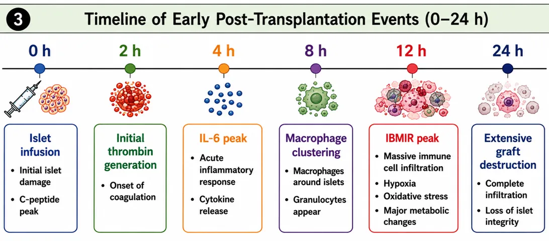
Figure: 8. Timeline of early Post-Transplantation events (0-24h).
METABOLIC PROFILE of POST-TRANSPLANTATION EVENTS
HRMAS NMR spectroscopy of liver tissue at 24 hours after islet transplantation. Each 1H HRMAS NMR spectrum exhibited good spectral resolution with sharply defined resonances allowing ready identification and quantification [15]. Sixteen individual metabolites were quantified to determine initial metabolic changes of post-transplantation events.
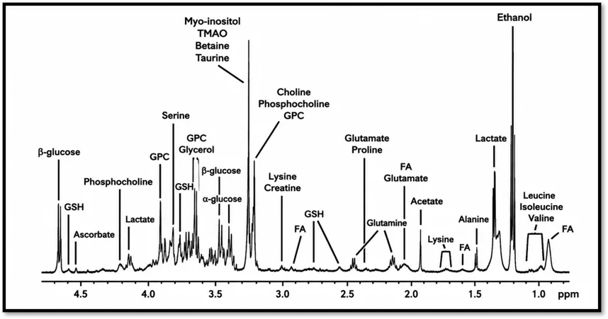
Figure 9. 1H HRMAS NMR spectrum in Carr-Purcell-Meiboom-Gill (CPMG) experiment of the non-diabetic rat liver tissue.
It noted resonances for a number of metabolic products across the spectral region; glucose, amino acids, choline, phospholipids, glutamic acid, taurine, glutamate, fatty acid, and a host of other metabolites are assigned. The large peak attributed to ethanol can be traced back to surgical contamination. Fatty acids; GPC, glycerophosphocholine; GSH, glutathione; TMAO, trimethylamine-N-oxide [40].
The analysis shows significant differences in the amounts of creatine, and fatty acids between the groups ND and D (P<0.05) while the concentrations of both glucose and glycogen in the serum are significantly higher compared with group ND (P < 0.05). It suggests that insulin treatment can sufficiently reduce or ameliorate metabolic disorders caused by the diabetes so only the impacts by the transplanted tissue on rats can be regarded as the main factors to consider in future studies.
Right after transplantation (time 0), the islet-transplanted group showed a big jump in glycogen compared to the bead group (p < 0.01). This matches earlier studies turns out, when you culture pancreatic islets in glucose-rich media, they soak up glycogen over time, especially as glucose concentration goes up [35]. So, the high glycogen levels most likely came from the islets themselves before they were transplanted.
The most dramatic metabolic shifts happened 12 hours after transplantation. When we ran partial least squares discriminant analysis (PLS-DA), it highlighted 12 hours as a key moment, with data showing a strong fit and solid predictive power (R²Y = 0.995; Q² = 0.937) [20].
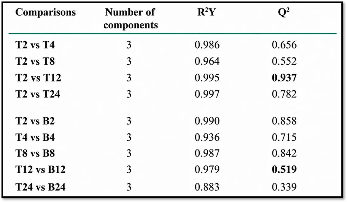
Table 2. Represents the Partial Least Squares-Discriminant Analysis (PLS-DA) to compare the metabolic profiles of livers after they received either pancreatic islets (T) or inert beads (B), checking in at set times from 2 up to 24 hours after transplantation.
Pairwise PLS-DA showed clear shifts in metabolism between groups, and the biggest change happened at 12 hours in the islet-transplanted livers. At that point, the model nailed both fit and predictive ability (T2 vs T12: R²Y = 0.995; Q² = 0.937). We picked the right number of model components with cross-validation. Here, R²Y tells you how well the model explains what’s going on it’s a measure of fit while Q² shows how well the model predicts new data, so it’s about reliability. The closer Q² gets to 1, the more you can trust those predictions. In all this, T stands for the islet-transplanted group, and B means the bead-infused controls [40].
Right now, glucose levels shot up in the islet group compared to the bead group (p < 0.01). This kind of spike usually means the tissue’s breaking down when macromolecules fall apart, things like glucose get set free and show up more in the NMR spectra [13]. People have pointed to interleukin-6 (IL-6) in the past, since it ties into how the body handles glucose during inflammation. But if you look at the timing, the IL-6 levels and the glucose jump don’t really line up. That makes it pretty clear that the glucose rise comes from islet damage itself, not from IL-6 or other cytokine-driven changes [21].
Digging deeper into the metabolism, researchers saw that alanine, aspartate, glutamate, and glutamine levels shot up in the islet-transplanted group after 12 hours. These four are pretty closely linked in how cells process energy, especially when there isn’t much oxygen around. Glutamate and aspartate can actually step in as backup energy sources when oxygen runs low [37]. Alanine tends to pile up when cells ramp up transamination basically, it shows that pyruvate is getting redirected, so instead of making lactate, it’s converted into alanine under low-oxygen conditions [36]. Altogether, these shifts make it clear that transplanted islets face hypoxic stress and adjust their metabolism to cope.
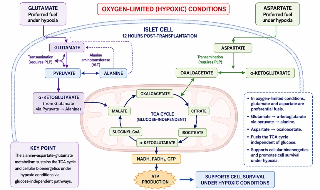
Figure: 10. Alanine-Aspartate-Glutamate metabolic pathway. 12hours after islet transplantation (Oxygen-Limited conditions).
When islets are transplanted, they lose their original blood supply, and the new vessels don't grow fast enough to keep up [8]. That leaves them short on oxygen. Pancreatic β-cells really need steady oxygen, so they start dying off early if they don't get it [39]. On top of that, IBMIR which triggers clotting can actually block the flow of oxygen right to the center of the islets. So, hypoxia ends up playing a big role in early graft failure [29].
Alongside changes in metabolism, glutathione levels shot up in the islet group after 12 hours (p < 0.05). That’s a clear sign the cells cranked up their antioxidant defenses. Glutathione built from glutamate acts as a major antioxidant inside cells and shields them from oxidative stress [38]. Now, the fact that they didn’t detect any oxidized glutathione (GSSG) probably comes down to the method they used. CPMG-based NMR tends to overlook protein-bound forms, so those just didn’t show up. Still, the rise in reduced glutathione (GSH) points to an adaptive response, likely because hypoxia and inflammation are both known to trigger oxidative stress [19].
Earlier studies back this up they’ve shown that nitric oxide (NO) production hits its peak around 12 hours after islet transplantation, which triggers toxic effects on β-cells [17]. Animals that only got beads transplanted didn’t show these changes, so it’s clear these shifts in metabolism are tied to the islet grafts themselves, not just the procedure.
What’s also clear is that these metabolic changes at 12hours don’t last. By 24hours, they’re gone. Immunohistochemistry showed a lot of graft damage by then, which tells us most of the islet tissue is already lost making it hard to pick up any unique metabolic patterns. Really, that 12-hour mark stands out as a key moment when inflammation, low oxygen, and oxidative stress all come together, setting off early graft failure.
CONCLUSION AND FUTURE PERSPECTIVES
Early graft loss is still a big challenge in islet transplantation for Type 1 Diabetes. A lot of transplanted islets get destroyed in just the first 24hours [5]. Evidence keeps piling up that this quick loss happens because of a tangle of inflammation, blood clots, low oxygen, and oxidative stress all mostly set off by what's called the instant blood-mediated inflammatory reaction [29].
Studies in living systems, using both immunohistochemistry and metabolomics, have shed light on what’s actually happening in these early moments. After islets are transplanted, thrombin starts showing up around them, which quickly draws in immune cells like macrophages and granulocytes [27]. These cells flood the area, turning it into a pretty hostile place for the islets. All this inflammation goes hand in hand with metabolic shifts that suggest low oxygen and oxidative stress both making it even harder for the transplanted β-cells to survive [39].
High-resolution magic angle spinning (HRMAS) NMR spectroscopy has become a great tool for real-time metabolic profiling in transplanted tissues [15]. You can actually catch biochemical changes as they happen seeing how the graft responds, whether it’s getting injured or adapting. That means you get a direct look at what’s going on right after the transplant, a window that used to be pretty tough to access [10].
All these findings really drive home the need to focus on early inflammatory and metabolic responses if we want better results after transplantation. When doctors work on controlling IBMIR, boosting oxygen delivery, and supporting antioxidant defenses, islet engraftment and function get a real chance to improve [29]. On top of that, combining metabolomics with immunology opens the door to discovering new biomarkers and therapies that actually predict and shape outcomes [11]. Bringing these different fields together can move islet transplantation forward and give people with Type 1 Diabetes much better long-term results [20].
ACKNOWLEDGEMENT
The authors would like to extend their profound thanks to the Department of Pharmacy, University College of Technology, Osmania University, Hyderabad, for the academic help and facilities provided for writing this review paper.
CONTRIBUTION OF AUTHOR’S
Each author has made substantial contributions to conception and design, acquisition of data, or analysis and interpretation of data; have been involved in drafting the manuscript or revising it critically for important intellectual content; agree to be accountable for all aspects of the work; have given final approval of the version to be published; and have agreed to submit their manuscript to the present journal.
REFERENCES

Gode Sunny Grace, Kandi Subhashini, Kotla Sahithi, Sravanthi Kuruvella, A Review on Early Post-Transplantation changes in Islet-Transplanted Livers Using 1H HRMAS NMR Spectroscopy and Histological Analysis, Int. J. of Pharm. Sci., 2026, Vol 4, Issue 10, 1287-1306. https://doi.org/10.5281/zenodo.23241486
 10.5281/zenodo.23241486