View Article

Abstract

Numerous advantageous substances that are useful in the treatment of various illnesses can be found in plants. Calotropis procera is a versatile plant that can be used for phytoremediation, medicinal, fodder, fuel, lumber, and fibre production. Many conditions, including fever, rheumatism, indigestion, colds, eczema, diarrhoea, boils, jaundice, asthma, and skin infections, have historically been treated using different sections of Calotropis procera. Cardenolides, steroids, glycosides, tannins, terpenoids, phenols, flavonoids, saponins, and alkaloids are among the many beneficial phytochemical substances that are abundant in Calotropis procera. A wide range of pharmacological activities, including analgesic, anti-asthmatic, anti-diabetic, anti-helminthic, anti-inflammatory, anti-microbial, antioxidant, anti-pyretic, anti-ulcer, anticancer, cardioprotective, hepatoprotective, hypolipidemic, immunomodulatory, larvicidal, antifertility, and wound healing activity, have been studied both in vitro and in vivo. This review article analyzes Calotropis procera's ability to heal wounds

Keywords

Calotropis procera, Medicinal plants, Pharmacological properties, Phytochemical Composition, Toxicity, Traditional Uses

Introduction

× Popup Image

Wounds are one of the most common health issues worldwide. Wounds are defined as damage or rupture of the skin tissues, and wound healing is a gradual dynamic process that is classified into four stages: haemostasis, inflammation, proliferation, and tissue maturation or remodelling [1,2,3,4,5]. Wounds are classified into two types based on their causative agents and duration of the healing phases, such as acute and chronic wounds [6,7]. One of the most common health issues in the world is wounds [2]. A wound is an injury or rupture of the skin's tissues, and wound healing is a slow process that includes blood clotting, infection eradication, and tissue repair. The four stages of wound healing, on the other hand, are haemostasis, inflammation, proliferation, and tissue maturation or remodelling [2,3,4,5]. Acute and chronic wounds are two categories of wounds based on the factors that cause them and how long the healing process takes [7].

Neutrophil-derived elastases and proteinase are the fundamental degeneration mediators of the cellular wound matrix, released at the wound site during the inflammation phase [8,9,10]. HNEs play the destructive role by cleaving the surface receptors, interfering with the cell

signalling for the release of the leukocytes at the wound site, and influencing the activation of the liberation of components that derive wound matrix for healing [11,12]. Human neutrophil elastases are essentially a serine proteinase polypeptide glycoprotein. Neutrophils and the pancreas generate and secrete elastase enzymes. HNEs are essential for immunity and resistance against microbial invasion in the human body [10]. The fundamental degeneration mediators of the cellular wound matrix are neutrophil-derived elastases and proteinase, which are produced at the wound site during the inflammatory phase. By cleaving surface receptors and interfering with cell signalling to release leukocytes at the wound site, HNEs play a damaging effect. They also affect the activation of the liberation of components that derive wound matrix for healing [11,12]. Newly regenerated or repaired epithelial cells at the wound site are shielded from excessive hydrolysis as the lesion heals.The use of medicinal plants is undoubtedly a traditional method of healing or treatment of various ailments, and many advancements in the pharmaceutical industry have made the clinical field a very sophisticated one with modern protocols of drug development [13]. The potential of the phytochemicals obtained from the medicinal plants is analysed by formulating the plant extract containing those specific phytochemical constituents for wound healing and using the use of in vivo and in vitro assay techniques. Only 6% of the therapeutic plants are evaluated by scientific biological screening, according to the researchers, who have researched about 15% of the plants for the analysis of vital phytochemical elements. The usage of medicinal plants is undoubtedly a traditional way of mending or treating a variety of illnesses, but recent developments in the pharmaceutical business have made the clinical sector quite complicated with contemporary drug development protocols [14]. By creating a plant extract with those particular phytochemical components for wound healing and using both in vitro and in vivo assay methods, the potential of the phytochemicals derived from medicinal plants is examined.Calotropis procera is a member of the plant family. Because of its pharmacological properties, Asclepiadaceae, commonly referred to as gigantic milk weed, has become more significant in the global health care system of underdeveloped nations. It is a woody, evergreen shrub that grows to a height of roughly three to five meters. This plant is frequently found in South West Asian countries and is widely dispersed in tropical regions [15]. Numerous phytochemical components found in Calotropis procera provide this plant the ability to treat a variety of illnesses. This plant is highly significant for medicinal applications because to the presence of numerous phytochemicals, including calactin, amyrin uscharidin, calotropin, coroglaucigenin, amyrin esters, calotropageni, frugoside, voruscharine, and calotoxin. Because of its therapeutic capabilities, Calotropis procera has thus shown to be an essential plant in the administration of health care. Nowadays, synthetic antimicrobial medications are undoubtedly useful in treating illnesses, but they are more expensive than herbal remedies and have significant adverse effects [16]. Calotropis procera extract possesses insecticidal and antibacterial qualities. Mixtures of flavonoids were found in the alcoholic extract of Calotropis procera leaves, according to physico-chemical examination. The plant's extract contains isolated flavonoids that prevent bacterial growth and shield grains and pulses from insecticidal attacks [17].

POTENTIAL BIOMEDICAL APPLICATIONS   OF      CALOTROPIS PROCERA


Table 1: Taxonomic classification

Kingdom

Plantae

Sub kingdom

Tracheobionta

Super division

Spermatophyta

Division

Magnoliophyte

Class

Magnoliopsida

Subclass

Asteriidae

Order

Gentianales

Family

Asclepiadaceae

Genus

Calotropis

Species

Calotropis procera


Antimicrobial activity:

CALE at minimum inhibitory concentration (MIC) suppressed the development of Escherichia coli, Aspergillus niger, Salmonella typhi, and Candida albicans. On the other hand, CALE had modest antibacterial action against Staphylococcus epidermidis and Escherichia coli, with zones of inhibition of 7.5 ±0.25 mm and 8.0 ±0.05 mm, respectively. The largest zone of inhibition against Candida tropicalis (14.5 ±0.80 mm), followed by Penicillium chrysogenum (12.5 ±0.65 mm), Saccharomyces cerevisiae (12.0 ±0.45 mm),

Candida albicans (11.5 ±0.60 mm), Aspergillus flavus (11.0 ±0.10 mm), and Aspergillus

niger (10.0 ±0.40 mm) [18].

Antidiabetic activity:

Globally, 463 million people were predicted to have diabetes in 2019. It is anticipated that the number of people with diabetes would increase to 578 million by 2030 and 700 million by 2045 [19]. A condition known as hyperglycemia occurs when blood glucose levels rise quickly as a result of intestinal α-glucosidases absorbing glucose and pancreatic α-amylase breaking down starch [20]. Therefore, inhibiting these enzymes that hydrolyse carbohydrates aids in lowering postprandial hyperglycemia and is an essential strategy for controlling diabetes mellitus [21]. In vitro, CALE significantly inhibited α-amylase at a dosage of 15.75 ± 1.05 mg/mL and α-glucosidase at a concentration of 3.25 mg/mL.

Anti-hyperbilirubinemia:

Similar to silymarin, which has been found to have hepatoprotective qualities, phenyl hydrazine and paracetamol-induced hyperbilirubinemia Wistar rat models treated with CALE have demonstrated a decrease in total serum bilirubin levels. The bilirubin-lowering characteristic  of  antioxidant  phytochemicals  in  CALE  is  responsible  for  its anti-hyperbilirubinemia effect, which stabilizes the hepatocyte plasma membrane [22].

Antioxidant Potential:

It is commonly recognized that the development of cancer is directly linked to oxidative damage to DNA [23]. Numerous studies have connected oxidative stress to a number of cardiovascular conditions, including ischemia, hypertension, atherosclerosis, congestive heart

failure, cardiomyopathy, and cardiac hypertrophy [24,25,26,27]. According to reports, oxidative stress leads to the development of ß-amyloid, a toxic peptide that is important in degenerative neurological diseases [28]. By raising NF-kappa B and AP-1 levels, oxidative stress even intensifies the inflammatory process, making pulmonary inflammatory illnesses more complicated [29]. Because oxidative stress produces reactive nitrogen and oxygen free radicals in and around the joints, it also contributes to the development of rheumatoid arthritis [30]. Oxidative stress causes kidney disorders as glomerulonephritis and tubulointerstitial, uraemia, nephritis, proteinuria, cataracts, and age-related macular degeneration [31,32]. By neutralizing these oxidative stressors, antioxidants can prevent a number of diseases [32].

PHYTOCHEMICAL CONSTITUENTS RELATED TO WOUND HEALING

The wound healing activity of Calotropis procera is associated with the presence of several biologically active phytochemicals. Different extracts of the plant contain flavonoids, tannins, alkaloids, triterpenoids, saponins, glycosides, and phenolic compounds [33].

Flavonoids are important antioxidants that neutralize reactive oxygen species generated during tissue injury. By reducing oxidative stress, these compounds protect cells and promote tissue repair [34]. Tannins contribute to wound contraction and stabilization of newly formed tissues by precipitating proteins and enhancing collagen formation [35]. Saponins and triterpenoids exhibit antimicrobial and anti-inflammatory activities that reduce microbial contamination and inflammation at the wound site [36]. The latex of Calotropis procera also contains proteolytic enzymes and glycosides that participate in the removal of necrotic tissue and support faster healing [37]. The synergistic action of these phytoconstituents contributes significantly to the therapeutic effect of the plant in wound management.


PHASES         OF      WOUND        HEALING     AND    ROLE OF            CALOTROPIS PROCERA

i.          Haemostasis Phase:

Immediately after injury, blood clotting occurs to prevent excessive blood loss. Platelets aggregate and release growth factors that initiate tissue repair. Although Calotropis procera does not directly influence coagulation, its bioactive constituents help protect the damaged tissue from infection and oxidative stress during the early stage of healing [38].

ii.         Inflammatory Phase:

Inflammation is characterized by migration of neutrophils and macrophages to the wound site for removal of debris and microorganisms. Excessive inflammation may delay healing and increase tissue damage. Studies indicate that Calotropis procera suppresses inflammatory mediators and reduces edema, thereby improving the healing environment [39].

iii.       Proliferative Phase:

During this phase, fibroblasts synthesize collagen and extracellular matrix proteins, while angiogenesis and epithelialization occur simultaneously. Experimental studies demonstrate that Calotropis procera increases fibroblast proliferation, granulation tissue formation, and collagen deposition, resulting in the faster and early wound contraction in the body [40].

iv.        Remodeling Phase:

In the final phase, collagen fibers are reorganized and tensile strength of the tissue improves. Histological studies have shown improved collagen alignment and reduced scar formation in wounds treated with Calotropis procera extracts [41].


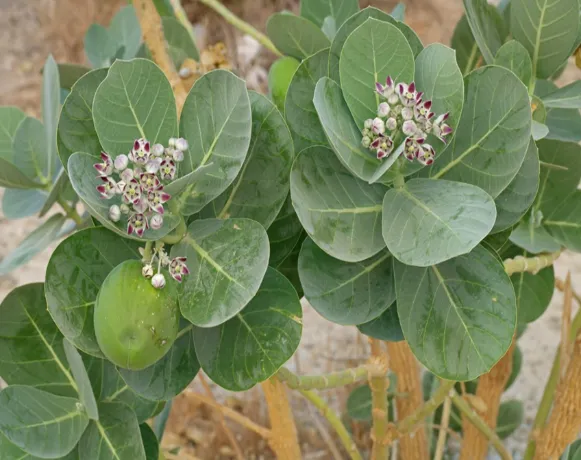
Fig 1: Calotropis procera [14]

MECHANISMS INVOLVED IN WOUND HEALING

i.          Antioxidant Mechanism:

Reactive oxygen species generated at the site of injury can damage cellular proteins, lipids, and DNA, thereby delaying healing. Antioxidant compounds present in Calotropis procera reduce oxidative stress by scavenging free radicals and protecting tissues from oxidative damage and thus preventing the body from further oxidative procedure inside the body [42].

ii.         Anti-inflammatory Mechanism:

Inflammation is necessary for wound repair, but prolonged inflammation can impair tissue regeneration. Calotropis procera inhibits inflammatory pathways and reduces the production of inflammatory mediators such as prostaglandins and cytokines [43]. Reduction in inflammation promotes faster tissue regeneration and minimizes tissue destruction.

iii.       Antimicrobial Mechanism:

Microbial infection is a major factor responsible for delayed wound healing. The plant exhibits antibacterial activity against common wound pathogens including Staphylococcus aureus and Pseudomonas aeruginosa. This antimicrobial effect helps maintain a clean wound environment and which helps to accelerates the tissue repair mechanism of the body [44].

iv.        Collagen Formation and Tissue Regeneration:

Collagen synthesis is essential for restoration of tissue strength and integrity. Studies have reported increased hydroxyproline content and enhanced collagen maturation in wounds treated with Calotropis procera extracts [45]. The plant also promotes angiogenesis and epithelialization, which are essential for complete wound closure.


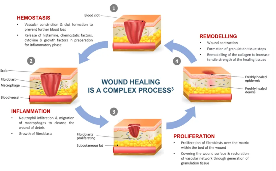
Fig 2: Wound healing Process [15]


EXPERIMENTAL EVIDENCE OF WOUND HEALING ACTIVITY

Several animal studies have confirmed the wound healing potential of Calotropis procera. Ethanolic leaf extracts have shown significant reduction in wound area and faster epithelialization in excision wound models [46]. Researchers observed improved granulation tissue formation and increased tensile strength in animals treated with topical preparations containing Calotropis procera latex [47]. Histopathological studies demonstrated increased fibroblast proliferation, collagen deposition, and neovascularization in treated wounds compared with untreated control groups [48]. Enhanced hydroxyproline content further confirmed the stimulation of collagen synthesis. In incision wound models, treated animals showed faster tissue remodeling and better wound contraction [49]. The therapeutic effectiveness of Calotropis procera has also been associated with reduction in microbial growth and oxidative tissue injury. These findings support the traditional application of the plant in wound care and tissue repair [50].

FORMULATION APPROACHES IN WOUND MANAGEMENT

Different formulations containing Calotropis procera have been investigated for wound treatment. Ointments, creams, gels, and latex-based preparations are commonly studied dosage forms. Topical application allows direct delivery of bioactive compounds to the wound site and enhances local therapeutic action [51]. Recent advances in herbal drug delivery systems have encouraged the development of hydrogel dressings, nanoparticle-based systems, and bioactive wound films containing plant extracts. These modern formulations may improve stability, controlled release, and penetration of active constituents [52].

Standardization of herbal formulations is important to ensure reproducible therapeutic effects and safety. Optimization of extraction methods and identification of active compounds may further improve the medicinal value of Calotropis procera in wound care applications.

SAFETY AND LIMITATIONS

Although Calotropis procera demonstrates promising wound healing activity, improper use of concentrated latex may produce skin irritation and toxicity due to the presence of cardiac glycosides [53]. Therefore, controlled topical application and proper formulation development are essential for safe therapeutic use. Another limitation is the lack of large-scale clinical studies evaluating the efficacy and safety of Calotropis procera in human subjects. Most available evidence is based on experimental animal studies. Additional pharmacological and toxicological investigations are required before widespread clinical application [54].

FUTURE PERSPECTIVES

Future research on Calotropis procera should focus on isolation and characterization of specific phytoconstituents responsible for wound healing activity. Molecular studies investigating the regulation of growth factors, cytokines, collagen synthesis, and angiogenesis may provide deeper understanding of the mechanisms involved [55].

Clinical trials are necessary to establish therapeutic efficacy and safety in human wound management. Integration of Calotropis procera into advanced wound dressings and herbal biomaterials may open new possibilities for cost-effective and efficient wound care products

[56].

CONCLUSION

Calotropis procera possesses remarkable wound healing properties supported by traditional knowledge and experimental evidence. The plant promotes tissue repair through antioxidant, antimicrobial, anti-inflammatory, and collagen-stimulating activities. Studies have demonstrated accelerated wound contraction, improved epithelialization, enhanced granulation tissue formation, and increased collagen deposition following treatment with Calotropis procera extracts and latex formulations. Despite encouraging results, further clinical studies and formulation standardization are necessary to establish its therapeutic safety and efficacy in humans. Calotropis procera remains a promising natural source for the development of modern herbal wound healing therapies.

REFERENCES

  1. Petrovska BB. Historical review of medicinal plants’ usage. Pharmacogn Rev. 2012;6(11):1-5.
  2. Zhu Y, Zhang J, Song J, Yang J, Xu T, Pan C, et al. Zwitterionic hydrogels promote skin wound healing. J Mater Chem B. 2016;4(31):5105-11.
  3. Thakur R, Jain N, Pathak R, Sandhu SS. Practices in wound healing studies of plants. Evid Based Complement Alternat Med. 2011; 2011:438056.
  4. Guo S, DiPietro LA. Factors affecting wound healing. J Dent Res. 2010;89(3):219-29.
  5. Broughton G 2nd, Janis JE, Attinger CE. Wound healing: an overview. Plast Reconstr Surg. 2006;117(7 Suppl):1e-S–32e-S.
  6. Percival NJ. Classification of wounds and their management. Surg Infect (Larchmt). 2002;20(2):114-7.
  7. Tottoli EM, Dorati R, Genta I, Chiesa E, Pisani S, Conti B. Skin wound healing process and new emerging technologies for skin wound care and regeneration. Pharmaceutics. 2020;12(8):735.
  8. Siedle B, Hrenn A, Merfort I. Natural compounds as inhibitors of human neutrophil elastase. Planta Med. 2007;53(5):401-20.
  9. Marinaccio L, Stefanucci A, Scioli G, Della Valle A, Zengin G, Minosi P, et al. Peptide human neutrophil elastase inhibitors from natural sources: an overview. Int J Mol Sci. 2022;23(5):2924.
  10. Mustafa Z, Zhanapiya A, Kalbacher H, Burster T. Neutrophil elastase and proteinase 3 cleavage sites are adjacent to the polybasic sequence within the proteolytic sensitive activation loop of the SARS-CoV-2 spike protein. ACS Omega. 2021;6(10):7181-5.
  11. Pham CT. Neutrophil serine proteases: specific regulators of inflammation. Nat Rev Immunol. 2006;6(7):541-50.
  12. Henriksen PA. The potential of neutrophil elastase inhibitors as anti-inflammatory therapies. Curr Opin Hematol. 2014;21(1):23-8.
  13. Shakya AK. Medicinal plants: future source of new drugs. Int J Herb Med. 2016;4(4):59-64.
  14. Khan H. Medicinal plants need biological screening: a future treasure as therapeutic agents. Biol Med. 2014;6(3):1-3.
  15. Boutraa T. Growth performance and biomass partitioning of the desert shrub Calotropis procera under water stress conditions. Res J Agric Biol Sci. 2010;6(1):20-6.
  16. Ali-Seyed M, Ayesha S. Calotropis-a multi-potential plant to humankind: special focus on its wound healing efficacy. Biocatal Agric Biotechnol. 2020; 28:101725.
  17. Mendki PS, Maheshwari VL, Kothari RM. Antimicrobial and insecticidal activities of flavonoid glycosides from Calotropis procera L. for post-harvest preservation of pulses. Biopestic Int. 2005;1(1-2):4-12.
  18. Nenaah EG, Ahmed ME. Antimicrobial activity of extracts and latex of Calotropis procera (Ait.) and synergistic effect with reference antimicrobials. Res J Med Plant. 2011;5(6):706-16.
  19. Saeedi P, Petersohn I, Salpea P, Malanda B, Karuranga S, Unwin N, et al. Global and regional diabetes prevalence estimates for 2019 and projections for 2030 and 2045: results from the International Diabetes Federation Diabetes Atlas, 9th edition. Diabetes Res Clin Pract. 2019; 157:107843.
  20. Deshpande MC, Venkateswarlu V, Babu RK, Trivedi RK. Design and evaluation of oral bioadhesive controlled release formulations of miglitol, intended for prolonged inhibition of intestinal alpha-glucosidases and enhancement of plasma glucagon like peptide-1 levels. Int J Pharm. 2009;380(1-2):16-24.
  21. Hirsh AJ, Yao SY, Young JD, Cheeseman CI. Inhibition of glucose absorption in the rat jejunum: a novel action of alpha-D-glucosidase inhibitors. Gastroenterology. 1997;113(1):205-11.
  22. Patil RA, Makwana AB. Anti-hyperbilirubinemia and wound healing activity of aqueous extract of Calotropis procera leaves in Wistar rats. Indian J Pharmacol. 2015;47(4):398-402.
  23. Valko M, Rhodes CJ, Moncol J, Izakovic M, Mazur M. Free radicals, metals and antioxidants in oxidative stress-induced cancer. Chem Biol Interact. 2006;160(1):1-40.
  24. Bahorun T, Soobrattee MA, Luximon-Ramma V, Aruoma OI. Free radicals and antioxidants in cardiovascular health and disease. Internet J Med Update. 2006;1(2):1-17.
  25. Droge W. Free radicals in the physiological control of cell function. Physiol Rev. 2002;82(1):47-95.
  26. Ceriello A. Possible role of oxidative stress in the pathogenesis of hypertension. Diabetes Care. 2008;31 Suppl 2: S181-4.
  27. Butterfield DA, Boyd-Kimball D. Amyloid beta-peptide (1-42) contributes to the oxidative stress and neurodegeneration found in Alzheimer disease brain. Brain Pathol. 2004;14(4):426-32.
  28. Hoshino Y, Mishima M. Redox-based therapeutics for lung diseases. Antioxid Redox Signal. 2008;10(4):701-4.
  29. Valko M, Leibfritz D, Moncol J, Cronin MT, Mazur M, Telser J. Free radicals and antioxidants in normal physiological functions and human disease. Int J Biochem Cell Biol. 2007;39(1):44-84.
  30. Galle        J.         Oxidative        stress   in chronic        renal    failure. Nephrol     Dial     Transplant. 2001;16(11):2135-7.
  31. Santosa S, Jones PJ. Oxidative stress in ocular disease: does lutein play a protective role? CMAJ. 2005;173(8):861-2.
  32. Bai N, He K, Roller M, Lai CS, Shao X, Pan MH, et al. Flavonoids and phenolic compounds from Rosmarinus officinalis. J Agric Food Chem. 2010;58(9):5363-7.
  33. Sharma R, Thakur GS. Pharmacological properties of Calotropis procera. Int J Pharm Sci Rev Res. 2012;15(2):1-6.
  34. Sen CK. Oxidants and antioxidants in wound healing. Wound Repair Regen. 2003;11(6):431-51.
  35. Singh N, Gupta P. Role of tannins in wound healing. Int J Pharm Pharm Sci. 2014;6(5):45-9.
  36. Yesmin MN, Uddin SN. Anti-inflammatory activity of Calotropis procera extracts. Bangladesh J Pharmacol. 2008;3(2):44-7.
  37. Seiber JN, Nelson CJ. Cardenolides in Calotropis procera latex. Phytochemistry. 1982;21(10):2343-8.
  38. Diegelmann RF, Evans MC. Wound healing: an overview of acute, fibrotic and delayed healing. Front Biosci. 2004; 9:283-9.
  39. Eming SA, Krieg T, Davidson JM. Inflammation in wound repair: molecular and cellular mechanisms. J Invest Dermatol. 2007;127(3):514-25.
  40. Gurtner GC, Werner S, Barrandon Y, Longaker MT. Wound repair and regeneration. Nature. 2008;453(7193):314-21.
  41. Clark RAF. Biology of dermal wound repair. Dermatol Clin. 1993;11(4):647-66.
  42. Dunnill C, Patton T, Brennan J, Barrett J, Dryden M, Cooke J, et al. Reactive oxygen species (ROS) and wound healing: the functional role of ROS and emerging ROS-modulating technologies for augmentation of the healing process. Int Wound J. 2017;14(1):89-96.
  43. Ricciotti E, FitzGerald GA. Prostaglandins and inflammation. Arterioscler Thromb Vasc Biol. 2011;31(5):986-1000.
  44. Mossa JS, Tariq M, Mohsin A, Ageel AM, Al-Yahya MA, Al-Said MS, et al. Pharmacological studies on aerial parts of Calotropis procera. Am J Chin Med. 1991;19(3-4):223-31.
  45. Jain AK, Basal E. Wound healing studies on Calotropis procera. J Nat Remedies. 2003;3(1):45-8.
  46. Patil MB, Jalalpure SS, Ali A. Topical wound healing activity of Calotropis procera latex. Int J PharmTech Res. 2011;3(2):986-90.
  47. Bowler PG, Duerden BI, Armstrong DG. Wound microbiology and associated approaches to wound management. Clin Microbiol Rev. 2001;14(2):244-69.
  48. Tonnesen MG, Feng X, Clark RA. Angiogenesis in wound healing. J Investig Dermatol Symp Proc. 2000;5(1):40-6.
  49. Midwood KS, Williams LV, Schwarzbauer JE. Tissue repair and the dynamics of the extracellular matrix. Int J Biochem Cell Biol. 2004;36(6):1031-7.
  50. Sies H, Jones DP. Reactive oxygen species (ROS) as pleiotropic physiological signalling agents. Nat Rev Mol Cell Biol. 2020;21(7):363-83.
  51. Noor S, Tajik N, Mahmood T, Nazir S, Iqbal A, Rana S, et al. Herbal formulations in wound management: recent advances and future perspectives. Front Pharmacol. 2021; 12:658987.
  52. Hu Q, Li W, Li X, Liu Q, Zhang X, Li Z, et al. Herbal nanoparticle systems for wound healing: a review. Int J Nanomedicine. 2020; 15:1907-27.
  53. Basak SK, Bhaumik A, Mohanta TK, Singhal P. Toxicological aspects of Calotropis procera latex: a review. Toxicol Int. 2009;16(1):1-5.
  54. Flora K, Hahn M, Rosen H, Benner K. Milk thistle (Silybum marianum) for the therapy of liver disease. Am J Gastroenterol. 1998;93(2):139-43.
  55. Torres M, Martinez FO, Gordon S. Molecular dynamics of wound healing. J Invest Dermatol. 2024;144(5): S123.
  56. Noor S, Tajik N, Mahmood T, Nazir S, Iqbal A, Rana S, et al. Herbal biomaterials in wound repair: mechanisms and applications. Front Pharmacol. 2021; 12:658987.

Reference

  1. Petrovska BB. Historical review of medicinal plants’ usage. Pharmacogn Rev. 2012;6(11):1-5.
  2. Zhu Y, Zhang J, Song J, Yang J, Xu T, Pan C, et al. Zwitterionic hydrogels promote skin wound healing. J Mater Chem B. 2016;4(31):5105-11.
  3. Thakur R, Jain N, Pathak R, Sandhu SS. Practices in wound healing studies of plants. Evid Based Complement Alternat Med. 2011; 2011:438056.
  4. Guo S, DiPietro LA. Factors affecting wound healing. J Dent Res. 2010;89(3):219-29.
  5. Broughton G 2nd, Janis JE, Attinger CE. Wound healing: an overview. Plast Reconstr Surg. 2006;117(7 Suppl):1e-S–32e-S.
  6. Percival NJ. Classification of wounds and their management. Surg Infect (Larchmt). 2002;20(2):114-7.
  7. Tottoli EM, Dorati R, Genta I, Chiesa E, Pisani S, Conti B. Skin wound healing process and new emerging technologies for skin wound care and regeneration. Pharmaceutics. 2020;12(8):735.
  8. Siedle B, Hrenn A, Merfort I. Natural compounds as inhibitors of human neutrophil elastase. Planta Med. 2007;53(5):401-20.
  9. Marinaccio L, Stefanucci A, Scioli G, Della Valle A, Zengin G, Minosi P, et al. Peptide human neutrophil elastase inhibitors from natural sources: an overview. Int J Mol Sci. 2022;23(5):2924.
  10. Mustafa Z, Zhanapiya A, Kalbacher H, Burster T. Neutrophil elastase and proteinase 3 cleavage sites are adjacent to the polybasic sequence within the proteolytic sensitive activation loop of the SARS-CoV-2 spike protein. ACS Omega. 2021;6(10):7181-5.
  11. Pham CT. Neutrophil serine proteases: specific regulators of inflammation. Nat Rev Immunol. 2006;6(7):541-50.
  12. Henriksen PA. The potential of neutrophil elastase inhibitors as anti-inflammatory therapies. Curr Opin Hematol. 2014;21(1):23-8.
  13. Shakya AK. Medicinal plants: future source of new drugs. Int J Herb Med. 2016;4(4):59-64.
  14. Khan H. Medicinal plants need biological screening: a future treasure as therapeutic agents. Biol Med. 2014;6(3):1-3.
  15. Boutraa T. Growth performance and biomass partitioning of the desert shrub Calotropis procera under water stress conditions. Res J Agric Biol Sci. 2010;6(1):20-6.
  16. Ali-Seyed M, Ayesha S. Calotropis-a multi-potential plant to humankind: special focus on its wound healing efficacy. Biocatal Agric Biotechnol. 2020; 28:101725.
  17. Mendki PS, Maheshwari VL, Kothari RM. Antimicrobial and insecticidal activities of flavonoid glycosides from Calotropis procera L. for post-harvest preservation of pulses. Biopestic Int. 2005;1(1-2):4-12.
  18. Nenaah EG, Ahmed ME. Antimicrobial activity of extracts and latex of Calotropis procera (Ait.) and synergistic effect with reference antimicrobials. Res J Med Plant. 2011;5(6):706-16.
  19. Saeedi P, Petersohn I, Salpea P, Malanda B, Karuranga S, Unwin N, et al. Global and regional diabetes prevalence estimates for 2019 and projections for 2030 and 2045: results from the International Diabetes Federation Diabetes Atlas, 9th edition. Diabetes Res Clin Pract. 2019; 157:107843.
  20. Deshpande MC, Venkateswarlu V, Babu RK, Trivedi RK. Design and evaluation of oral bioadhesive controlled release formulations of miglitol, intended for prolonged inhibition of intestinal alpha-glucosidases and enhancement of plasma glucagon like peptide-1 levels. Int J Pharm. 2009;380(1-2):16-24.
  21. Hirsh AJ, Yao SY, Young JD, Cheeseman CI. Inhibition of glucose absorption in the rat jejunum: a novel action of alpha-D-glucosidase inhibitors. Gastroenterology. 1997;113(1):205-11.
  22. Patil RA, Makwana AB. Anti-hyperbilirubinemia and wound healing activity of aqueous extract of Calotropis procera leaves in Wistar rats. Indian J Pharmacol. 2015;47(4):398-402.
  23. Valko M, Rhodes CJ, Moncol J, Izakovic M, Mazur M. Free radicals, metals and antioxidants in oxidative stress-induced cancer. Chem Biol Interact. 2006;160(1):1-40.
  24. Bahorun T, Soobrattee MA, Luximon-Ramma V, Aruoma OI. Free radicals and antioxidants in cardiovascular health and disease. Internet J Med Update. 2006;1(2):1-17.
  25. Droge W. Free radicals in the physiological control of cell function. Physiol Rev. 2002;82(1):47-95.
  26. Ceriello A. Possible role of oxidative stress in the pathogenesis of hypertension. Diabetes Care. 2008;31 Suppl 2: S181-4.
  27. Butterfield DA, Boyd-Kimball D. Amyloid beta-peptide (1-42) contributes to the oxidative stress and neurodegeneration found in Alzheimer disease brain. Brain Pathol. 2004;14(4):426-32.
  28. Hoshino Y, Mishima M. Redox-based therapeutics for lung diseases. Antioxid Redox Signal. 2008;10(4):701-4.
  29. Valko M, Leibfritz D, Moncol J, Cronin MT, Mazur M, Telser J. Free radicals and antioxidants in normal physiological functions and human disease. Int J Biochem Cell Biol. 2007;39(1):44-84.
  30. Galle        J.         Oxidative        stress   in chronic        renal    failure. Nephrol     Dial     Transplant. 2001;16(11):2135-7.
  31. Santosa S, Jones PJ. Oxidative stress in ocular disease: does lutein play a protective role? CMAJ. 2005;173(8):861-2.
  32. Bai N, He K, Roller M, Lai CS, Shao X, Pan MH, et al. Flavonoids and phenolic compounds from Rosmarinus officinalis. J Agric Food Chem. 2010;58(9):5363-7.
  33. Sharma R, Thakur GS. Pharmacological properties of Calotropis procera. Int J Pharm Sci Rev Res. 2012;15(2):1-6.
  34. Sen CK. Oxidants and antioxidants in wound healing. Wound Repair Regen. 2003;11(6):431-51.
  35. Singh N, Gupta P. Role of tannins in wound healing. Int J Pharm Pharm Sci. 2014;6(5):45-9.
  36. Yesmin MN, Uddin SN. Anti-inflammatory activity of Calotropis procera extracts. Bangladesh J Pharmacol. 2008;3(2):44-7.
  37. Seiber JN, Nelson CJ. Cardenolides in Calotropis procera latex. Phytochemistry. 1982;21(10):2343-8.
  38. Diegelmann RF, Evans MC. Wound healing: an overview of acute, fibrotic and delayed healing. Front Biosci. 2004; 9:283-9.
  39. Eming SA, Krieg T, Davidson JM. Inflammation in wound repair: molecular and cellular mechanisms. J Invest Dermatol. 2007;127(3):514-25.
  40. Gurtner GC, Werner S, Barrandon Y, Longaker MT. Wound repair and regeneration. Nature. 2008;453(7193):314-21.
  41. Clark RAF. Biology of dermal wound repair. Dermatol Clin. 1993;11(4):647-66.
  42. Dunnill C, Patton T, Brennan J, Barrett J, Dryden M, Cooke J, et al. Reactive oxygen species (ROS) and wound healing: the functional role of ROS and emerging ROS-modulating technologies for augmentation of the healing process. Int Wound J. 2017;14(1):89-96.
  43. Ricciotti E, FitzGerald GA. Prostaglandins and inflammation. Arterioscler Thromb Vasc Biol. 2011;31(5):986-1000.
  44. Mossa JS, Tariq M, Mohsin A, Ageel AM, Al-Yahya MA, Al-Said MS, et al. Pharmacological studies on aerial parts of Calotropis procera. Am J Chin Med. 1991;19(3-4):223-31.
  45. Jain AK, Basal E. Wound healing studies on Calotropis procera. J Nat Remedies. 2003;3(1):45-8.
  46. Patil MB, Jalalpure SS, Ali A. Topical wound healing activity of Calotropis procera latex. Int J PharmTech Res. 2011;3(2):986-90.
  47. Bowler PG, Duerden BI, Armstrong DG. Wound microbiology and associated approaches to wound management. Clin Microbiol Rev. 2001;14(2):244-69.
  48. Tonnesen MG, Feng X, Clark RA. Angiogenesis in wound healing. J Investig Dermatol Symp Proc. 2000;5(1):40-6.
  49. Midwood KS, Williams LV, Schwarzbauer JE. Tissue repair and the dynamics of the extracellular matrix. Int J Biochem Cell Biol. 2004;36(6):1031-7.
  50. Sies H, Jones DP. Reactive oxygen species (ROS) as pleiotropic physiological signalling agents. Nat Rev Mol Cell Biol. 2020;21(7):363-83.
  51. Noor S, Tajik N, Mahmood T, Nazir S, Iqbal A, Rana S, et al. Herbal formulations in wound management: recent advances and future perspectives. Front Pharmacol. 2021; 12:658987.
  52. Hu Q, Li W, Li X, Liu Q, Zhang X, Li Z, et al. Herbal nanoparticle systems for wound healing: a review. Int J Nanomedicine. 2020; 15:1907-27.
  53. Basak SK, Bhaumik A, Mohanta TK, Singhal P. Toxicological aspects of Calotropis procera latex: a review. Toxicol Int. 2009;16(1):1-5.
  54. Flora K, Hahn M, Rosen H, Benner K. Milk thistle (Silybum marianum) for the therapy of liver disease. Am J Gastroenterol. 1998;93(2):139-43.
  55. Torres M, Martinez FO, Gordon S. Molecular dynamics of wound healing. J Invest Dermatol. 2024;144(5): S123.
  56. Noor S, Tajik N, Mahmood T, Nazir S, Iqbal A, Rana S, et al. Herbal biomaterials in wound repair: mechanisms and applications. Front Pharmacol. 2021; 12:658987.

Photo
Kasak Gupta
Corresponding author

Research Scholar PG, M.pharm (Pharmacology), BIU COP Bareilly International University, Pilibhit Bypass Road, Bareilly, Uttar Pradesh (U.P.) - 243006, India.

Photo
Dr Aseem Tewari
Co-author

Associate Professor, BIU COP Bareilly International University, Pilibhit Bypass ?Road, Bareilly, Uttar Pradesh (U.P.) - 243006, India.

Photo
Dr. Pushpendra Kannojia
Co-author

Principal BIU COP Bareilly International University, Pilibhit Bypass ?Road, Bareilly, Uttar Pradesh (U.P.) - 243006, India.

Photo
Suman
Co-author

Assistant professor, BIU COP Bareilly International University, Pilibhit Bypass ?Road, Bareilly, Uttar Pradesh (U.P.) - 243006, India.

Kasak Gupta, Aseem Tewari, Dr Pushpendra Kannojia, Suman, A Review on Pharmacological Potential of Calotropis Procera In Wound Healing, Int. J. of Pharm. Sci., 2026, Vol 4, Issue 615- 623, https://doi.org/10.5281/zenodo.20509467

More related articles
RP-HPLC Method Development & Validation for The Si...
Yashasvi Kale, N.S. Bhajipale, L.R. Gandhi...
Development and Validation of RP-HPLC Method for t...
Govind Soni, Rakesh Jatav, Vinita Mathankar, Krishana Alanse...
Extraction Techniques in Modern Research: Conventional Methods, Emerging Technol...
Manas Jyoti Kapil, Chayanika Talukdar, Suman Basak, Ananya Saha, Rishika Choudhury, Bipul Nath, Nee...
Establishment of Animal Research Facilities in Drug Development/Research...
Muthuramu T., Ayisha Nidha V., Niya Thressia, Nabeel V .N., Alwin Antony, Risana Rafi ...
Novel Targeted Drug Delivery Strategies for Combating Antimicrobial Resistance i...
Dr. Shiv Shankar Hardenia, Dr. Dinesh Kumar Jain, Shivani Singh Solanki...
Related Articles
Anti-Diabetic Efficacy of Persimmon Polyphenols...
Ananya Dhane, Vaishnavi Patil , Shraddha Bhosale , Sangita Shelar...
A Research on Synthesis Characterization and Comparative Study of Imidazole Deri...
Ambhore Pravin, Patil Koushal, Sarode Tanvi, Auti Vaishnavi, Mate Trupti...
Formulation And Evaluation of Herbal Antiseptic Cream Containing Tridax Procumb...
Giri Sanket , Dr. Rahane Rahulkumar , Dr. Kadam V. N , Patil Bhavesh , Najan Suraj , Nagare Pratik...
Evaluation of Knowledge about Dietary Preventive Measures of Cardiovascular Dise...
Dr. Md Sayeed Anwar, Dr. Shyam lal Yadav, Dr. Robin Singh, Dr. Sonu, Dr. Piyush Ranjan Gupta, Dr. La...