

We use cookies to ensure our website works properly and to personalise your experience. Cookies policy
Department of Pharmacology, Visveswarapura Institute of Pharmaceutical Sciences, BSK II Stage, Bengaluru, Karnataka - 560070.
Gastrointestinal motility represents a complex interplay of coordinated muscular contractions, neural regulation, and mechanical forces that governs the movement of contents throughout the gastrointestinal tract. Disturbances in this finely regulated process are associated with a wide spectrum of gastrointestinal disorders, highlighting the need for reliable and sensitive methods for its evaluation. This review explores the evolving landscape of experimental and non-invasive approaches used to assess and investigate gastrointestinal motility. Conventional in vitro systems, including hydraulic, piston/probe-driven, and pneumatic models, provide controlled platforms for reproducing intestinal contractions, mixing, and peristaltic patterns. In vivo methods such as phenol red–based transit assessment, carmine red dye assay, and intragastric balloon pressure recording enable quantitative evaluation of gastrointestinal transit and gastric motor activity. Emerging video-based approaches combine digital imaging with computational motion tracking to characterize spontaneous intestinal contractions with greater precision. Furthermore, transcutaneous electrical stimulation offers a non-invasive means of influencing gastrointestinal function through peripheral neural and autonomic pathways, while gastrointestinal ultrasonography provides real-time visualization of gastric and intestinal movements. Together, these approaches demonstrate the transition from conventional endpoint-based measurements toward dynamic, quantitative, and minimally invasive assessment of gastrointestinal motility. Understanding the principles, applications, and limitations of these methods is essential for selecting appropriate models and advancing the investigation of gastrointestinal motility disorders and their potential therapeutic interventions.
Over the past decade, numerous in vitro gastrointestinal (GI) models have been developed to reproduce different aspects of the human digestive process. Depending on the design and intended application, these models incorporate physiological and biochemical characteristics of the GI tract to varying degrees. Among the different physiological processes that must be reproduced, GI motility remains one of the most technically demanding aspects because of the complex and highly coordinated nature of human gastrointestinal movements.[1,3]
GI motility is essential for the normal functioning of the digestive system. It facilitates the forward movement of gastrointestinal contents, promotes their mixing with digestive secretions, supports digestion and absorption, and contributes to the removal of waste materials. Disturbances in normal GI motility, commonly referred to as GI dysmotility, can have important clinical consequences and have been associated with conditions such as feeding intolerance, malnutrition, gastroesophageal reflux, bacterial overgrowth, dyspepsia, and irritable bowel syndrome .[41,42,43]
The contractile activity of the gastrointestinal muscles also has a major influence on the physical environment within the gut. Variations in muscular contractions can modify luminal pressure, fluid movement, shear forces, mixing intensity, and overall flow patterns, thereby influencing the digestion of food and the transport of digestive contents . Consequently, an effective dynamic in vitro digestion system should not only reproduce the biochemical conditions of the GI tract but should also adequately represent its characteristic motility patterns and the associated biomechanical and hydrodynamic events.[21]
A detailed understanding of human GI motility is therefore essential for developing physiologically relevant in vitro models. This review first summarizes the major findings and experimental approaches used to investigate human gastrointestinal motility and describes the principal characteristics of gastric and small intestinal motor activity. Subsequently, existing dynamic in vitro gastric and small intestinal models are classified according to the engineering principles used to reproduce GI motility, including hydraulic, piston- or probe-driven, roller-driven, and pneumatic systems.
The different model categories are further examined with respect to their ability to reproduce physiological motility patterns and the resulting biomechanical and hydrodynamic conditions relevant to digestion. Particular attention is given to parameters such as luminal pressure, shear forces, shear rate, and flow behavior, as these factors contribute to the mechanical environment experienced by food within the GI tract.[41]
Unlike previous reviews that have primarily focused on general in vitro digestion systems ,this review emphasizes the engineering aspects of GI motility simulation. It considers how motility is reproduced in currently available models, compares the major engineering approaches in terms of their advantages and limitations, and examines the extent to which simulated motility patterns and their associated biomechanical and hydrodynamic effects correspond to those observed in vivo. Such an analysis may provide useful insights for the future development of dynamic in vitro GI models with greater physiological relevance, predictive capability, and applicability to food and digestive research.[43]
In vivo assessment of gastrointestinal (GI) motility is essential for understanding the normal physiological movement of gastrointestinal contents and for identifying abnormalities in digestive function. GI motility can be evaluated by measuring parameters such as gastric emptying, intestinal transit, intraluminal pressure, muscular contractions, and overall movement of gastrointestinal contents. Several techniques have been developed to assess these functions in humans, ranging from imaging-based methods to direct measurement of pressure and motor activity.[25]
Commonly used approaches include gastric emptying scintigraphy, gastrointestinal manometry, breath tests, radiographic techniques, magnetic resonance imaging (MRI), ultrasonography, and wireless motility capsules. Each method provides information about specific aspects of GI function and differs in terms of invasiveness, accuracy, physiological relevance, and clinical applicability. Scintigraphy is widely used for quantitative assessment of gastric emptying and gastrointestinal transit, while manometry provides information about the pressure patterns and contractile activity of the gut.[1,3,22]
Methods For Determining Gi Motility
In Vitro Methods
1. Hydraulic Systems
Hydraulic systems reproduce gastrointestinal (GI) muscle contractions by generating controlled pressure changes around flexible gut-like walls. Among the earliest and most established examples is the TIM (TNO Intestinal Model) system, developed at TNO Nutrition and Food Research, which mimics GI motility through alternating water pressure applied to flexible silicone walls. Different configurations have been developed to model specific regions and applications of the GI tract, including TIM-1 for the upper GI tract, TinyTIM for simplified and high-throughput studies, TIM-2 for colonic fermentation, and TIMagc for gastric processes. Importantly, the TIM platform can be adapted to represent different physiological stages, including infancy, adulthood, and ageing, as well as pathological conditions such as gastric hyperacidity and pancreatic insufficiency.
Similarly, the computer-controlled dynamic simulator of the GI tract (SIMGI) employs pressure-driven deformation of flexible silicone gastric walls to reproduce peristaltic mixing. Another example is the bionic gastrointestinal reactor (BGR), which generates GI-like contractions by regulating the pressure of circulating water between the vessel and an inner flexible silicone wall. Collectively, these hydraulic platforms provide controlled and reproducible approaches for investigating GI transit, mixing, digestion, and mechanical interactions under simulated physiological conditions.[18,19]
TIM-1 and Tiny-TIM
The TIM-1 and Tiny-TIM systems are dynamic in vitro models developed to simulate the physiological conditions and mechanical activity of the upper gastrointestinal tract. In these models, the small intestinal compartments representing the duodenum, jejunum, and ileum are housed within glass jackets containing water. Gastrointestinal motility is reproduced by alternating the pressure of circulating water between the outer glass jacket and an inner flexible silicone membrane, causing controlled deformation of the intestinal compartments. This pressure-driven movement provides a mechanical representation of intestinal contractions and mixing, allowing the systems to investigate gastrointestinal transit and digestion under controlled experimental conditions. Tiny-TIM represents a simplified configuration of the TIM platform and was developed to facilitate more streamlined and higher-throughput experimental studies.[19]
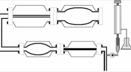
TIM-1 and Tiny TIM
2.Piston/Probe-Driven Systems
Piston- or probe-driven systems simulate gastrointestinal motility through the controlled reciprocating movement of a piston or probe within a digestion chamber. The mechanical action generates compression, displacement, and mixing of the test material, thereby reproducing aspects of gastric contractions. In several models, a texture analyzer is used to control the movement and force applied by the probe, allowing precise adjustment of contraction conditions.
Different configurations have been developed to reproduce specific aspects of gastric motility. The simple gastric model described by used cyclic probe movement within a food chamber to generate fluid motion. The dynamic gastric simulating model (DGSM) similarly employed a texture analyzer, with the contraction force regulated by changing the distance between the probe and chambe . The dynamic gastric model (DGM) incorporated an elastic membrane to represent gastric accommodation, while piston movement through the membrane simulated antral contractions.The engineered stomach and small intestine (ESIN) model used opposing pistons to generate gastric contractions and incorporated separate pathways to reproduce the differential emptying of liquids and solid particles. Likewise, the in vitro mechanical gastric system (IMGS) reproduced peristaltic activity through sequential movement of paired pistons toward the pylorus.
Overall, piston/probe-driven systems offer a relatively simple and controllable approach for studying mechanical digestion and GI motility. Their movement speed, force, geometry, and probe–chamber distance can be modified to regulate mixing and flow patterns. However, because many of these models employ simplified and symmetrical chamber geometries, their ability to fully reproduce the complex anatomical and physiological characteristics of the human gastrointestinal tract requires further validation.[1]
Dynamic Gastric Model (DGM)
The Dynamic Gastric Model (DGM) is an in vitro system designed to reproduce the mechanical conditions of the human stomach, including gastric accommodation and antral mixing. The model consists of a cone-shaped stomach body, an antral region, and a valve connecting the two compartments, with an approximate working volume of 800 mL. Gentle contractions in the main gastric body are generated by controlled changes in water pressure, producing distension and compression of the flexible gastric wall. In contrast, stronger mechanical forces are generated in the antrum by the reciprocating movement of a piston through an elastic annulus at approximately 3 cycles per minute. This arrangement creates shear and mixing forces that mimic the mechanical action of antral contractions and allows investigation of food breakdown and gastric processing under controlled in vitro conditions [16]
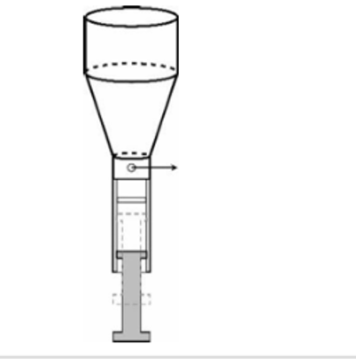
DGM
3. Pneumatic Systems
Pneumatic systems reproduce gastrointestinal motility by using controlled air pressure to inflate and deflate flexible components, thereby generating localized compression and relaxation of the simulated gastrointestinal wall. The small intestine model (SIM) developed at the University of Birmingham used two inflatable cuffs that alternately compressed and released an inner intestinal tube to reproduce segmentation movements. Similarly, the human duodenum model (HDM) employed a series of inflatable finger cots arranged along the intestinal tube, producing evenly distributed constrictions that mimic small-intestinal segmentation.[38,39,40]
Pneumatic actuation has also been applied to gastric models. The gastric simulation model (GSM) used multiple pneumatically operated pistons to reproduce antral contraction waves. Coordinated activation of the piston sets allowed different motility patterns to be generated, including sequential contractions that produced distal propagation resembling peristalsis. More recently, the soft robotic gastric simulator (SoGut) employed seven independently controlled air chambers. Inflation compressed localized regions of the gastric wall, whereas deflation allowed relaxation. By coordinating the inflation sequence and varying the pressure applied to individual chambers, the model reproduced propagating peristaltic contractions and differences in contraction strength between the fundus and antrum.[36]
Overall, pneumatic systems provide flexible control over the location, strength, and sequence of gastrointestinal contractions, making them useful for reproducing complex motility patterns. However, their construction can be relatively complex because multiple actuators and interconnected pneumatic components may be required. In addition, the materials used for the simulated gastrointestinal wall must withstand repeated compression and friction during operation.[3]
Human Duodenum Model (HDM)
The human duodenum model (HDM) is a pneumatic in vitro system designed to reproduce the segmentation movements of the duodenum. The model consists of a sigmoidal duodenal chamber with an inner semipermeable membrane enclosed within an outer acrylic cylinder measuring approximately 5.2 cm in internal diameter and 57 cm in length. Segmentation is generated by the alternating inflation and deflation of finger cots secured to stainless-steel rings along the intestinal chamber. Sequential activation of these pneumatic elements produces localized constrictions and relaxation of the simulated intestinal wall, thereby mimicking the rhythmic segmentation pattern of the human duodenum.[3]
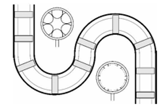
Human duodenal model
Gastric Simulation Model (GSM)
The Gastric Simulation Model (GSM)developed is a pneumatic in vitro model designed to reproduce gastric contraction and peristaltic activity. It consists of a handcrafted J-shaped latex stomach representing the cardia, fundus, body, antrum, and pylorus, with a tapered antral region and an approximate working volume of 600 mL. Gastric contractions are generated by pneumatically driven syringes that compress the inner gastric wall. A total of 60 syringes are arranged circumferentially along the model and activated sequentially, producing coordinated contractions that propagate from the fundus toward the pylorus. This arrangement enables the GSM to reproduce different regional gastric contractions and generate peristaltic waves that mimic the mechanical activity of the human stomach.[1,3]
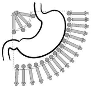
Gastric stimulation model
In Vivo Method
1. The colestipol–phenol red complex method:
It provides a quantitative approach for evaluating gastrointestinal transit of solid food in experimental animals. Following oral administration, the marker is recovered from different gastrointestinal segments and quantified spectrophotometrically, allowing assessment of gastric emptying and intestinal transit.[33,35,37]
Materials required
Experimental rats (male Sprague–Dawley rats), Colestipol, Phenol red,5% charcoal suspension in 0.25% methylcellulose, Distilled water,1 N HCl and 1 N NaOH, Chloroform, Centrifuge, Homogenizer/ mixer, Spectrophotometer, Syringes/oral administration equipment
Atropine sulfate, when evaluating the effect of reduced GI motility
The colestipol–phenol red method is a quantitative technique used to evaluate gastrointestinal (GI) transit of solid food in experimental animals. The method involves preparing a complex of colestipol and phenol red, which is administered orally to fasted rats. After predetermined time intervals, the animals are sacrificed, and the gastrointestinal tract is removed.32,33,34] The stomach and small intestine are processed separately, and the marker present in different GI segments is extracted and quantified. The tissue contents are homogenized and centrifuged, followed by treatment with suitable reagents, and the absorbance of the resulting solution is measured spectrophotometrically at 560 nm. The amount and distribution of phenol red are used to determine gastric emptying and intestinal transit. Atropine may be used to induce delayed GI motility and validate the sensitivity of the method. Thus, the method provides a useful quantitative assessment of solid-food gastrointestinal transit. [2]
2. Carmine Red Dye Assay :
Materials: Swiss-Webster mice, carmine red dye, 0.5% methylcellulose, 1 mL syringe with feeding needle, and a sterile container for collecting fecal pellets.
Method: A 6% carmine red solution in 0.5% methylcellulose was prepared and sterilized. Each mouse was administered 150 µL of the solution orally by gavage, and the exact administration time was recorded. After gavage, fresh fecal pellets were collected at regular intervals and examined for the appearance of red coloration. If no red color was observed, monitoring was continued at approximately 20–30 min intervals until a red pellet appeared. The time between carmine administration and the first appearance of red dye in feces was considered the whole-gut transit time.
The assay generally requires 2–8 hours, depending on the mouse model. Food and water may be provided during the test, and abdominal pressure should not be applied because stress can influence intestinal transit. The experimental timing should also be kept consistent between groups because gut transit can vary with the time of day and light–dark cycle.[9]
3.Balloon Method:
Animal Preparation and Experimental Procedures:
Animal experiments were conducted using adult male Wistar rats weighing approximately 310–390 g. The animals were anesthetized using urethane following brief induction with halothane. During surgery, anesthesia was maintained with low-concentration halothane as required. The trachea was cannulated and artificial ventilation was provided, while end-tidal CO₂, body temperature, arterial blood pressure, and heart rate were continuously monitored and maintained within the experimental range. Femoral arterial and venous cannulation was performed to facilitate cardiovascular monitoring and administration of fluids or drugs, respectively. Anesthetic depth was assessed throughout the experiment and supplemented when necessary.[31]
Assessment of Gastric Motility:
Gastric motor activity was evaluated by direct measurement of changes in intragastric pressure. Following a midline abdominal incision, a compliant balloon was introduced into the pyloric region through a small opening in the duodenum. The balloon was connected to a pressure transducer through a catheter, enabling continuous detection of pressure fluctuations generated by gastric contractions. These pressure changes were subsequently amplified and recorded continuously using a polygraph recording system, providing an objective measure of gastric motor activity. In addition, a separate catheter was positioned within the duodenum adjacent to the balloon catheter to allow drainage of intestinal secretions and minimize interference with the gastric pressure measurements. Thus, the balloon-pressure recording technique permitted continuous assessment of the frequency and magnitude of gastric contractions under different experimental stimulation conditions.[6]
4. Video Assessment:
Materials: Excised ileal smooth muscle tissue from three healthy wild-type mice, M199 culture medium supplemented with fetal bovine serum, glucose, L-glutamine, and antibiotic-antimycotic solution, Incubator maintained at 37°C with 95% air and 5% CO₂, Olympus DP22 digital camera, Computer with MATLAB (R2022b), Video recordings captured at 15 frames per second with a resolution of 1920 × 1440 pixels.
The ileal smooth muscle tissues were first isolated from three healthy wild-type mice following ethical approval and prepared for ex vivo motility analysis. The tissues were pinned at their four corners, washed with supplemented M199 medium, and maintained for 3 hours at 37°C under 95% air and 5% CO₂ to preserve tissue viability. Spontaneous tissue movements were then recorded using an Olympus DP22 digital camera positioned perpendicular to the tissue surface at 15 frames/s and a resolution of 1920 × 1440 pixels. The recorded videos were processed in MATLAB, where individual frames were converted to grayscale and enhanced using histogram equalization. A region of interest covering most of the tissue was manually selected, and naturally occurring tissue features or corner points were identified using the Minimum Eigenvalue feature-detection algorithm. These features were subsequently tracked across successive video frames, with only continuously detectable features retained for analysis. The selected tissue area was divided into 50 × 50 pixel grids, and the movement of tracked features within each grid was grouped to characterize local tissue motion. [30]Feature displacement was calculated and filtered, and Fast Fourier Transform (FFT) analysis was applied to determine the frequency of spontaneous contractions. Further analysis provided measurements of contraction frequency, amplitude, spatial coordination, and overall motility index using density-based clustering. Finally, the feature-tracking approach was validated by comparing the calculated movements with direct measurements of tissue-wall displacement, demonstrating its ability to quantitatively characterize intestinal smooth muscle motility.[12,14,15]
5. Transcutaneous Electrical Stimulation
Transcutaneous electrical stimulation (TES) has emerged as a potential non-invasive therapeutic approach for gastrointestinal (GI) motility disorders. Treatment is generally administered for less than 60 minutes per session, with protocols ranging from alternate-day to twice-daily stimulation. Major TES approaches include transcutaneous peripheral nerve stimulation (TPNS), transcutaneous electrical acustimulation (TEA), transcutaneous interferential current (IFC), and transcutaneous electrical nerve stimulation (TENS).[29]
Transcutaneous Peripheral Nerve Stimulation [TPNS]
TPNS delivers low-intensity electrical currents through surface electrodes positioned over specific peripheral nerves.[28] Important applications include transcutaneous vagal nerve stimulation (tVNS), transcutaneous sacral nerve stimulation (tSNS), and transcutaneous tibial nerve stimulation (tTNS). tVNS can be applied either through the auricular branch of the vagus nerve (taVNS) or through the cervical region (tcVNS), providing a non-invasive alternative to surgically implanted vagal stimulation. Depending on the stimulation parameters, tVNS may influence vagal and sympathetic autonomic activity. In contrast, tSNS and tTNS are primarily investigated for lower GI dysfunction, with stimulation of the sacral and tibial nerves proposed to modulate neural pathways involved in bowel and defecatory function.
Transcutaneous Electrical Acustimulation [TEA]
TEA is another needle-free TES modality derived from conventional acupuncture and electroacupuncture. It applies electrical stimulation to selected acupuncture points using surface electrodes rather than needles. Points such as Neiguan (PC6) and Zusanli (ST36) are frequently targeted in GI applications. Overall, TES techniques may improve GI function by modulating autonomic and peripheral neural pathways, thereby influencing gastrointestinal motility, visceral sensation, and secretion. These approaches therefore represent promising non-invasive strategies for the management of GI motility disorders.
Transcutaneous Interferential Current [TIC]
Transcutaneous interferential current (IFC) is a non-invasive electrical stimulation technique that has been used clinically since the 1950s.[28] Initially developed for genitourinary conditions, IFC has subsequently demonstrated potential benefits in the management of gastrointestinal motility disorders. The technique involves the application of two medium-frequency sinusoidal electrical currents that intersect within the targeted tissue, producing an interference current capable of reaching deeper neural structures. Carrier frequencies in the range of 4–10 kHz have been investigated, with approximately 4 kHz reported to provide better patient tolerance and effective penetration into deeper tissues. These characteristics make IFC a promising modality for influencing gastrointestinal neuromuscular activity.[7,8]
Transcutaneous Electrical Nerve Stimulation [TENS]
Transcutaneous electrical nerve stimulation (TENS) is a non-invasive peripheral neuromodulation technique widely used for the management of chronic pain. Electrodes are generally positioned over painful muscles, specific dermatomes, or, in some applications, acupuncture points.[28] Compared with transcutaneous electrical acustimulation (TEA), TENS typically covers a broader treatment area and commonly uses pulse widths below 0.3 ms. Recent evidence has highlighted its potential application in functional gastrointestinal disorders, with reported improvements in GI motility. TENS is thought to act primarily by stimulating large-diameter, non-nociceptive sensory afferent fibers, thereby reducing nociceptive signaling and neural sensitization. Its therapeutic effects may also involve enhanced local blood flow and modulation of several neurochemical pathways, including acetylcholine, noradrenaline, serotonin, γ-aminobutyric acid, nitric oxide, and opioid receptors. In addition, alterations in vasoactive intestinal peptide and inflammatory mediators such as interleukin may contribute to its effects on GI function.[4,5,6]
6. Gastrointestinal Ultrasonography (GIUS)
GI ultrasonography is a non-invasive, real-time imaging method used to assess gastrointestinal anatomy and movement, including gastric emptying, gastric motility, intestinal motility, bowel-wall characteristics and transpyloric flow. Both low- and high-frequency ultrasound probes are used for examining the small intestine, colon and mesentery.[26]
Material: Gastrointestinal ultrasonography was performed using ultrasound equipment with low-frequency (3–8 MHz) and high-frequency linear (7–17 MHz) probes for examination of the stomach, small intestine, colon and mesentery.
Method: Patients were examined after more than 6 hours of fasting, initially in the supine position. A low-frequency probe was first used to identify gastrointestinal anatomy, followed by a high-frequency probe for detailed examination. The gastric antrum was assessed in a semi-sitting position using a curvilinear probe. Real-time ultrasound imaging was used to evaluate gastrointestinal movement, peristalsis, gastric emptying and intestinal motility.[17,18,19]
RESULTS
The review identified several in vitro, ex-vivo, in vivo, and non-invasive methods used for the assessment of gastrointestinal (GI) motility. In vitro models such as hydraulic, piston/probe-driven, pneumatic, and dynamic gastric systems were found to provide controlled and reproducible conditions for simulating intestinal and gastric contractions. Models including TIM, Tiny-TIM, DGM, HDM, and GSM were able to reproduce different aspects of GI movement, mixing, segmentation, and peristaltic activity. However, some systems have limitations in reproducing the complete anatomical and physiological complexity of the human GI tract. [24]
In vivo methods such as the colestipol–phenol red complex method and carmine red dye assay provide quantitative measurements of gastric emptying and whole-gut transit time. The balloon-pressure technique allows continuous measurement of gastric contraction frequency and magnitude. Ex vivo video-based analysis of isolated ileal tissue provides a quantitative assessment of contraction frequency, amplitude, spatial coordination, and overall motility index using digital image processing and feature tracking.[11,12]
Non-invasive techniques, particularly transcutaneous electrical stimulation and gastrointestinal ultrasonography, offer promising approaches for evaluating or modulating GI motility without invasive procedures. Overall, the reviewed methods demonstrate that combining mechanical models, animal-based approaches, quantitative video analysis, and non-invasive imaging can provide complementary information about gastrointestinal motility and may support the development and evaluation of treatments for GI motility disorders.[10]
DISCUSSION
Gastrointestinal motility is a complex physiological process involving coordinated muscular contractions, mechanical mixing, propulsion, and neural regulation. The methods described in this review demonstrate that no single technique can reproduce or measure all aspects of gastrointestinal motility. Instead, different approaches provide information at different levels, ranging from controlled mechanical simulation in vitro to direct physiological assessment in animals and non-invasive evaluation in humans.
In vitro systems such as hydraulic, piston/probe-driven, and pneumatic models provide useful platforms for reproducing the mechanical component of gastrointestinal motility under controlled experimental conditions. Hydraulic systems, including the TIM models, reproduce intestinal movements through controlled pressure changes and can be adapted to represent different physiological conditions. Their major advantage is the ability to provide reproducible control over pressure, movement, mixing, and transit. However, these models remain simplified representations of the gastrointestinal tract and cannot completely reproduce the neural, hormonal, vascular, and biochemical factors involved in physiological motility.[15,16]
Piston- and probe-driven systems provide relatively simple and controllable methods for generating compression and mixing. Parameters such as movement speed, applied force, geometry, and probe position can be modified according to the experimental requirement. The Dynamic Gastric Model is particularly useful because it combines gastric accommodation with stronger antral contractions, thereby providing a closer representation of the mechanical processing occurring in the stomach. Nevertheless, the simplified geometry used in several piston-based systems may limit their ability to reproduce the complex anatomical structure and physiological behaviour of the human gastrointestinal tract.
Pneumatic models provide greater flexibility in reproducing localized and sequential contractions. The ability to independently control the pressure, location, strength, and timing of contractions allows models such as the Human Duodenum Model and Gastric Simulation Model to reproduce segmentation and peristaltic patterns. These systems therefore provide a useful bridge between simple mechanical models and more physiologically representative motility simulations. However, their construction and operation can be relatively complex because they require multiple actuators and interconnected components, and repeated compression may also place mechanical demands on the artificial gastrointestinal wall.
In vivo methods provide a different level of assessment because gastrointestinal transit and motor activity are measured within a living organism. The colestipol–phenol red method allows quantitative estimation of gastric emptying and intestinal transit by measuring the distribution of the marker along different gastrointestinal segments. Similarly, the carmine red assay provides a simple assessment of whole-gut transit time based on the appearance of the marker in feces. These methods are relatively straightforward and useful for screening changes in intestinal transit; however, they may provide limited information regarding the detailed pattern, frequency, and strength of individual contractions.[23]
The balloon-pressure method provides more direct information about gastric motor activity by continuously recording intragastric pressure changes produced by contractions. Unlike transit-marker methods, it allows assessment of contraction frequency and magnitude over time. This makes it particularly useful when the objective is to evaluate changes in gastric motor activity rather than only overall transit. However, the requirement for anesthesia, surgical intervention, catheter placement, and continuous physiological monitoring makes the technique invasive and technically demanding.
Video-based assessment represents a more recent quantitative approach for evaluating intestinal smooth muscle activity. By combining digital imaging, feature tracking, spatial analysis, and Fast Fourier Transform analysis, spontaneous contractions can be assessed in terms of frequency, amplitude, spatial coordination, and overall motility. This approach has the advantage of generating objective quantitative data from tissue movement and may reduce some of the subjectivity associated with visual assessment. However, because the method uses excised tissue, it does not fully represent the integrated physiological environment of the intact gastrointestinal tract.[13]
Transcutaneous electrical stimulation differs from the other methods because it is primarily concerned with modulating gastrointestinal motility rather than simply measuring it. Techniques such as transcutaneous peripheral nerve stimulation, transcutaneous electrical acustimulation, interferential current, and TENS offer non-invasive approaches for influencing neural pathways involved in gastrointestinal function. Their potential to alter autonomic and peripheral neural activity makes them particularly relevant for the study and management of gastrointestinal motility disorders. However, the response may depend on stimulation site, intensity, frequency, and the underlying physiological condition, indicating the need for further standardization and validation.[4]
Gastrointestinal ultrasonography provides an important non-invasive alternative for evaluating motility in humans. Real-time ultrasound can assess gastric emptying, peristalsis, intestinal movement, bowel-wall characteristics, and transpyloric flow without requiring invasive instrumentation. The use of different ultrasound probes also permits examination of different regions of the gastrointestinal tract. Its non-invasive nature and ability to provide real-time observations make GI ultrasonography particularly valuable for clinical assessment. However, appropriate patient preparation, operator expertise, probe selection, and standardised interpretation are important for obtaining reliable results.[17,19,20]
Overall, the methods reviewed demonstrate a progression from simplified mechanical simulation toward increasingly quantitative, physiological, and non-invasive approaches. In vitro models are particularly valuable for studying mechanical digestion, transit, and the influence of controlled mechanical forces, whereas in vivo methods provide information about gastrointestinal function within an intact biological system. Video analysis and imaging techniques add quantitative and real-time assessment, while electrical stimulation provides opportunities for therapeutic modulation of motility. Therefore, the selection of a method should be based on the specific objective of the study, the gastrointestinal region being investigated, the required level of physiological relevance, and whether the purpose is measurement, simulation, or therapeutic intervention.
CONCLUSION
The evaluation of gastrointestinal motility requires a combination of complementary experimental and clinical approaches because GI motility involves complex mechanical and physiological processes that cannot be completely represented by a single method. Hydraulic, piston/probe-driven, and pneumatic models provide controlled and reproducible platforms for studying mechanical aspects of gastrointestinal movement, while in vivo methods allow assessment of gastric emptying, intestinal transit, and contractile activity under biological conditions. Video-based analysis offers quantitative assessment of tissue movement, whereas transcutaneous electrical stimulation provides a non-invasive approach for modulating gastrointestinal function. GI ultrasonography further expands the available techniques by enabling real-time, non-invasive assessment of gastric and intestinal motility in humans.
Taken together, these methods provide complementary information and can be selected according to the specific objectives of research or clinical evaluation. Future development should focus on improving physiological similarity, standardizing experimental parameters, increasing quantitative accuracy, and integrating multiple measurement techniques. Such improvements may provide a more comprehensive understanding of gastrointestinal motility and support the development of better experimental models, diagnostic approaches, and therapeutic strategies for GI motility disorders..
REFERENCES

Samhitha J, Spoorthy B S, Srinithya S, Jhanhavi S, Manoj L, Shrivatsa S H, A Symphony in Motion: Decoding Gastrointestinal Motility in vivo and in vitro models, Int. J. of Pharm. Sci., 2026, Vol 4, Issue 9, 1647-1661. https://doi.org/10.5281/zenodo.22741929
 10.5281/zenodo.22741929