Novel vesicular drug delivery systems have gained significant attention for their ability to provide controlled, sustained, and site-specific drug delivery while minimizing systemic side effects. Among these systems, novasomes represent an advanced vesicular carrier composed of non-phospholipid paucilamellar structures capable of encapsulating both hydrophilic and hydrophobic drugs with high efficiency. Novasomes consist of multiple concentric bilayers surrounding a large amorphous core, enabling high drug loading, sustained release, and improved stability over a wide range of pH and temperature conditions. This review highlights the evolution of vesicular drug delivery systems, classification of vesicular carriers, and the unique structural and functional features of novasomes. Various methods of novasome preparation, including thin film hydration, ether injection, microfluidization, reverse phase evaporation, multiple membrane extrusion, and sonication, are discussed. In addition, key evaluation parameters such as morphology, particle size, zeta potential, entrapment efficiency, drug loading efficiency, and in-vitro drug release are summarized. Owing to their biocompatibility, stability, high encapsulation efficiency, and versatility, novasomes show promising potential in pharmaceutical, cosmetic, and vaccine delivery applications, making them an effective platform for targeted and controlled drug delivery.
Keywords
Vesicular drug delivery systems (VDDS); Novasomes; Drug encapsulation; In-vitro drug release
Introduction
×
Novel vesicular drug delivery systems are developed to ensure controlled and sustained release of drugs in accordance with physiological requirements during therapy, while also facilitating site specific delivery of the active pharmaceutical ingredient. Vesicular structures of biological origin were first identified in 1965 by Bingham, who described them as Bingham bodies1. In recent years, several advanced vesicular drug delivery systems have been developed for use through different routes of administration to achieve controlled and site-specific drug delivery2. The concept of targeted drug delivery was first introduced by Paul Ehrlich in 1909, who demonstrated the possibility of directing therapeutic agents specifically to diseased cells3.Targeted drug delivery is a method in which a drug is directed to the specific site of action in the body, allowing higher drug concentration at the target area while reducing its presence in non-target tissues. This approach improves therapeutic effectiveness and lowers the risk of systemic side effects4.Various pharmaceutical carriers, including polymeric micelles, particulate systems, and macro and micromolecular carriers, have been developed as novel drug delivery systems to achieve targeted drug delivery. Among these, particulate carriers also referred to as colloidal carrier systems comprise lipid based particles, micro and nanoparticles, micro and nanospheres, polymeric micelles, and vesicular drug delivery systems. Vesicular systems encompass liposomes, niosomes, transfersomes, aquasomes, ufasomes, novasomes, and related carrier systems5 as showm in Table 1.
Types of vesicular drug delivery system
Lipoidal biocarrier
Nonlipodidal biocarrier
Table 1: Types of vesicular drug delivery system
Category
Biocarrier System
Brief Description
Lipoidal biocarrier systems
Liposomes
Liposomes are small lipid vesicles with one or more layers that surround a water-filled center and carry drugs6.
Niosomes
Niosomes are drug carrying vesicles that release medicines slowly and help deliver the drug to the required site in the body7.
Transfersomes
Transferosomes are highly flexible vesicles that penetrate deep into the skin and deliver drugs more effectively than other carriers8.
Aquasomes
Aquasomes are self assembled, three layered nanoparticulate carriers consisting of a solid nanocrystalline core coated with an oligomeric film that adsorbs bioactive molecules9.
Ufasomes
Aquasomes are three layered, self-assembled nanoparticles with a solid nanocrystalline core and an oligomeric coating that carries bioactive molecules10.
Novasomes
Novasomes are proprietary multi-bilayered vesicles developed by IGI–Novavax that can carry large amounts of active ingredients within a small size11.
Lipid-based particles
Lipid-based nanoparticles such as liposomes, SLNs, and NLCs are low toxic carriers that deliver both hydrophilic and hydrophobic drugs with controlled release12.
Non-lipoidal biocarrier systems
Polymeric micelles
Polymeric micelles are stable, tiny structures formed when amphiphilic polymers naturally assemble in solution, useful for carrying drugs and other molecules13.
Polymeric micro- and nanoparticles
Polymeric micro- and nanoparticles are small biodegradable carriers that help deliver drugs more effectively and safely by improving targeting and controlled release14.
Micro- and nanospheres
Polymer-based spherical carriers providing sustained and localized drug release15.
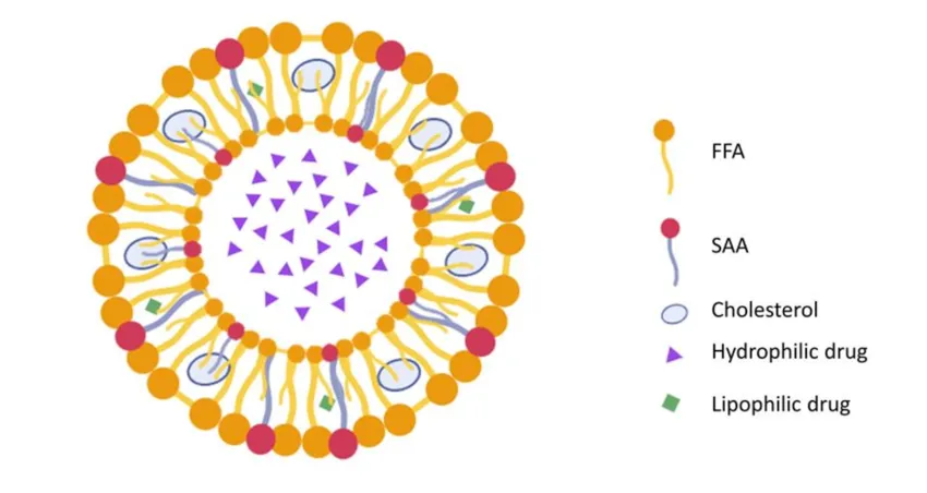
Among the various vesicular drug delivery systems, novasomes are considered modified liposomal or niosomal carriers composed of monoesters of polyoxyethylene fatty acids, cholesterol, and free fatty acids in a ratio of 74:22:4. These non-phospholipid paucilamellar vesicles generally exhibit a particle size range of 0.1–1.0 µm16. Novasome technology is a patented encapsulation approach originally developed by Novavax in collaboration with IGI Laboratories to facilitate the effective delivery of a wide range of therapeutic agents17. Novasomes consist of 2–7 concentric bilayer membranes enclosing a large, unstructured amorphous core, which allows the simultaneous incorporation of both hydrophilic (water-soluble) and hydrophobic (water-insoluble) drug substances18 as shown in Fig 1. The encapsulation efficiency of novasomes typically ranges from 78-88% for aqueous compounds and 99-100% for lipid-based compounds19. The major portion of the vesicle is occupied by the amorphous core, which can entrap finely dispersed insoluble particles such as diamond and titanium dioxide, along with water-soluble substances20. Novasome microvesicles exhibit inherent stability over a wide temperature range, extending from liquid nitrogen to temperatures above the boiling point of water, and across a broad pH range of 2-13. These systems provide sustained drug release and possess bilayer membranes that lack a rigid or ordered structure. Novasomes can be formulated with varying surface charges, enabling effective interaction with negatively charged biological surfaces such as skin, mucosa, and hair, and they demonstrate good adhesion to hair shafts. Furthermore, novasomes enhance therapeutic efficacy while minimizing adverse effects21. Owing to their biocompatibility and non-cytotoxic nature, novasomes have found extensive applications in cosmetics, personal care, nutrition, chemicals, agrochemicals, and pharmaceuticals, and have also been employed as adjuvants in human vaccines22.
Fig. 1: Diagrammatic representation of novasomes23.
Types of novasomes:
Pro-novasomes (ProNovs): are dry, free-flowing particulate systems with a dispersion structure that rapidly convert into novasomal suspensions upon hydration. Compared to conventional novasomes, ProNovs exhibit superior drug absorption. Their solid-state nature enhances physical stability while preserving the inherent properties of novasomal systems. ProNovs are typically obtained by lyophilization and are developed to improve the stability and handling of novasomes23.
Selinium novasomes: Selenium-based novasomes are an advanced form of conventional novasomal carriers designed to improve stability and therapeutic performance. While conventional novasomes enhance membrane diffusion and passive tumor targeting, their rapid clearance limits effectiveness. Incorporation of selenium improves plasma stability and prolongs circulation time of the vesicles. Selenium also contributes inherent antioxidant and anticancer properties, enhancing cellular uptake and promoting apoptosis in tumor cells. Overall, selenium-modified novasomes offer a synergistic nanocarrier system with improved pharmacokinetics and enhanced anticancer efficacy24.
Characteristics of novasomes:
Novasomes are multilamellar vesicles with a large central core.
Their surface charge may be positive, negative, or neutral.
The inner amorphous core can encapsulate up to 80–85% of the drug.
They can be prepared in a controlled and specific size range.
Novasomes can adhere to the skin or hair shaft depending on vesicle surface charge and skin conditions.
They allow incorporation of a high amount of active ingredient in a small volume.
They provide a predictable and sustained release of active substances, reducing dosing frequency.
They are capable of carrying and releasing large amounts of water-soluble ingredients25.
Mechanism of action:
The particles migrate within the bilayers through random movement between internal channels, resulting in continuous and controlled drug release. The surface charge of the microvesicles can be adjusted to be positive, negative, or neutral. The drug is securely entrapped within the core, enabling effective and efficient delivery. This protected encapsulation also enhances the stability and storage properties of the microvesicles26.
ADVANTAGES OF NOVASOMES:
Novasomes can simultaneously deliver hydrophilic and hydrophobic drugs, either individually or in combination, within a single formulation.
Incompatible drugs can be incorporated into different bilayers, thereby minimizing drug-drug interactions.
Surface charge modulation enables site-specific drug release.
They exhibit high drug loading efficiency (up to ~80%), allowing delivery of large amounts of active ingredients and reducing dosing frequency.
Novasomes possess good adhesion to the skin and hair shaft, making them suitable for topical and cosmetic formulations.
They enhance skin penetration and deposition due to their elastic and flexible vesicular structure.
Encapsulation in novasomes improves drug stability, safety, and therapeutic performance in topical applications.
Novasomes have potential applications in wound healing by enabling effective topical delivery of hormones and bioactive agents.
In addition to drug delivery, novasomes can act as vaccine adjuvants by enhancing immunogenicity and protective immune responses.
They are stable at broad range of pH range between 2 and 13.
Compared to liposomes preparation of novasomes is more affordable27.
Formulation of novasomes:
Novasomes are generally formulated by combining a non-ionic surface-active agent (such as Span 60, Span 80, or Tween 80), a free fatty acid (e.g., oleic acid or stearic acid), and cholesterol as a bilayer-stabilizing component. The cholesterol content is usually kept constant, while the type of surfactant and free fatty acid and their ratios (commonly 2:1 or 1:1) are varied to obtain vesicles with acceptable stability, suitable consistency, and desirable physicochemical characteristics for further characterization and application28.
METHOD OF PREPARATIONS:
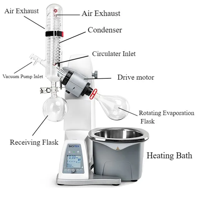
Thin film hydration method:The thin-film hydration method is a commonly employed technique for the preparation of novasomes. In this method, accurately weighed quantities of surfactant, free fatty acid, and cholesterol are transferred into a round-bottom flask and dissolved in a suitable organic solvent. The flask is then attached to a rotary evaporator, as shown in Fig 2, and the organic solvent is removed under reduced pressure for about 30 minutes, resulting in the formation of a uniform thin lipid film on the inner wall of the flask. The formed film is subsequently hydrated with an aqueous drug solution at a controlled temperature, and the flask is reattached to the rotary evaporator and rotated for approximately 60 minutes to facilitate complete hydration and vesicle formation. The obtained dispersion is then subjected to gentle magnetic stirring for 15 minutes to improve homogeneity, followed by probe sonication for 1–2 minutes to reduce vesicle size. Finally, the dispersion is centrifuged at 13,500 rpm for 30 minutes, leading to the formation of a pellet consisting of drug-loaded novasomes29.
Fig. 2: Rotary Evaparator
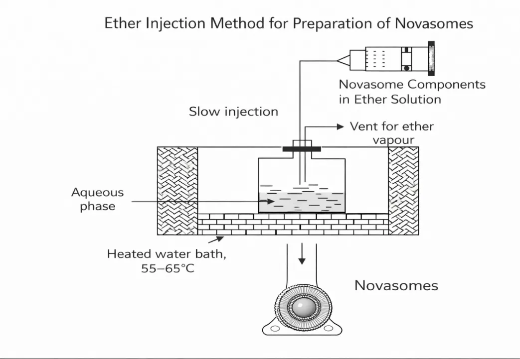
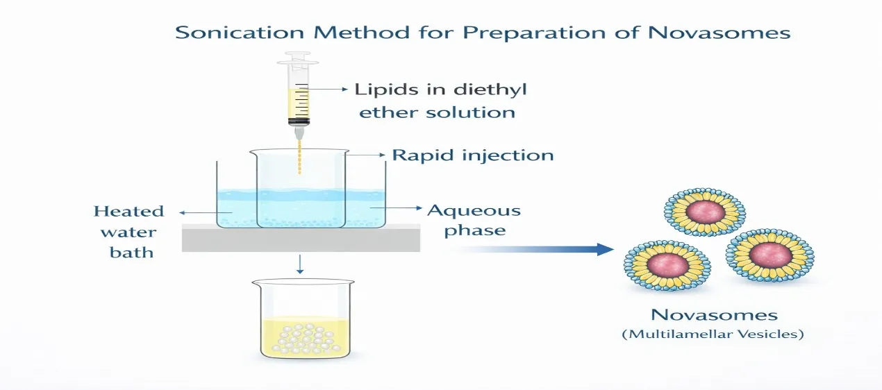
Ether injection method: In the ether injection method, the nonionic surfactants, cholesterol, and free fatty acids, are dissolved in ether or an ether-methanol mixture and slowly injected into an aqueous phase containing the drug to be encapsulated. The aqueous phase is maintained at an elevated temperature (approximately 55–65 °C) to facilitate evaporation of the organic solvent. Progressive removal of ether, often under reduced pressure, promotes self-assembly of the formulation constituents into paucilamellar vesicles, leading to the formation of Novasomes as shown in Fig 3. This process allows the production of vesicular dispersions with relatively high drug entrapment efficiency. However, the method may result in vesicles with a broad size distribution and involves exposure of the active agent to organic solvents and elevated temperatures, which may influence formulation stability30.
Fig. 3: Diagramatic Representation Of Ether Injection Method.
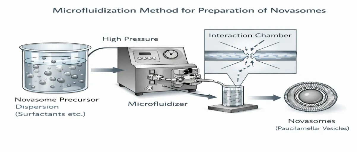
Microfluidization method: In the microfluidization method, the formulation constituents are first dispersed in an aqueous medium to obtain a coarse vesicular dispersion. This dispersion is then processed through a microfluidizer, where it is subjected to high pressure and forced through precisely engineered microchannels within an interaction chamber. Inside the chamber, opposing fluid streams collide at very high velocities following the submerged jet principle, generating intense shear and impact forces. These forces reduce vesicle size and promote the formation of uniformly distributed Novasomes as illustrated in Fig 4. The dispersion may be passed through the microfluidizer for multiple cycles to achieve the desired size and homogeneity31.
Fig 4: Microfluidization method of preparation of novasomes.
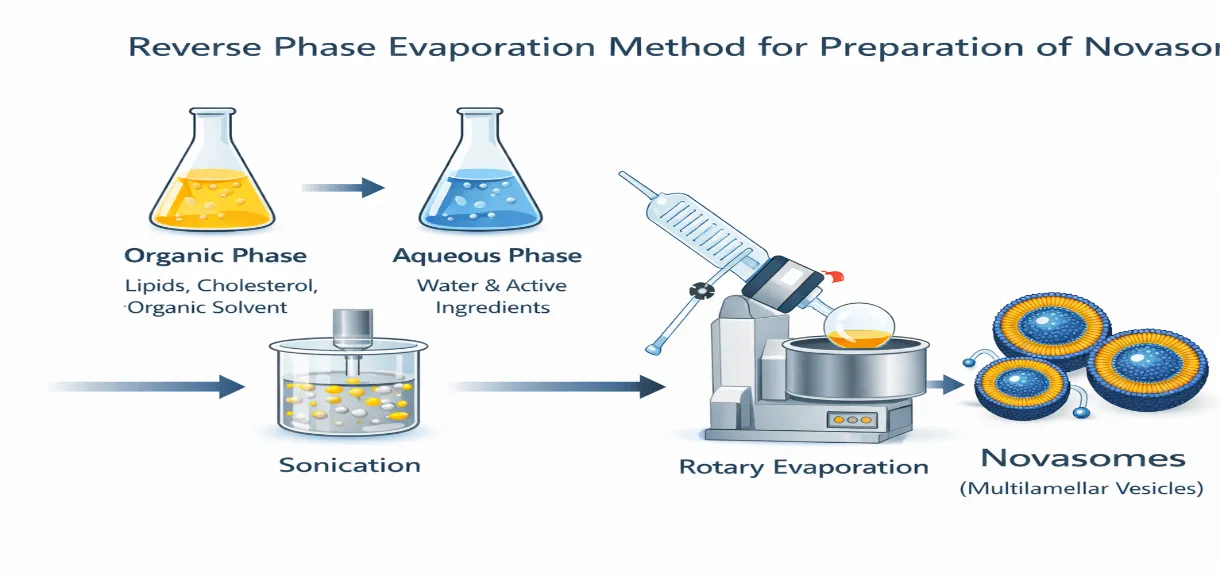
Reverse phase evaporation method: In the reverse phase evaporation method, the formulation is prepared using two phases. In the organic phase, cholesterol, surfactant, and free fatty acid in an appropriate ratio are dissolved in a mixture of ether and chloroform. The aqueous phase is prepared separately by dissolving the drug in an aqueous medium. The organic phase is then slowly added to the aqueous phase and the mixture is sonicated at 4-5 °C to obtain a stable emulsion. As illustrated in Fig 5, after the formation of a clear gel, further sonication is performed, followed by the addition of a small quantity of phosphate buffer. The organic solvents are removed under reduced pressure at about 40 °C. The resulting suspension is diluted with phosphate buffer and heated in a water bath at 60 °C for 10 minutes, which leads to the formation of novasomes32 .
Fig. 5: Reverse phase evaporation method of preparation of novasome
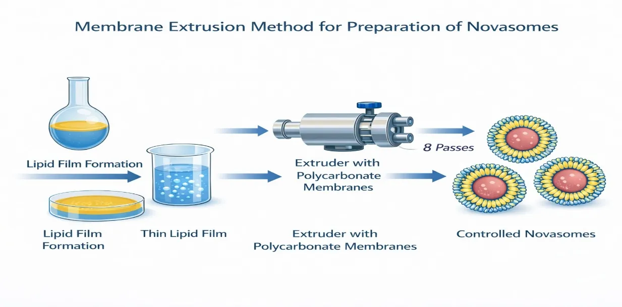
Multiple membrane extrusion method: In the multiple membrane extrusion method, a mixture of surfactant, cholesterol, and free fatty acid dissolved in chloroform is first evaporated to form a thin lipid film. The film is then hydrated with an aqueous drug solution to produce a vesicular suspension as illustrated in Fig 6. This suspension is extruded through polycarbonate membranes arranged in series for eight successive passes, allowing precise control of novasome size and uniformity33.
Fig. 6: Multiple membrane extrusion method
Sonication method: In the sonication method, all components of the novasomes, including cholesterol, non-ionic surfactants, active pharmaceutical ingredients and other excipients, are dispersed in an appropriate aqueous buffer. The dispersion is then subjected to sonication at around 60 °C for 15-30 minutes to achieve uniform particle size and efficient drug encapsulation. After sonication, the dispersion is cooled, and the resulting Novasomes as shown in Fig 7, then the novasomes are collected for further use or characterization34.
Fig. 7: Sonication Method Of Preparation Novasomes
.CHARACTERIZATION OF NOVASOMES:
Morphology: Morphology refers to the physical structure, shape, and surface characteristics of vesicular systems. In novasome based drug delivery, morphological analysis provides essential information about vesicle size, shape, and surface features. Techniques such as Dynamic light scattering (DLS) and Scanning electron microscopy (SEM) are commonly used to visualize novasomes at the micro and nanoscale. These methods help confirm vesicle integrity, uniformity, and dispersion, as well as identify aggregation tendencies. Morphological evaluation is important for understanding the stability, drug encapsulation efficiency, and biological interaction of novasome formulations35.
Scanning electron microscopy (SEM): The surface morphology of novasomes was examined using a scanning electron microscope operated under cryogenic conditions to maintain the hydrated structure of the vesicles. A small quantity of the novasome dispersion was placed on a suitable specimen holder and rapidly frozen before analysis. The sample was then observed at an accelerating voltage in the range of 0.1–30 kV to evaluate vesicle shape, surface characteristics, and size.
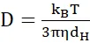
Dynamic light scattering (DLS): The particle size of novasomes was determined by dynamic light scattering. The novasome dispersion was suitably diluted with distilled water or buffer and analyzed at 298.15 K and pH 7.4. The hydrodynamic diameter of novasomes was calculated from their Brownian motion using the Stokes–Einstein equation:
D=kBT3πηdH
Where, D is the translational diffusion coefficient, kB is the Boltzmann constant, T is the absolute temperature, 𝞰 is the viscosity of the dispersion medium, and dH is the hydrodynamic diameter of the vesicles.
Fourier transform infrared spectroscopy: Fourier Transform Infrared Spectroscopy (FTIR) is used to analyze the chemical composition and molecular structure of novasome formulations by studying their interaction with infrared radiation. The technique identifies functional groups and chemical bonds present in novasomes and their components through characteristic absorption peaks. By comparing the FTIR spectra of novasomes with those of the pure drug and excipients, possible interactions, compatibility, and structural changes can be evaluated. FTIR is particularly useful for confirming vesicle formation and detecting molecular interactions that may affect the stability and performance of novasome-based drug delivery systems36.
Zeta potential: The surface charge of novasomes is evaluated by measuring the zeta potential using a laser light scattering instrument with an electrophoretic mobility analyzer, where vesicle movement under an applied electric field is analyzed and converted into zeta potential values. Prior to measurement, the novasome dispersion is diluted with a low-conductivity medium to reduce ionic interference. Zeta potential values indicate the electrostatic stability of the formulation, with values greater than +30 mV or less than −30 mV reflecting good physical stability due to strong electrostatic repulsion between vesicles, while values between −30 mV and +30 mV suggest a tendency toward aggregation. The sign and magnitude of the zeta potential also influence vesicle interaction with biological membranes and play an important role in the stability and performance of novasome-based drug delivery systems37.
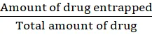
Entrapment efficiency: The entrapment efficiency of novasomes is determined by separating the unentrapped drug from the vesicular dispersion using techniques such as centrifugation or gel filtration. The collected novasomes are then subjected to complete vesicle disruption to release the entrapped drug. The released drug is quantified using an appropriate analytical method. Entrapment efficiency is calculated using the following formula38:
Entrapment efficiency (%) = Amount of drug entrappedTotal amount of drug
× 100
Drug loading efficiency: The loading efficiency of novasomes was determined by extracting the entrapped drug using an acidic medium. Accurately weighed novasomes (50 mg) were dispersed in 50 mL of 0.1 M hydrochloric acid and stirred continuously until complete disruption of the vesicular structure and release of the drug occurred. The resulting solution was filtered through a millipore membrane filter to remove any undissolved material. The filtrate was suitably diluted and the drug content was analyzed using UV-visible spectrophotometry against an appropriate blank. The amount of drug present in the novasomes was calculated using a previously established calibration curve.
Drug loading Efficiency (L %) = QnWn×100
Where,
Qn= amount of drug present in the novasomes.
Wn= total weight of novasomes39.
In-Vitro drug release: The in-vitro drug release of novasomes was carried out using the semipermeable membrane diffusion technique. A suitable semipermeable membrane was selected to ensure negligible drug adsorption and unrestricted diffusion of the drug. A known volume of novasome suspension was filled into the membrane, securely sealed, and immersed in the receptor compartment containing an appropriate dissolution medium. The system was maintained under continuous magnetic stirring at controlled temperature, and sink conditions were preserved throughout the study. At predetermined time intervals, samples were withdrawn from the receptor medium and analyzed for drug content using a suitable analytical method. An equal volume of fresh dissolution medium was added after each sampling to maintain a constant volume40.
APPLICATIONS
Used for controlled and targeted drug delivery to improve bioavailability and therapeutic efficacy.
Act as nanocarriers for encapsulation and sustained release of drugs.
Enhance penetration of active ingredients into deeper skin layers in dermal and skincare products.
Used in shampoos, creams, lotions, and moisturizers for improved conditioning and hydration.
Applied for controlled delivery of pesticides, herbicides, and fertilizers.
Used as carriers for controlled release and stabilization of chemical agents.
Employed for encapsulation and protection of flavors, nutrients, and bioactive compounds.
Other applications includes in dermatology, veterinary products, and specialized industrial uses27.
CONCLUSION
Vesicular drug delivery systems have significantly advanced modern pharmaceutical research by offering improved drug stability, controlled release, and enhanced therapeutic performance. Among these systems, novasomes have emerged as a promising carrier due to their unique paucilamellar structure, which consists of multiple concentric bilayers surrounding a large amorphous core. This structural organization allows novasomes to encapsulate both hydrophilic and hydrophobic drugs efficiently, making them suitable for a wide range of therapeutic applications.
The versatility of novasomes is further supported by their high drug loading capacity, sustained release behavior, and excellent physicochemical stability across broad pH and temperature ranges. Various preparation techniques enable control over vesicle size, surface charge, and composition, thereby optimizing their performance for topical, transdermal, and systemic delivery. Comprehensive evaluation studies, including morphology, particle size, zeta potential, entrapment efficiency, and in-vitro drug release, play a critical role in ensuring formulation quality and reproducibility.
Overall, novasomes represent a robust and adaptable vesicular drug delivery platform with considerable potential in pharmaceutical, cosmetic, and vaccine applications. Their biocompatibility, non-cytotoxic nature, and ability to enhance therapeutic efficacy while minimizing adverse effects highlight their importance as an emerging carrier system. Continued research and optimization of novasome based formulations are expected to further expand their clinical and industrial applications in the future.
REFERENCES
Kamboj S, Saini V, Maggon N, Bala S, Vikas Jhawat. Vesicular drug delivery systems: A novel approach for drug targeting. International Journal of Drug Delivery. 2013 Jul 6;5(2):121-30.
Myneni G S, Radha G V, Soujanya G V R L. Novel vesicular drug delivery systems: A review. Indo American Journal of Pharmaceutical Research. 2024; 11(04): 2231-76.
Yadav D, Imran M, Dubey A, Kushwaha D. Pharmacosomes: In targeted drug delivery system. Asian Journal of Pharmaceutical Research and Development. 2024; 12(5): 41-45.
Gupta M, Sharma V. Targeted drug delivery system: A review. Research Journal of Chemical Sciences. 2011;1(2):135-138.
Jain S, Jain V, Mahajan S C. Lipid based vesicular drug delivery system. Advances in Pharmaceutics. 2014;1:1-12.
Ping L, Chen G, Zhang J. A review of liposomes as a drug delivery system: Current status of approved products, regulatory environments and future perspectives. Molecules. 2022;27(1372):1-23.
Kaur P, Rani R, Singh A P, Singh A P. An overview of niosomes. Journal of Drug Delivery and Therapeutics. 2024; 14(3): 437-446.
Kodi S R, Reddy M S. Transfersomes: A novel topical approach. Journal of Drug Delivery and Therapeutics. 2023; 13(2): 126-131.
Jain S S, Jagtap P S, Dand N M, Jadhav K R, Kadam V J. Aquasomes: A novel drug carrier. Journal of Applied Pharmaceutical Science. 2012; 2(1): 184-192.
Athira N. J., Aswathi K., Akhitha G., Athira P. P., Sreeja N. C. Ufasomes: A potential phospholipid carrier as a novel pharmaceutical formulation. International Research Journal of Pharmacy. 2014; 5(4): 250-253.
Mosallam S, Ragab M H, Moftah N H, Elshafey A H, Abdelbary A A. Use of novasomes as a vesicular carrier for improving the topical delivery of itraconazole:In vitro characterization, in vivo assessment and exploratory clinical experimentation. International Journal of Nanomedicine. 2021; 16: 119-132.
García-Pinel B, Porras-Alcalá C, Ortega-Rodríguez A, Sarabia F, Prados J, Melguizo C, et al. Lipid-Based Nanoparticles: Application and Recent Advances in Cancer Treatment. Nanomaterials. 2019;9(4):638-60.
Perumal S, Atchudan R, Lee W. A review of polymeric micelles and their applications. Polymers. 2022; 14:2510-29.
Hussein A S, Abdulla N, Ahmadu F I. Polymeric micro- and nanoparticles as promising drug delivery vehicles:A review. International Journal of Nanoparticles.2019;4(4):316-337.
Wang H, Leeuwenburgh S C G, Li Y, Jansen J A. The use of micro- and nanospheres as functional components for bone tissue regeneration. Tissue Engineering.2012;18(1):24-39.
Fatima I, Rasul A, Shah S, Saadullah M, Islam N, Khames A, et al. Novasomes as nano-vesicular carriers to enhance topical delivery of fluconazole: A new approach to treat fungal infections. Molecules. 2022;27(9):2936-49.
Abd-Elal R M A, Shamma R N, Rashed H M, Bendas E R. Trans-nasal zolmitriptan novasomes: in-vitro preparation, optimization and in-vivo evaluation of brain targeting efficiency. Drug Delivery. 2016 May 17;23(9):3374-86.
Gulshan S, Shah S, Shah P A, Safan M, Saadullah M, Abbas G, Hanaf M, Rasul A, Ahmad N, et al. Development and pharmacokinetic evaluation of novasome for the trans-nasal delivery of fluvoxamine using arachidonic acid–chitosan conjugate. Pharmaceutics. 2023;15:2259-76.
Mustafa MA, Rasheed N, Ayesha M, Ain Q ul, Mughal M, Muhammad Fahad, et al. Novasomes: A Versatile Drug Delivery System for Enhanced Therapeutic Efficacy. Journal of Young Pharmacists. 2025 Jan 20;17(1):36-45.
Swapnil Waghmare, Patil A, Patil P. Novasome: Advance in Liposome and Niosome. Deleted Journal. 2016;5(5):34-38.
Rahiman A A C A, Krishnan K, Sreelekshmi A S, et al. Novasome: A pioneering advancementin vesicular drug delivery. International Journal of Applied Pharmaceutics. 2021;13(1):59–64.
Elkomy MH, El F, Kharshoum RM, Abdeltwab AM, Hussein S, Hamad DS, et al. Innovative pulmonary targeting of terbutaline sulfate-laded novasomes for non-invasive tackling of asthma: statistical optimization and comparative in vitro/in vivo evaluation. Drug Delivery. 2022 Jul 8;29(1):2058-71.
Elnady RE, Mortagi Y. A comprehensive review on novasomes as lipid-based drug carriers. Sinai Int Sci J. 2025 Jul;2(1):21-35.
Aboud HM, Hussein AK, Zayan AZ, Tarek Saad Makram, Sarhan MO, El-Sharawy DM. Tailoring of Selenium-Plated Novasomes for Fine-Tuning Pharmacokinetic and Tumor Uptake of Quercetin: In Vitro Optimization and In Vivo Radiobiodistribution Assessment in Ehrlich Tumor-Bearing Mice. Pharmaceutics. 2022;14(4):875-85.
Singh A, Malviya R, Sharma PK. Novasome-a breakthrough in pharmaceutical technology a review article. Adv Biol Res. 2011;5(4):184-189.
Agarwal S, Kamala Kumari PV. Advances in novasome technology- a review. Int J Appl Pharm. 2013;5(1):1-4
Illastria R A, Sagita E, Iskandarsyah I. Novasome: Combining Ufasome and Niosome for Excellent Vesicular Drug Delivery System. Sciences of Pharmacy. 2023;2(1):35-49.
Fatima I, Rasul A, Shah S, Saadullah M, Islam N, Khames A, et al. Novasomes as Nano-Vesicular Carriers to Enhance Topical Delivery of Fluconazole: A New Approach to Treat Fungal Infections. Molecules. 2022;27(9):2936-49.
Thabet Y, Elsabahy M, Eissa NG. Methods for Preparation of Niosomes: a Focus on Thin-Film Hydration Method. Methods. 2021 May;199-206.
Lombardo D, Kiselev MA. Methods of Liposomes Preparation: Formation and Control Factors of Versatile Nanocarriers for Biomedical and Nanomedicine Application. Pharmaceutics [Internet]. 2022 Feb 28;14(3):543-91.
Javaid S, Rana T, Naeem Z, Sajid N. Exploring Niosomes: A Comprehensive Review of their Structure, Formulation, and Biomedical Applications. Currents in Pharmaceutical Research. 2024 Jun 15;2(1):01-34.
John C H , Sailaja A K. A curcumin loaded niosomes as novel drug delivery system by ether injection method. Drug Discovery. 2023 Jun 15;17(39):1–6.
Mandal S, Sarkar S, Nandi J, Dan A, Mahata R K. Niosomes: A promising target specific vesicular drug delivery carrier in modern Pharmaceutical approach. IJRAR. 2024;11(2):944-61.
Ababei-Bobu A, Bianca-Ștefania Profire, Andreea-Teodora Iacob, Oana-Maria Chirliu, Lupașcu FG, Lenuța Profire, et al. Niosomes as Vesicular Carriers: From Formulation Strategies to Stimuli-Responsive Innovative Modulations for Targeted Drug Delivery. Pharmaceutics. 2025;17(11):1473-83.
Maharana P K, Kumar A, Pandey A, Sakarkar S N, Bisen P, et al. advancing niosomal drug delivery: Manufacturing, characterization, and therapeutic applications - a comprehensive review. WJPLS. 2024;10(3):92-108.
Judy E, Lopus M, Kishore N. Mechanistic insights into encapsulation and release of drugs in colloidal niosomal systems: biophysical aspects. RSC Advances. 2021;11(56):35110–26.
Pradhan B, Kumar N, Saha S, Roy A. Liposome: method of preparation, advantages, evaluation and its application. 2015;3(3):1–8.
Yadav JD, Kulkarni PR, Vaidya KA, Shelke GT. Niosomes: A Review. J Pharm Res. 2011;4(3):632-36.
Umbarkar M, Thakare S, Surushe T, Giri A, Chopade V, Formulation and Evaluation of Liposome by Thin Film Hydration Method, Journal of Drug Delivery and Therapeutics. 2021; 11(1):72-76.
Laouini A, Jaafar-Maalej C, Limayem-Blouza I, Sfar S, Charcosset C, Fessi H. Preparation, characterization and applications of liposomes: state of the art. Colloid Sci Biotechnol. 2012;1(2):147-168
Reference
Kamboj S, Saini V, Maggon N, Bala S, Vikas Jhawat. Vesicular drug delivery systems: A novel approach for drug targeting. International Journal of Drug Delivery. 2013 Jul 6;5(2):121-30.
Myneni G S, Radha G V, Soujanya G V R L. Novel vesicular drug delivery systems: A review. Indo American Journal of Pharmaceutical Research. 2024; 11(04): 2231-76.
Yadav D, Imran M, Dubey A, Kushwaha D. Pharmacosomes: In targeted drug delivery system. Asian Journal of Pharmaceutical Research and Development. 2024; 12(5): 41-45.
Gupta M, Sharma V. Targeted drug delivery system: A review. Research Journal of Chemical Sciences. 2011;1(2):135-138.
Jain S, Jain V, Mahajan S C. Lipid based vesicular drug delivery system. Advances in Pharmaceutics. 2014;1:1-12.
Ping L, Chen G, Zhang J. A review of liposomes as a drug delivery system: Current status of approved products, regulatory environments and future perspectives. Molecules. 2022;27(1372):1-23.
Kaur P, Rani R, Singh A P, Singh A P. An overview of niosomes. Journal of Drug Delivery and Therapeutics. 2024; 14(3): 437-446.
Kodi S R, Reddy M S. Transfersomes: A novel topical approach. Journal of Drug Delivery and Therapeutics. 2023; 13(2): 126-131.
Jain S S, Jagtap P S, Dand N M, Jadhav K R, Kadam V J. Aquasomes: A novel drug carrier. Journal of Applied Pharmaceutical Science. 2012; 2(1): 184-192.
Athira N. J., Aswathi K., Akhitha G., Athira P. P., Sreeja N. C. Ufasomes: A potential phospholipid carrier as a novel pharmaceutical formulation. International Research Journal of Pharmacy. 2014; 5(4): 250-253.
Mosallam S, Ragab M H, Moftah N H, Elshafey A H, Abdelbary A A. Use of novasomes as a vesicular carrier for improving the topical delivery of itraconazole:In vitro characterization, in vivo assessment and exploratory clinical experimentation. International Journal of Nanomedicine. 2021; 16: 119-132.
García-Pinel B, Porras-Alcalá C, Ortega-Rodríguez A, Sarabia F, Prados J, Melguizo C, et al. Lipid-Based Nanoparticles: Application and Recent Advances in Cancer Treatment. Nanomaterials. 2019;9(4):638-60.
Perumal S, Atchudan R, Lee W. A review of polymeric micelles and their applications. Polymers. 2022; 14:2510-29.
Hussein A S, Abdulla N, Ahmadu F I. Polymeric micro- and nanoparticles as promising drug delivery vehicles:A review. International Journal of Nanoparticles.2019;4(4):316-337.
Wang H, Leeuwenburgh S C G, Li Y, Jansen J A. The use of micro- and nanospheres as functional components for bone tissue regeneration. Tissue Engineering.2012;18(1):24-39.
Fatima I, Rasul A, Shah S, Saadullah M, Islam N, Khames A, et al. Novasomes as nano-vesicular carriers to enhance topical delivery of fluconazole: A new approach to treat fungal infections. Molecules. 2022;27(9):2936-49.
Abd-Elal R M A, Shamma R N, Rashed H M, Bendas E R. Trans-nasal zolmitriptan novasomes: in-vitro preparation, optimization and in-vivo evaluation of brain targeting efficiency. Drug Delivery. 2016 May 17;23(9):3374-86.
Gulshan S, Shah S, Shah P A, Safan M, Saadullah M, Abbas G, Hanaf M, Rasul A, Ahmad N, et al. Development and pharmacokinetic evaluation of novasome for the trans-nasal delivery of fluvoxamine using arachidonic acid–chitosan conjugate. Pharmaceutics. 2023;15:2259-76.
Mustafa MA, Rasheed N, Ayesha M, Ain Q ul, Mughal M, Muhammad Fahad, et al. Novasomes: A Versatile Drug Delivery System for Enhanced Therapeutic Efficacy. Journal of Young Pharmacists. 2025 Jan 20;17(1):36-45.
Swapnil Waghmare, Patil A, Patil P. Novasome: Advance in Liposome and Niosome. Deleted Journal. 2016;5(5):34-38.
Rahiman A A C A, Krishnan K, Sreelekshmi A S, et al. Novasome: A pioneering advancementin vesicular drug delivery. International Journal of Applied Pharmaceutics. 2021;13(1):59–64.
Elkomy MH, El F, Kharshoum RM, Abdeltwab AM, Hussein S, Hamad DS, et al. Innovative pulmonary targeting of terbutaline sulfate-laded novasomes for non-invasive tackling of asthma: statistical optimization and comparative in vitro/in vivo evaluation. Drug Delivery. 2022 Jul 8;29(1):2058-71.
Elnady RE, Mortagi Y. A comprehensive review on novasomes as lipid-based drug carriers. Sinai Int Sci J. 2025 Jul;2(1):21-35.
Aboud HM, Hussein AK, Zayan AZ, Tarek Saad Makram, Sarhan MO, El-Sharawy DM. Tailoring of Selenium-Plated Novasomes for Fine-Tuning Pharmacokinetic and Tumor Uptake of Quercetin: In Vitro Optimization and In Vivo Radiobiodistribution Assessment in Ehrlich Tumor-Bearing Mice. Pharmaceutics. 2022;14(4):875-85.
Singh A, Malviya R, Sharma PK. Novasome-a breakthrough in pharmaceutical technology a review article. Adv Biol Res. 2011;5(4):184-189.
Agarwal S, Kamala Kumari PV. Advances in novasome technology- a review. Int J Appl Pharm. 2013;5(1):1-4
Illastria R A, Sagita E, Iskandarsyah I. Novasome: Combining Ufasome and Niosome for Excellent Vesicular Drug Delivery System. Sciences of Pharmacy. 2023;2(1):35-49.
Fatima I, Rasul A, Shah S, Saadullah M, Islam N, Khames A, et al. Novasomes as Nano-Vesicular Carriers to Enhance Topical Delivery of Fluconazole: A New Approach to Treat Fungal Infections. Molecules. 2022;27(9):2936-49.
Thabet Y, Elsabahy M, Eissa NG. Methods for Preparation of Niosomes: a Focus on Thin-Film Hydration Method. Methods. 2021 May;199-206.
Lombardo D, Kiselev MA. Methods of Liposomes Preparation: Formation and Control Factors of Versatile Nanocarriers for Biomedical and Nanomedicine Application. Pharmaceutics [Internet]. 2022 Feb 28;14(3):543-91.
Javaid S, Rana T, Naeem Z, Sajid N. Exploring Niosomes: A Comprehensive Review of their Structure, Formulation, and Biomedical Applications. Currents in Pharmaceutical Research. 2024 Jun 15;2(1):01-34.
John C H , Sailaja A K. A curcumin loaded niosomes as novel drug delivery system by ether injection method. Drug Discovery. 2023 Jun 15;17(39):1–6.
Mandal S, Sarkar S, Nandi J, Dan A, Mahata R K. Niosomes: A promising target specific vesicular drug delivery carrier in modern Pharmaceutical approach. IJRAR. 2024;11(2):944-61.
Ababei-Bobu A, Bianca-?tefania Profire, Andreea-Teodora Iacob, Oana-Maria Chirliu, Lupa?cu FG, Lenu?a Profire, et al. Niosomes as Vesicular Carriers: From Formulation Strategies to Stimuli-Responsive Innovative Modulations for Targeted Drug Delivery. Pharmaceutics. 2025;17(11):1473-83.
Maharana P K, Kumar A, Pandey A, Sakarkar S N, Bisen P, et al. advancing niosomal drug delivery: Manufacturing, characterization, and therapeutic applications - a comprehensive review. WJPLS. 2024;10(3):92-108.
Judy E, Lopus M, Kishore N. Mechanistic insights into encapsulation and release of drugs in colloidal niosomal systems: biophysical aspects. RSC Advances. 2021;11(56):35110–26.
Pradhan B, Kumar N, Saha S, Roy A. Liposome: method of preparation, advantages, evaluation and its application. 2015;3(3):1–8.
Yadav JD, Kulkarni PR, Vaidya KA, Shelke GT. Niosomes: A Review. J Pharm Res. 2011;4(3):632-36.
Umbarkar M, Thakare S, Surushe T, Giri A, Chopade V, Formulation and Evaluation of Liposome by Thin Film Hydration Method, Journal of Drug Delivery and Therapeutics. 2021; 11(1):72-76.
Laouini A, Jaafar-Maalej C, Limayem-Blouza I, Sfar S, Charcosset C, Fessi H. Preparation, characterization and applications of liposomes: state of the art. Colloid Sci Biotechnol. 2012;1(2):147-168
Keerthana C.
Corresponding author
Department of Pharmaceutics, T. John College of Pharmacy, Bangalore, Karnataka.
Ganesh N. S.
Co-author
Department of Pharmaceutics, T. John College of Pharmacy, Bangalore, Karnataka.
J. Adlin Jino Nesalin
Co-author
Department of Pharmaceutics, T. John College of Pharmacy, Bangalore, Karnataka.
Vineeth Chandy
Co-author
Department of Pharmaceutics, T. John College of Pharmacy, Bangalore, Karnataka.
Keerthana C.*, Ganesh N. S. , J. Adlin Jino Nesalin, Vineeth Chandy, Advances In Vesicular Drug Delivery. Novasomes As Next Generation, Int. J. of Pharm. Sci., 2026, Vol 4, Issue 8, 3895-3908. https://doi.org/ 10.5281/zenodo.22079469





 10.5281/zenodo.22079469