
We use cookies to ensure our website works properly and to personalise your experience. Cookies policy
1,2 Department of Pharmaceutics, BLDEA’s SSM college of Pharmacy and Research Centre, Vijayapur, Karnataka.
3 Department of Pharmaceutics, JSS college of Pharmacy, Mysore, Karnataka
Nanotechnology has brought a major shift in the field of pharmaceutical sciences by offering new and efficient ways to deliver drugs. It focuses on the use of materials at a very small scale (1–100 nm), where they show unique properties such as increased surface area, better solubility, and improved interaction with biological systems. These features help in enhancing the performance of drugs that otherwise face problems like poor solubility, low bioavailability, and lack of targeted action. A variety of nanocarrier systems such as nanoparticles, liposomes, niosomes, nanosponges, cubosomes, polymeric nanoparticles, dendrimers, carbon nanotubes, quantum dots, ethosomes, and transferosomes have been developed to improve drug delivery. These systems are capable of protecting drugs from degradation, delivering them to specific sites, and releasing them in a controlled manner, which ultimately improves therapeutic outcomes and reduces side effects. Although nanotechnology offers many advantages, certain challenges like high cost, complex preparation methods, stability issues, and safety concerns still exist. However, ongoing research is continuously working to overcome these limitations. Overall, nanotechnology-based drug delivery systems show great promise in improving modern treatment approaches and have the potential to significantly enhance patient care in the future.
Nanotechnology involves the design, development, and application of materials that exist at an extremely small scale, generally extending from 1 to 100 nanometers. At this nanoscale level, materials exhibit exclusive physical, chemical, and biological characteristics not seen in their larger forms. These belongings include a significantly larger surface area, enhanced reactivity, improved solubility, and better interaction with biological systems. Because of these special characteristics, Nanotechnology has drawn a lot of interest in many scientific fields, particularly in pharmaceutical sciences. In recent years, it has become especially important in the development of advanced drug delivery systems aimed at improving therapeutic efficiency and patient results. 1
Traditional or conventional drug delivery systems often face several limitations that reduce the effectiveness of many therapeutic agents. These problems include poor water solubility of drugs, chemical and physical instability, low bioavailability, rapid elimination of drugs from the body, and the inability to deliver drugs specifically to the target site. As a result, higher doses of drugs may be required to achieve the chosen therapeutic effect, which can increase the risk of side effects and toxicity. To address these challenges, researchers have developed nanotechnology-based drug delivery systems that can enhance the overall performance of pharmaceutical formulations. These systems are capable of improving drug solubility, protecting drugs from premature degradation, enabling controlled or sustained drug release, and facilitating targeted delivery of drugs to specific tissues, cells, or organs within the body.2
A variability of nanocarriers have been investigated and developed for pharmaceutical applications. These include polymeric nanoparticles, liposomes, niosomes, nanosponges, cubosomes, and microspheres. Each of these nanocarrier systems possesses unique structural and functional characteristics that allow them to encapsulate drugs and deliver them more effectively to the desired site of action. By improving drug stability, bioavailability, and targeting ability, these nanocarriers can significantly enhance therapeutic outcomes while minimizing adverse effects associated with conventional drug therapy.
With the continuous development of science and technology, researchers are gradually focusing on the development of drug delivery systems that are not only active but also safer, cost-effective, and environmentally sustainable. There is also growing awareness regarding the economic feasibility and long-term sustainability of modern technologies, particularly in view of the rapid depletion of natural resources worldwide. Therefore, the development of efficient and sustainable pharmaceutical technologies has become an important research priority. In this context, the present review focuses on the fundamental concepts of nanotechnology, various types of nanocarriers used in drug delivery, their pharmaceutical applications, as well as their advantages, limitations, and potential future prospects in modern medicine.3
Advantages 3,4
Disadvantages 4

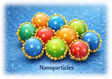
1.Nanoparticles
Introduction
Nanoparticles are extremely small solid particles, usually ranging from 1to 100 nanometers in size. Because of their very small dimensions, nanoparticles behave differently from larger particles and show unique physical, chemical, and biological properties. One of the most important features of nanoparticles is their large surface area, which allows better interaction with drugs and biological systems. This makes them extremely useful in pharmaceutical drug delivery. 5
In drug delivery submissions, nanoparticles act as carriers that can hold drugs either on their surface or within their structure. The drug may be dissolved, entrapped, adsorbed or chemically linked to the nanoparticles matrix. By doing so, nanoparticles help protect drugs from degradation, recover the solubility of poorly water soluble drugs, and enhance their absorption in the body6. They also allow drugs to be release in a controlled manner and, in some cases, delivered directly to the target site, which reduces unwanted side effects.
Based on their structure, nanoparticles are mainly secret into nanosphere and nanocapsules. Nanospheres consist of a solid matrix in which the drug is uniformly distributed, whereas nanocapsules have a core- shell structure where the drug is enclosed within a cavity surrounded by a polymeric membrane. This structural difference plays an significant role in causal the drug release behavior.
Nanoparticles are commonly prepared using biodegradable and biocompatible materials such as polymers, lipids, and inorganic substances. Polymers like chitosan, PLGA, and alginate are frequently used due to their safety and versatility. Several preparation methods, including solvent evaporation, nanoprecipitation, emulsification, and ionic gelation, are employed to obtain nanoparticles with desired size, stability, and drug-loading efficiency.5
In pharmaceutical sciences, nanoparticles have been commonly investigated for various routes of administration such as oral, parenteral, ocular, and transdermal delivery. They are especially important in cancer therapy, vaccine delivery, gene therapy, and the conduct of chronic diseases. Although nanoparticles offer many advantages, challenges related to toxicity, large scale manufacturing, and regulatory approval still exist. Nevertheless, nanoparticles continue to be one of the most promising and widely studied systems in modern drug delivery research
Advantage of Nanoparticles
Nanoparticles offer several important benefits in drug delivery. Their small size tolerates them to increase the solubility of poorly water soluble drugs and improve drug adsorption. They protect the drug from chemical and enzymatic degradation, which enhances drug stability. Nanoparticles can be planned to release drugs in a controlled or sustained manner, reducing dosing frequency and enlightening patient compliance. In addition, targeted delivery using nanoparticles helps concentrate the drug at the disease site while minimizing unwanted side effects. These advantages make nanoparticles highly effective for modern therapeutic application. 6
Disadvantage of nanoparticles
Despite their benefits, nanoparticles have a number of disadvantages. The preparation process can be costly and difficult, especially in extensive production. Certain nanoparticles may be toxic or cause unwanted immune reactions if they are not properly planned. Particle collection and medication leakage are examples of stability issues that can occur during storage. Furthermore, obtaining regulatory approval for products based on nanoparticles is interesting due to the strict safety and quality requirements. 6
Applications
Nanoparticles are widely used in many pharmaceutical applications. They play a dangerous role in targeted drug delivery, particularly in the treatment of cancer, where they help deliver anticancer drugs directly to tumour cells. Nanoparticles are also used to recover oral bioavailability of poorly soluble drugs, as well as drug penetration in ocular and transdermal delivery. Additionally, they are employed in diagnostic imagination, gene therapy, and vaccine delivery. They can be used to treat both chronic illnesses and infectious infections due to their flexibility.5
Assessment of Nanoparticles
To guarantee the efficacy, safety, and quality of nanoparticles, evaluation is crucial. Measurements of particle size and size delivery are used to verify the formulation's stability and uniformity. Physical stability is predicted by evaluating surface charge, which is often expressed as zeta potential. To evaluate how much drug is successfully incorporated into the nanoparticles, drug loading and entrapment efficiency are calculated. To realize the drug's release pattern, in vitro drug release investigations are carried out. Particle form and surface structure are investigated through the use of microscopic methods. To assess the performance and shelf life of nanoparticles under various storage circumstances, stability tests are carried out.6
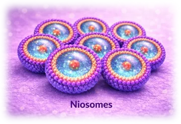
2. Niosomes
Definition
When hydrated in an aqueous solution, niosomes vesicular drug delivery systems mostly made of non-ionic surfactants and cholesterol—form locked bilayer structures. These vesicles are supple carriers in pharmaceutical drug delivery because they can encapsulate both lipophilic and hydrophilic medications. Niosomes have drawn a lot of interest as a liposome extra since of their stability and biocompatibility.
Summary7
In an aqueous environment, non-ionic surfactants self-assemble to produce vesicular carriers called niosomes, which are typically stabilized by cholesterol. The system can transport both hydrophilic and lipophilic medications because these surfactants organize into bilayer structures that encircle an internal aqueous core. Niosomes are becoming a vital tool in regulated and targeted drug delivery studies due to their dual drug-loading capability.
They were created to get around some of the drawbacks of liposomes, namely their expensive price and chemical instability. Large-scale pharmaceutical applications find niosomes appealing due to their relative stability, ease of storage, and affordability. Growing interest in their usage in a variety of therapeutic areas has been fueled by their capacity to increase medication bioavailability, extend medicinal activity, and lessen side effects 7,9
Niosome Types
The number of bilayers and vesicle size are the primary criteria used to categorize niosomes:
Applications and Uses of Niosomes
Niosome Evaluation Parameters
Niosomes must be well characterized in order to guarantee their stability and functionality.
1) Size Distribution and Vesicle Size- Drug release, stability, and biodistribution are all impacted by particle size. Dynamic light trickle methods are frequently used to measure it.
2) Morphology - Vesicle shape and lamellar structure can be ascertained using microscopic methods like transmission electron microscopy. 9
3) Efficiency of Entrapment - This measure, which is essential for assessing formulation efficiency, shows how much medication was successfully absorbed into the vesicles.
4) Zeta Potential - Higher absolute values often imply stronger resistance to aggregation, and surface charge assessment aids in the prediction of physical stability.
5) Drug Release studies in vitro - These investigations ascertain the release pattern and verify whether the technology offers sustained or controlled drug administration. 8
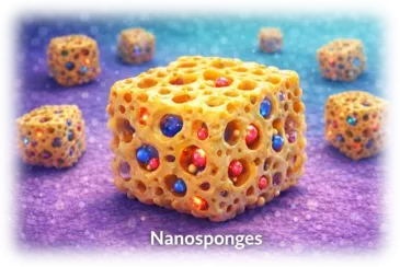
3. NANOSPONGES
Overview
A more recent class of porous, nanoscale drug carriers called nanosponges was created to enhance the delivery of medicinal medicines. In terms of structure, they are networks of three-dimensional cross-linked polymers with microscopic cavities that can hold medicinal molecules. Nanosponges are extremely adaptable carriers in pharmaceutical applications because of these cavities, which enable them to carry both hydrophilic and lipophilic medicines
The majority of medicinal nanosponges are made by cross-linking cyclodextrins with appropriate agents to create stable, sponge-like structures with interior pores. Drug entrapment, degradation protection, and controlled release at the target site are made possible by their porous architecture. Nanosponges improve medication solubility, stability, and bioavailability due to their insignificant size and large surface area. 10
Nanosponges offer a versatile platform for contemporary drug administration since they may be added to a variety of dosage forms, including tablets, capsules, hydrogels, topical creams, and parenteral systems. 10
Different Types of Nanosponges
Nanosponges can be categorized according to their content and structure
a) Nanosponges Based on Cyclodextrin -These are the most extensively researched. They are created by cross-linking cyclodextrins with substances like dianhydrides or carbonyl compounds. Their hydrophilic exterior and internal hydrophobic holes enable them to efficiently encapsulate medications that are unwell soluble in water. 11
b) Nanosponges made of polymers - These are made with biodegradable polymers that create a network that can release drugs under controlled conditions. The polymer-to-crosslinker ratio can be changed to alter the drug release rate. 12
c) Nanosponges, both crystalline and paracrystalline)
Uses for Nanosponges 12
Nanosponges are widely used in the biomedical and pharmaceutical industries.
a) Improvement of Solubility - By creating inclusion complexes inside their cavities, they greatly increase the solubility & rate of dissolution of medications that are weakly soluble in water. Better bioavailability results from this
b) Sustained and Regulated Drug Release - Because of their porous structure, drugs can diffuse gradually, prolonging therapeutic efficacy and lowering the frequency of doses.
c) Topical Medication Administration - By keeping medications on the skin's surface and releasing them gradually, nanosponges lessen irritation and enhance the local therapeutic effect. They have been investigated for anti-inflammatory and antifungal medications.
d) Delivery of Anticancer Drugs - Anticancer medications can be delivered specifically using nanosponges, increasing drug concentration at tumor locations while reducing systemic side effects.
e) Delivery of Proteins and Enzymes - They aid in preserving the stability of delicate biomolecules during delivery, such as proteins and enzymes.
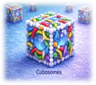
4. CUBOSOMES
INTRODUCTION:
Cubosomes are nano-sized, self-assembled liquid crystalline particles generated from specific lipids in the occurrence of water and stabilizers. In terms of structure, they have a special bicontinuous cubic phase in which two distinct but connected aqueous channels are formed when a lipid bilayer folds into a three-dimensional periodic structure. Because of this configuration, cubosomes have a remarkably high interior surface area and can load medications with various solubility properties. 13
Cubosomes are thermodynamically stable and can hold their structure for prolonged periods of time, in contrast to traditional vesicular systems. They are a very flexible nanocarrier system in contemporary pharmaceutical research because of their internal honeycomb-like structure, which enables them to entrap hydrophilic, lipophilic, and amphiphilic medicines. Cubosomes are especially appealing for topical, transdermal, oral, and controlled drug delivery applications because of their nanoscale size and bioadhesive properties.
Different Cubosome Preparation Techniques 14
1. Top-Down Approach- This method starts by combining a lipid (often glyceryl monooleate) with water to create a bulk cubic phase gel. High-energy procedures like probe sonication or high-pressure homogenization are then used to break down this viscous phase into nanoparticles. To stop aggregation, stabilizers like Poloxamer 407 are used. This approach is extensively used because it provides stable dispersions with repeatable particle size.
2. Bottom-Up Method - Using molecular precursors, cubosomes are formed. Hydrotropes are used to dissolve lipids, and then water is carefully diluted to create cubosomes. This method offers for more control over particle creation and uses less energy than the top-down approach. It is a more modern and effective method of producing cubosomes.
ADVANTAGE 15, 16
1. High Drug Loading Capacity - Cubosomes possess a vast internal surface area with both aqueous channels and lipid domains, allowing them to encapsulate hydrophilic, lipophilic, and amphiphilic medicines efficiently.
2. Sustained and Regulated Drug Release - Drug diffusion is slowed by the intricate, winding interior structure, which prolongs therapeutic efficacy and lowers dosage frequency.
3. Low Toxicity and Biocompatibility - Cubosomes are typically safe and well tolerated because they are frequently made from biodegradable lipids like glyceryl monooleate.
4. Nature of Bioadhesion - Particularly in topical and mucosal distribution, their ability to stick to biological membranes prolongs their residence time at the site of action.
APPLICATION
1. Oral Medication Administration- By promoting breakdown and shielding medications from deterioration in the gastrointestinal tract, cubosomes increase the bioavailability of poorly water-soluble medications. Additionally, their lipid-based composition may facilitate lymphatic absorption.
2. Systems of Controlled and Sustained Release- Cubosomes release medications gradually over time due to their intricate internal channels. This characteristic lowers the occurrence of dosage and aids in maintaining consistent medication levels in the body.
3. Drug Delivery in the Eyes - Their bioadhesive and controlled-release features make cubosomes appropriate for eye formulations, where prolonged contact time promotes drug absorption.
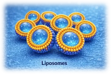
5. LIPOSOMES
Overview
Liposomes are spherical vesicles with an aqueous core encircled by one or more phospholipid bilayers. They are frequently utilized as medication delivery systems and as models for membrane research because of their structural similarity to actual cell membranes.
Liposomes are highly adaptable carriers in medicine, cosmetics, and research because they can carry both fat-soluble medications (inside the lipid bilayer) and water-soluble drugs (within the core). 16
Liposome Types 16,17
Cubosomes are typically prepared by two major approaches:
The size and quantity of bilayers (lamellae) are the primary criteria used to categorize liposomes:
1. Lamellarity-based
2. Considering Function and Composition
Methods of Preparation 17
The goal of liposome production techniques is to create stable lipid vesicles that effectively entrap drugs.
1. Traditional Techniques
Typically, organic solvents are used in these:
These techniques might leave behind solvent residues, which could lead to problems with stability and toxicity.
2. Innovative/Solvent-Free Techniques
Designed to steer clear of dangerous solvents:
These techniques are safer and more suited for medications that are more sensitive, such as proteins and vaccinations.
Uses for Liposomes 18
There are numerous applications for liposomes:
1. Drug Administration
2. Delivery of Vaccines - serve as adjuvants to strengthen the immune system.
3. Gene Transfer -DNA and RNA are delivered into cells by cationic liposomes
4. Examination - utilized as imaging carriers and contrast agents
5. Makeup - Vitamin and antioxidant delivery to the skin
Benefits of Liposomes 17,18
Liposome drawbacks
6. Polymeric nanoparticles
Introduction
Polymeric nanoparticles (PNPs) are colloidal systems made of natural or synthetic polymers that are submicron in size, usually ranging from 10 to 1000 nm. To increase medication stability, solubility, bioavailability, and targeted administration, they are frequently utilized in drug delivery systems. The drug may dissolve, entrap, encapsulate, or adhere to the matrix of the nanoparticle. Polymeric nanoparticles can be either nanospheres (matrix system) or nanocapsules (core–shell system), depending on their structure.
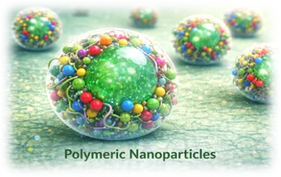
Because of their capacity to improve therapeutic efficacy while lowering toxicity, shield labile pharmaceuticals from degradation, and provide regulated drug release, polymeric nanoparticles have drawn a lot of interest 19. They are especially helpful in vaccine delivery systems, gene delivery, and cancer treatment.20
TYPES OF POLYMERIC NANOPARTICLES
In general, polymeric nanoparticles are categorized as:
A. Structure-Based 19
1. Nanospheres
2. Nanoparticles
B. Based on Polymer Type 21
1. Natural Polymers
2. Synthetic Polymers
APPLICATIONS 19,20
Numerous biomedical uses exist for polymeric nanoparticles:
1. Delivery of Drugs
2. Therapy for Targeted Cancer
3. Delivery of Genes
4. Delivery of Vaccines
5. Uses for Diagnostics
ADVANTAGES 21
DISADVANTAGES
7. CARBON NANOTUBES
INTRODUCTION
Carbon nanotubes (CNTs) are among the most promising carbon nanomaterials for biological applications. which are cylindrical nanostructures made of coiled graphene sheets. Since their discovery by Iijima in 1991, carbon nanotubes (CNTs) have garnered attention because of their outstanding mechanical strength, high surface area, electrical conductivity, chemical stability, and distinctive optical properties.22 Their ultra-small dimensions (diameter in the nanometer range) and high surface area-to-mass ratio allow them to adsorb or conjugate with a variety of therapeutic molecules, including drugs, proteins, DNA,& antibodies. Because of these characteristics, they can be used in tissue engineering, imaging, medication delivery, and diagnostic applications. But issues with toxicity and exposure at work continue to be major obstacles. 23

Types of Carbon Nanotubes 22,23
The quantity of graphene layers, or walls, determines the classification of carbon nanotubes:
1. Carbon nanotubes with a single wall (SWCNTs)
2. DWCNTs, or double-walled carbon nanotubes
3. Carbon nanotubes with multiple walls (MWCNTs)
APPLICATIONS 23
A. Delivery of Drugs
B. Delivery of Genes
C. Uses for Diagnostics
D. Tissue Engineering
E. Applications of Vaccines and Immunotherapy
Benefits of CNTs 22,23
Risks and Drawbacks of CNTs
8. QUANTUM DOTS
Introduction-
Zero-dimensional carbon-based nanomaterials called carbon quantum dots (CQDs) are famous for their exceptional photoluminescence, chemical stability, water solubility, and biocompatibility. They are usually smaller than 10 nm. They were identified as a novel class of fluorescent nanomaterials after being initially found during the purification of carbon nanotubes.
CQDs are structurally composed of an amorphous or graphitic carbon core encircled by several surface useful groups, including amino (-NH?), carboxyl (-COOH), and hydroxyl (-OH) groups. Their optical properties, quantum yield, and chemical reactivity are all greatly influenced by these surface states. Because CQDs are metal-free and less toxic than conventional semiconductor quantum dots (CdSe, CdTe), they are more suitable for use in biomedical applications. 24
Although the precise emission process is still being studied, their fluorescence is caused by emissive traps, surface imperfections, and quantum confinement phenomena. 25

Types of Carbon Quantum Dots 24,26
1. Quantum dots of carbon (CQDs) - Size-dependent fluorescence in quasi-spherical nanoparticles with mixed sp²/sp³ carbon structures
2. Quantum dots of graphene (GQDs)The optical characteristics are influenced by tiny pieces of crystalline graphene sheets with strong edge effects.
3. CNDs, or carbon nanodots -Amorphous carbon nanoparticles in which surface defect states are the primary source of fluorescence
4. Doped Quantum Dots of Carbon - Heteroatoms like nitrogen, sulfur, or phosphorus can be added to CQDs to improve their conductivity, catalytic activity, and fluorescence intensity.
Methods of Synthesis 25,26
There are two main methods used to synthesis CQDs:
1. Top-Down Techniques -These entail dissolving bulk carbon compounds using methods including electrochemical oxidation, arc discharge, and laser ablation.26
2. Bottom-Up Approaches- These entail employing hydrothermal, solvothermal, microwave-assisted, or pyrolysis techniques to carbonize tiny organic compounds.24
Better control over particle size and surface functionalization is possible with bottom-up methods.25
APPLICATION 24,25
1. Bioimaging- CQDs are appropriate for cellular imaging and diagnostic applications because of their bright, consistent fluorescence and low cytotoxicity.
2. Delivery of Drugs - They can conjugate with medications and targeting ligands for regulated and targeted delivery because of their nanosize and functional surface groups.
3. Chemical Sensing and Biosensing - Metal ions, proteins, glucose, and environmental contaminants can all be found using CQDs as fluorescent probes.
4. Solar power - In solar cells and other energy devices, CQDs enhance light gathering, charge transport, and overall efficiency.
5. Devices That Emit Light - CQDs are utilized in LEDs and display technologies because of their adjustable emission wavelengths.
6. Environmental Applications and Catalysis
Because of their electron transfer capabilities, CQDs participate in redox reactions and improve the photocatalytic destruction of contaminants.
Benefits 24,25
Drawbacks 26
1. A lack of knowledge on the mechanisms underlying fluorescence
2. The challenge of attaining a consistent size distribution
3. Differences in quantum yield across batches
4. Difficulties in large-scale manufacturing
9. DENDRIMERS
Introduction
Dendrimers are three-dimensional, monodisperse, highly branching macromolecules having a distinct structure and nanoscale dimensions. Since of their tree-like construction, the word "dendrimer" arises from the Greek words dendron (tree) and meros (part).
They were initially created by Vögtle in 1978, and then by Hawker & Fréchet (convergent synthesis) and Tomalia (divergent synthesis). 27
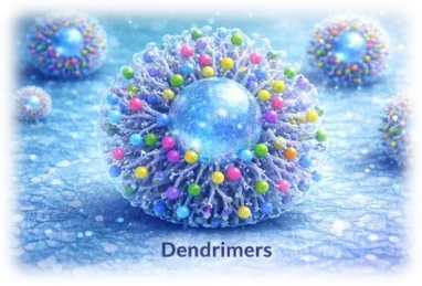
The components of dendrimers are:
1. Core
2. Repeating Branching units (generations G0–G12)
3. Surface functional groups
They typically have : 27,28
1. Their molecular weight is usually between 5,000 and 500,000 g/mol.
2. Minimal polydispersity
3. Globular shape
4. High functionality of the surface
Structure of Dendrimers
There are mainly 3 structure 29
1. Core
2. Interior Layers, or Branches
3. Functional Groups at the Terminal
Types of Dendrimers 27,28,29
In accordance with Chemical Composition
1. Polyamidoamine, or PAMAM
2. Polypropylene Imine, or PPI
3. PLL, or poly-L-lysine
4. Dendrimers made of polyester
B. Modern/Advanced Types
Methods of synthesis 28,31
1. Divergent Method (Tomalia)
2. Convergent Method (Hawker and Frechet)
3. Click Chemistry
Advantage 28,30
Disadvantage 27,29
Applications of Dendrimers 28,29,30
10.ETHOSOMES
Ethosomes are a modern and highly current drug delivery system designed to enhance the penetration of therapeutic agents through the skin. They are soft, flexible vesicular carriers primarily composed of phospholipids, a high concentration of ethanol, and water. Unlike conventional liposomes, ethosomes contain a significant amount of ethanol, which plays a crucial role in improving their ability to deliver drugs across the skin barrier.
The concept of ethosomes was introduced to get around the drawbacks of conventional transdermal medication delivery methods, particularly the difficulty of crossing the stratum corneum—the outermost layer of the skin that acts as a strong protective barrier. Ethanol, being a key component, disrupts the lipid structure of the skin, increasing its permeability. At the same time, it imparts flexibility to the vesicles, allowing them to deform and penetrate deeper layers of the skin more effectively.32
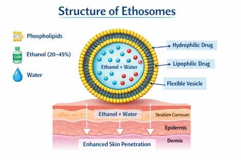
Structurally, ethosomes are vesicles formed by phospholipid bilayers that encapsulate both hydrophilic and lipophilic drugs. The presence of ethanol not only enhances drug solubility but also reduces the size of vesicles and increases their stability. This dual action significantly improves drug loading and delivery efficiency.
One of the major advantages of ethosomes is their ability to deliver drugs to deeper skin layers and even into systemic circulation, making them suitable for both local and systemic therapy. They have been effectively explored for the delivery of a wide range of drugs, including anti-inflammatory agents, antifungal drugs, antiviral drugs, and hormones. Additionally, ethosomes have shown promising results in cosmetic applications such as anti-aging and skin hydration treatments.
Ethosomes provide a number of advantages over traditional medication delivery methods, such as enhanced drug permeation, enhanced bioavailability, non-invasive administration, and better patient compliance. However, they also have some limitations, such as potential skin irritation due to high ethanol content and challenges in large-scale production.
In conclusion, ethosomes represent a significant advancement in transdermal drug delivery technology. Their unique composition and mechanism of action make them a promising carrier system for improving the therapeutic effectiveness of various drugs. Ongoing research continues to explore their potential in overcoming skin barrier limitations and expanding their applications in pharmaceutical and cosmetic fields.
Advantage of Ethosomes 33
Disadvantage of Ethosomes
Methods of Preparation of Ethosomes 33,34
Ethosomes can be prepared using different techniques, but the most commonly used methods are the cold method and the hot method. These methods are designed to produce soft, flexible vesicles with high drug-loading capacity and enhanced skin penetration ability.
1. Cold method
The cold method is the simplest and most frequently used technique for preparing ethosomes.
2. Hot method
The hot method is used when better control over vesicle formation is needed or when dealing with certain formulation conditions.
3. Mechanical Dispersion method
This method is similar to traditional liposome preparation but modified by incorporating ethanol.
Application of Ethosomes
Transdermal drug delivery
Topical drug delivery
Antiviral drug delivery
Antifungal drug delivery
Anti-inflammatory drug delivery
Cosmetic applications
Delivery of large molecules
Hair and scalp treatment
11. Transferosomes
Transferosomes are highly flexible and deformable vesicular drug delivery systems designed to enhance the transport of drugs through the skin. They are also known as ultra-deformable liposomes because of their ability to squeeze through very small pores in the skin without breaking.
These vesicles are mainly composed of:
The presence of edge activators is the key feature that differentiates transferosomes from conventional liposomes. These agents destabilize the lipid bilayer slightly, making it highly flexible and elastic.

Transferosomes work by utilizing the natural hydration gradient of the skin. They move from the dry outer layer (stratum corneum) to the deeper hydrated layers, carrying the drug along with them 35
Composition
Method of preparation 36
A. Thin film hydration method
B. Modified Hand Shaking
C. Reverse Phase Evaporation Method
A. Thin film hydration method
B. Modified Hand Shaking
C. Reverse Phase Evaporation Method
Advantages 37
Disadvantages
Application 38
CONCLUSION
Nanotechnology has emerged as a powerful tool in improving drug delivery systems and overcoming a number of restrictions related to traditional formulations. The development of various nanocarriers such as nanoparticles, liposomes, niosomes, nanosponges, cubosomes, polymeric nanoparticles, dendrimers, carbon nanotubes, quantum dots, ethosomes, and transferosomes has opened new possibilities in pharmaceutical research.
Each of these systems offers specific advantages, including improved drug solubility, enhanced stability, better bioavailability, targeted delivery, and controlled release of drugs. These benefits not only increase the effectiveness of therapy but also help in reducing side effects and improving patient compliance.
Nevertheless, despite their encouraging promise, difficulties like as toxicity risks, high production costs, formulation complexity, and difficulties in large-scale manufacturing still need to be addressed. With continuous advancements in technology and research, these issues are probable to be minimized in the future.
In conclusion, nanotechnology-based drug delivery systems represent an important advancement in modern medicine. They have the potential to make treatments more precise, effective, and safer, and are likely to play a significant role in the future of healthcare and personalized medicine.
REFERENCES

Shashank Namannavar, Sneha Jakaraddi, Raksha Galagali, An Overview : Nanotechnology Based Drug Delivery System in Pharmaceutical Science, Int. J. of Pharm. Sci., 2026, Vol 4, Issue 6, 2906-2930. https://doi.org/10.5281/zenodo.20636585
 10.5281/zenodo.20636585