

We use cookies to ensure our website works properly and to personalise your experience. Cookies policy
1,2,3,5 Department of Pharmacy Practice, Ezhuthachan College of Pharmaceutical Sciences, Marayamuttom Neyyattinkara, Thiruvananthapuram.
4Consultant Physician and Diabetologist in General medicine Department, Nims Medicity Neyyattinkara, Thiruvananthapuram.
This case report describes a 21yearold female diagnosed with Behcet’s disease who developed severe drug-induced pancytopenia secondary to azathioprine therapy, compounded by an acute scrub typhus and left lower limb cellulitis. The patient presented with symptoms such as gingival hypertrophy, and haematological abnormalities. Diagnostic workups, including bone marrow aspiration and biopsy. Management involved immediate cessation of azathioprine. This case highlights, Behcet’s disease complicated by severe azathioprine-induced pancytopenia within three months of therapy, further aggravated by concurrent scrub typhus infection and lower limb cellulitis. Bone marrow examination demonstrated selective myeloid and megakaryocytic suppression with relative erythroid hyperplasia rather than classical trilineage aplasia. Early recognition, discontinuation of azathioprine, hematopoietic growth factor support, eltrombopag, corticosteroids, and targeted antimicrobial therapy resulted in successful haematological recovery
Behcet's disease (BD) is a chronic, relapsing, multisystem inflammatory vasculitis of unknown etiology that affects arteries and veins of all sizes.¹ Initially described by Hulusi Behçet in 1937 as a clinical triad of recurrent oral ulcers, genital ulcers, and uveitis, the disease is now recognized as a complex systemic inflammatory disorder with a broad spectrum of mucocutaneous, ocular, vascular, neurological, gastrointestinal, and musculoskeletal manifestations.² The pathogenesis is multifactorial and is thought to result from an interaction between genetic susceptibility, particularly HLA-B51, environmental triggers, and dysregulated innate and adaptive immune responses, leading to persistent inflammation and endothelial dysfunction.³
The management of Behcet's disease is guided by the severity and extent of organ involvement, with the primary goals of suppressing inflammation, preventing irreversible organ damage, and reducing disease recurrence.⁴ Geographic variation in disease prevalence and clinical phenotype has been well documented, with the highest burden occurring along the historic Silk Road.⁵ Recent advances have further improved the understanding of the epidemiology, clinical spectrum, and therapeutic approaches to Behcet's disease.⁶ Azathioprine remains one of the most widely used immunosuppressive agents because of its proven efficacy in controlling mucocutaneous, ocular, and systemic manifestations while serving as an effective steroid-sparing therapy.⁸
Although azathioprine is generally well tolerated, it is associated with potentially serious adverse effects, of which bone marrow suppression is among the most clinically significant.¹⁰ Drug-induced myelosuppression may manifest as leukopenia, anemia, thrombocytopenia, or severe pancytopenia and can occur secondary to dose-dependent toxicity or individual variations in thiopurine metabolism, emphasizing the need for regular haematological monitoring during treatment.¹¹ Drug-induced pancytopenia also requires prompt recognition because delayed diagnosis may result in life-threatening infections and haemorrhagic complications.¹⁴ Scrub typhus, caused by Orientia tsutsugamushi, is an important endemic rickettsial infection that may itself produce haematological abnormalities, including thrombocytopenia and pancytopenia, thereby complicating the evaluation of cytopenias in immunosuppressed patients.¹² The coexistence of azathioprine-induced pancytopenia with acute scrub typhus infection is rare and poses significant diagnostic and therapeutic challenges.¹³ We report a young woman with Behcet's disease who developed severe azathioprine-induced pancytopenia complicated by concurrent scrub typhus infection and left lower limb cellulitis, highlighting the importance of early recognition of drug-induced haematological toxicity, careful exclusion of alternative causes of pancytopenia, and timely multidisciplinary management.
CASE PRESENTATION
A 21-year-old female with a history of Behçet’s disease diagnosed in December 2025—previously maintained on azathioprine, hydroxychloroquine, and steroids—presented with left lower limb cellulitis and an ulcer over the dorsum of the foot. Her clinical presentation was further complicated by systemic symptoms including hair fall, nail discoloration, epistaxis, and gingival hypertrophy. Initial evaluations revealed profound haematological suppression, prompting extensive inpatient diagnostic workups and multidisciplinary interventions.
Laboratory Investigations
Demonstrated microcytic hypochromic anaemia, moderate leukopenia with relative lymphocytosis, and thrombocytopenia, amounting to pancytopenia.
Showed a hypocellular marrow for age with prominent erythroid hyperplasia and a marked reduction in myeloid cells and megakaryocytes, favouring drug-induced suppression alongside co-existing iron deficiency anemia.
Acute leukaemia panel analysis of the bone marrow displayed no definitive evidence of blasts, with normal distribution of T-lymphocytes, B-lymphocytes, and NK cells.
Antiphospholipid antibodies (Beta-2 Glycoprotein 1 IgG/IgM and Cardiolipin IgG) were negative, while infectious screening indicated concurrent acute Scrub Typhus and positive Herpes Simplex Virus serology.
Liver function tests, renal function tests, electrolyte panels, and thyroid function parameters remained largely within normal limits, while C-reactive protein showed mild elevation.
Upon hospitalization from March 27, 2026, to April 14, 2026, the patient's therapeutic regimen was systematically modified. Azathioprine was immediately held and permanently discontinued due to confirmed drug-induced bone marrow toxicity. She received targeted antibiotic therapy for the acute scrub typhus infection, alongside supportive management comprising packed red blood cell (PRBC) transfusions, platelet management, and granulocyte colony-stimulating factor (Filgrastim) support to recover from severe myelosuppression. Anti-inflammatory and immunosuppressive management was adjusted using steroid regimens, including methylprednisolone and dexamethasone. She showed steady clinical improvement and was discharged with recommendations for regular rheumatology follow-up and future consideration of hydroxychloroquine resumption under specialist supervision.
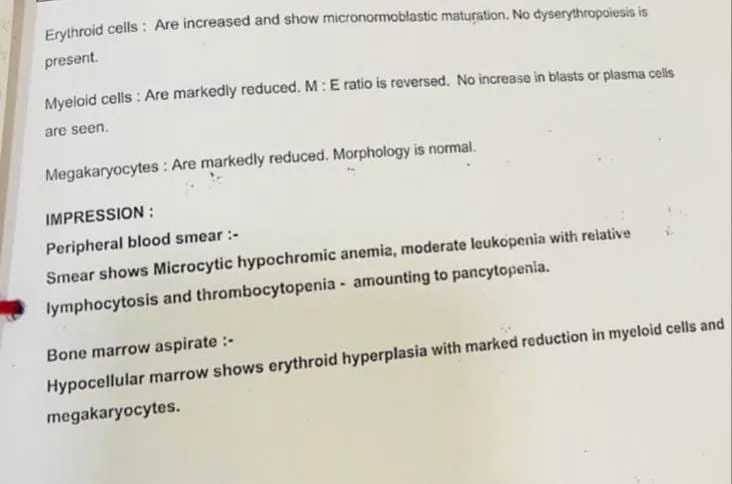
Fig 1:Peripheral blood smear
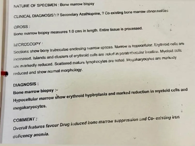
Fig 2: bone marrow biopsy
DISCUSSION
Behcet's disease is an intricate chronic vasculitis that demands careful, long-term immunomodulatory therapy. Azathioprine is a cornerstone purine analogue widely utilized as a steroid-sparing agent in managing systemic vasculitis and autoimmune conditions. However, its utilization carries inherent risks of severe adverse events, most notably dose-dependent or idiosyncratic bone marrow suppression resulting in leukopenia, thrombocytopenia, and pancytopenia.
In this case, the patient developed severe pancytopenia following maintenance therapy, which diagnostic bone marrow biopsy and aspirate confirmed as drug-induced suppression showing marked myeloid and megakaryocytic depletion alongside erythroid hyperplasia. Complicating this presentation was a concurrent acute Scrub Typhus infection and lower limb cellulitis, which heightened systemic inflammation and infection risk. The diagnostic challenge lay in differentiating drug toxicity from a primary haematological malignancy or flare of the underlying systemic disease. Flow cytometric analysis successfully ruled out acute leukaemia by confirming the absence of blast populations.
Management necessitated the prompt withdrawal of azathioprine alongside aggressive supportive care, G-CSF administration, blood component transfusions, and targeted anti-infective treatment for scrub typhus, mirroring protocols established for drug-induced myelosuppression. This case underscores the vital necessity for rigorous, routine monitoring of complete blood counts in patients undergoing chronic azathioprine therapy to mitigate life-threatening cytopenias.
The present case demonstrates several distinctive clinical and pathological features that differentiate it from previously reported cases of azathioprine-induced severe myelosuppression. While most published reports describe this adverse reaction in patients with systemic lupus erythematosus, inflammatory bowel disease, or renal transplant recipients, our patient had underlying Behçet’s syndrome, an uncommon indication associated with severe azathioprine-induced bone marrow toxicity. Bone marrow examination revealed an atypical pattern of lineage-specific suppression rather than classical pan-aplasia. There was marked suppression of the myeloid and megakaryocytic series, evidenced by a profound reduction in granulocytic precursors and megakaryocytes, whereas the erythroid lineage showed compensatory hyperplasia with micronormoblastic maturation and no evidence of dyserythropoiesis. This finding suggests preservation of erythropoiesis, likely driven by co-existing iron deficiency anemia, rather than uniform failure of all hematopoietic cell lines. Another unique aspect of this case was the concurrent presence of scrub typhus, confirmed by positive IgM serology. Unlike previously reported infectious complications that predominantly involve opportunistic bacterial or fungal infections secondary to neutropenia, the coexistence of an endemic vector-borne infection added further complexity to the clinical presentation and necessitated prompt initiation of doxycycline alongside management of severe marrow suppression. The patient also exhibited extensive dermatological and mucosal manifestations, including diffuse alopecia consistent with anagen effluvium, lower limb cellulitis with a non-healing foot ulcer, gingival hyperplasia, and epistaxis, all developing within approximately three months of initiating azathioprine. These findings indicate rapid cytotoxic injury affecting hair follicles, mucosal tissues, and hematopoietic cells, features that are infrequently reported together in the literature. Furthermore, successful management was achieved using a comprehensive non-transfusional rescue strategy comprising immediate discontinuation of azathioprine, early administration of eltrombopag to promote megakaryopoiesis, granulocyte colony-stimulating factor to accelerate neutrophil recovery, a short course of high-dose dexamethasone, and targeted antimicrobial therapy with doxycycline for scrub typhus and piperacillin–tazobactam for bacterial coverage. The favorable haematological recovery observed with this multidisciplinary approach highlights the importance of early recognition, prompt withdrawal of the offending drug, aggressive infection control, and individualized supportive therapy in managing severe azathioprine-induced bone marrow toxicity.
CONCLUSION
This case illustrates a complex presentation of Behcet’s disease complicated by azathioprine-induced pancytopenia and acute scrub typhus infection. Timely diagnostic confirmation via bone marrow examination, immediate discontinuation of the offending immunosuppressant, and comprehensive supportive and antimicrobial management resulted in successful haematological and clinical recovery. Vigilant monitoring and individualized patient evaluation remain essential for optimizing outcomes in chronic autoimmune disorders managed with potent cytotoxic therapies.
REFERENCES


Alfeena N, Renjitha M S, Grace Raju, Jim Litton, Shaiju Dharan, Drug-Induced Pancytopenia in Behcet's Disease with Coexisting Scrub Typhus Infection: A Case Report, Int. J. of Pharm. Sci., 2026, Vol 4, Issue 8, 1692-1697, https://doi.org/10.5281/zenodo.21870838
 10.5281/zenodo.21870838