

We use cookies to ensure our website works properly and to personalise your experience. Cookies policy
Department of Pharmacology, PES’s Modern College of Pharmacy, Nigdi, Pune.
Objectives Non-alcoholic fatty liver disease (NAFLD) is an increasingly prevalent global health concern, closely associated with obesity, abnormal lipid profiles, and impaired insulin sensitivity. The need for safer alternatives is highlighted by the restricted and sometimes harmful nature of current treatments. This study assessed the hepatoprotective effects of Nyctanthes arbor-tristis leaf extract against NAFLD in Wistar rats produced by a high-cholesterol, high-fat diet (HCHFD). MethodsNAFLD was induced by HCHFD administration for 4 weeks. Rats were divided into normal control, disease control, and treatment groups receiving N. arbor-tristis leaf extract at 50, 100, and 200 mg/kg body weight alongside HCHFD. Biochemical assessments included serum lipid profile (LDL, HDL, triglycerides), liver function markers (ALT, AST, ALP), body and liver weights, TNF-? levels, and antioxidant enzymes (SOD, CAT). Histopathology was performed to assess hepatic steatosis and inflammation.ResultsTreatment significantly improved lipid profile, reduced ALT, AST, and ALP, restored SOD and CAT activities, and lowered TNF-? levels in contrast to the disease control group. Histological analysis confirmed reduced lipid accumulation, steatosis, and inflammation in treated groups, with the 200 mg/kg dose exhibiting the the most pronounced effect.ConclusionsN. arbor-tristis leaf extract exerts hepatoprotective, antioxidant, and anti-inflammatory effects in HCHFD-induced NAFLD. These findings suggest its potential as a safe, plant-based therapeutic option for NAFLD management.
Non-alcoholic fatty liver disease, which comprises a variety of associated disorders ranging from simple hepatic steatosis to steatohepatitis, advanced fibrosis, and cirrhosis, is defined as the buildup of fat in the hepatic tissue that surpasses 5% to 10% of its weight in the absence of significant ethanol consumption. The accumulation of fatty liver and hepatic triglycerides plays a crucial part in the development of various metabolic diseases, including diabetes mellitus, obesity, insulin resistance, hypertension, and dyslipidemia, highlighting the critical role that NAFLD management plays in promoting health.(1)
The conventional categorization for nonalcoholic fatty liver disease (NAFLD) is the presence of fatty liver (>5% of hepatocytes with steatosis) accompanied by no other hepatic disorders, such as viral hepatitis or excessive alcohol consumption (2). Given that NAFLD can coexist with other liver illnesses, this suggests that NAFLD is only present after other pathologies have been ruled out, which is misleading (3). Since metabolic dysfunction is the primary cause of this type of liver disease, the alternative term metabolic-associated fatty liver disease (MAFLD) was recently proposed (2).
As obesity and metabolic problems have become more prevalent worldwide, so too has the prevalence of NAFLD. According to recent estimates, 25 to 30 percent of people worldwide suffer from NAFLD, with notable regional differences. (2). As the primary cause of chronic liver disease globally, nonalcoholic fatty liver disease (NAFLD) places a significant strain on healthcare systems because of its progression-related consequences, which include cardiovascular disease, which is the primary. As obesity and metabolic problems have become more prevalent worldwide, so too has the prevalence of NAFLD. According to recent estimates, 25 to 30 percent of people worldwide suffer from NAFLD, with notable regional differences. (2). As the primary cause of chronic liver disease globally, nonalcoholic fatty liver disease (NAFLD) places a significant strain on healthcare systems because of its progression-related consequences, which include cardiovascular disease, which is the primary cause of death for NAFLD patients (4).
Despite its high prevalence and clinical significance, there are currently no FDA-approved pharmacological treatments for NAFLD. Management primarily focuses on lifestyle interventions, including weight loss, dietary modifications, and increased physical activity, which have been shown to improve liver histology and metabolic parameters (5).
However, the heterogeneous nature of NAFLD and the complex interplay between genetic, epigenetic, and environmental factors highlight the need for novel therapeutic strategies and precision medicine approaches (6).
Natural remedies have a long history of being used safely and effectively in traditional medicine throughout many countries. Recent studies on the role of nutrition in the pathophysiology of fatty liver have concentrated on assessing how herbal extracts and supplements might work as functional food ingredients to stop the buildup of hepatic lipids.(7)Nyctanthes arbor-tristis has long been recognized in traditional medicine for its hepatoprotective properties. However, the specific effects of its leaf extract on non-alcoholic fatty liver disease (NAFLD) have not been extensively investigated. NAFLD is characterized by hepatic lipid accumulation, oxidative stress, and inflammation, which contribute to liver damage and disease progression. These contributing factors are targeted by phytochemicals such as flavonoids, alkaloids, and terpenoids, as well as active constituents like nyctanthic acid, β-sitosterol, and arborside A, B, and C present in Nyctanthes arbor- tristis leaf extract. These phytocompounds are known to modulate oxidative stress, regulate lipid metabolism, and exert anti-inflammatory effects. (8)
The present study have been undertaken with the objective to evaluate the potential protective effect of ethyl acetate leaf extract of NAT in a wistar rat model of NAFLD.
2. Materials and Methods:
2.1. Plant material and extraction:
2.1.1 Collection, identification and authentication of Nyctanthes arbor-tristis leaves
Fresh leaves of Nyctanthes arbor-tristis Linn. were collected during the month of January from Pune region, Maharashtra, India. Plant identification and authentication was done at Botanical Survey of India, Western Regional Centre, Pune. By Dr. A. Benniamin and the Plant species was identified and confirmed to be Nyctanthes arbor-tristis linn. From Oleaceae family. Specimen no. BSI/WRC/PI Id.2025/AM/08.
2.1.2 Extract Preparation
Leaves were washed, and shade-dried for 7–10 days, ensuring minimal exposure to direct sunlight to prevent photodegradation of phytoconstituents. The dried leaves were then coarsely powdered and subjected to maceration extraction using ethyl acetate as the solvent for 72 hrs, at the end of the extraction period, the mixture was filtered through Whatman filter paper to separate the liquid extract from the solid residue. The clear filtrate, containing flavonoids and other lipophilic components, was then concentrated by evaporating the ethyl acetate at room temperature. (9) Until further usage, the extract was kept at 4°C in sealed containers.
2.2 Phytochemical screening tests
The primary phytoconstituents (glycosides, alkaloids, tannins, saponins, terpenoids, carbohydrates, cardiac glycosides, anthraquinones glycosides, flavonoids, and phenols) contained in the extracts were identified through qualitative phytochemical screening of the extracts using color reactions.(10)
2.3 Experimental Animals:
Wistar albino male rats weighing 150–200 g were purchased from Crystal Bio Lab, Pune, Maharashtra, India. Animals were housed in standard polypropylene cages under controlled conditions (temperature 22 ± 2°C, relative humidity 50–60%, and 12 h light/dark cycle). They are allowed to access to standard laboratory chow and water ad li libtum. All experimental procedures were approved by the Institutional Animal Ethics Committee (IAEC) and conducted in accordance with CCSEA.
2.4 High Cholesterol High Fat Diet:
Rats were fed a high-cholesterol, high-fat diet (HCHFD) to induce NAFLD for a period of 4 weeks. The diet consisted of standard rat chow enriched with cholesterol (2%), cholic acid (0.5%), and corn oil (20%), a suspension was prepared using sodium CMC (0.5%) as a suspending agent & administered via oral gavage in the concentration of 10 ml/kg once daily for the duration of induction period to induce NAFLD(11).(12) Animals were observed for parameters such as food intake, water intake, and body weight changes. Table 1 represents composition of animal diets.
Table 1. Compositions of animal diets.
|
Component |
Normal Control Diet (%) |
HC-HF Diet (%) |
Purpose |
|
Standard chow |
100 |
77.5 |
Base diet for all groups |
|
Cholesterol |
– |
2.0 |
To induce hypercholesterolemia |
|
Cholic acid |
– |
0.5 |
To enhance cholesterol absorption |
|
Corn oil |
– |
20.0 |
High-fat source to induce hepatic steatosis |
|
Nyctanthes arbor-tristis extract |
– |
50, 100, and 200 mg/kg body weight (Groups IV–VI, respectively) |
Experimental treatment for evaluating hepatoprotective effect |
2.5. Experimental Design:
A total of 36 rats had been divided into six groups at random, each consisting of six individuals (n = 6). The study was conducted over a period of 60 days, with the first 30 days allocated for disease induction, followed by 30 days of treatment. The grouping and treatment protocols were as further: Group I (Normal Control) received a standard diet for 30 days to establish baseline parameters. Group II (Disease Control) was fed a high cholesterol high fat diet (HCHFD), consisting of standard chow supplemented with 2% cholesterol, 0.5% cholic acid, and 20% corn oil, once daily for 30 days to induce NAFLD. Group III received Pioglitazone (3 mg/kg/day) for 30 days following NAFLD induction, serving as the standard drug treatment group. (13)Groups IV, V, and VI were administered N. arbor-tristis leaf extract at doses of 50, 100, and 200 mg/kg/day, respectively, for 30 days after NAFLD induction to evaluate the extract’s dose- dependent therapeutic efficacy. Except for the Normal Control group, all other groups received the HCHFD for the first 30 days to induce NAFLD. Treatment regimens were initiated on day 31 and continued until day 60. Using a gavage technique, the extract was taken orally once daily. Throughout the study period, body weight and food intake were routinely recorded.
After the experiment was completed, the animals were put unconscious after receiving anesthesia. A cardiac puncture was used to draw blood for biochemical analysis of the serum, and liver samples were taken for histopathological evaluation, oxidative stress parameter assessment, and lipid analysis.
2.6 Procedure:
Male Wistar rats (150–180 g) were housed under standard laboratory conditions and fed a high- cholesterol high fat diet (HCHFD) for 4 weeks to induce non-alcoholic fatty liver disease (NAFLD). Fresh leaves of Nyctanthes arbor-tristis were collected, shade-dried, and subjected to maceration using ethyl acetate as a solvent. The extract was concentrated, stored at 4°C, and used for treatment. After 4 weeks on the HCHFD, rats were divided up into six groups at random. (n = 6): normal, disease control, three treatment groups receiving N. arbor-tristis extract at 50, 100 and 200 mg/kg orally, and a standard group receiving pioglitazone (3 mg/kg). Treatments were given once daily for 4 weeks. The rats were sedated for a cardiac puncture during the end of the experiment after fasting for the entire night.The collected blood samples were processed to separate the serum, which was then analyzed for various biochemical parameters to assess liver function and metabolic status. The evaluations included measurements of liver weight, liver enzymes (ALT and AST), lipid profile components (total cholesterol, triglycerides, LDL, and HDL), hepatic fat content and indicators of oxidative stress and inflammation, such as Sodium dismutase (SOD), catalase (CAT), reduced glutathione (GSH) and tumor necrosis factor-alpha (TNF-α).
2.7 Determination of body weight, liver weight, food & water intake
Body weight, food intake & water intake was recorded at the interval of day 0, 30 and day 60. At the conclusion of the study, liver weight was measured and compared to normal controls.
2.8 Evaluation of Blood and Serum Biochemical Parameters
Following four weeks of HCHFD feeding, each rat's body weight was measured after an overnight fast. Diethyl ether (3-5 ml) was used to anaesthetize them. Each rat had five milliliters of blood extracted using the retroorbital technique. The serum was used to measure the levels of triglycerides (TG), total cholesterol (TC), low-density lipoprotein (LDL), high-density lipoprotein (HDL), very low-density lipoprotein (VLDL), alanine transaminase (ALT), alkaline phosphatase (ALP), and aspartate transaminase (AST) using standard kits (Erba kits).
2.9 Determination of Oxidative stress & inflammatory biomarkers
To determine antioxidant effect of EANAT extract the systemic Oxidative stress levels of rats were determined The SOD and CAT assays were carried out at SCITELSA Navi Mumbai, India, (14)the Inflammatory mediator was determined from serum by detecting TNF-α levels in rat serum and the tests were carried out at SCITELSA Navi Mumbai, India.
2.10 Oral Glucose Tolerance Test
Rats received glucose (1.5 g/kg body weight) orally after fasting for 6 hours following the beginning of the light cycleTail vein blood samples were obtained at baseline and at the designated intervals (15, 30, 60, 90, and 120 minutes) after glucose treatments. We used a diabetic monitoring strip (Lifescan One Touch, IN) to measure blood glucose levels. (15)
2.1 Estimation of liver fat content
Hepatic fat percentage was estimated using the Folch method, involving homogenization of ~100 mg liver tissue in methanol:chloroform (2:1), followed by sonication, overnight incubation, and centrifugation. The lipid-containing phase was collected, dried, and weighed, with lipid content normalized to the initial tissue weight.(16)
2.12 Histopathological Examinations
After being separated, the sample of animal tissue was placed in a 10% neutral buffer formalin solution. The specimens were fixed in paraffin after being cut into slices that ranged in thickness from 3 to 5 μm. Hematoxylin-eosin (H&E) stain was applied to serial slices (3 μm) that had been cut with a microtome (Leica Biosystems, Germany). A microscope (Motic Microscopes, Tri-County Pkwy, Schertz, TX 78154, United States) was used to view the prepared slides. NAD = No Abnormality Detected, Minimal changes (+1), Mild changes (+2), Moderate changes (+3), and Severe alterations (+4) were the classifications used to indicate the severity of the identified lesions.
2.13 Statistical Analysis
GraphPad Prism Version 10 was used for statistical analysis. The mean ± SEM was used to express the data. Group differences were analyzed using a two-way ANOVA and Dunnett's multiple comparisons test, with p < 0.05 designated as the statistical significance level.
3.Results:
3.1 Acute toxicological investigation
The preliminary toxicological study revealed that EANAT did not cause adverse effects at a high dosage of 2000 mg/kg, with do not observed unwanted effects or mortality in the animals during the 14-day monitoring period.
3.2 Qualitative assessment of phytoconstituents
Preliminary identification of EANAT demonstrated the occurrence of important bioactive secondary compounds. Tests confirmed the presence of flavonoids (Shinoda test), glycosides, phenolic acids, and tannins (Ferric chloride test), terpenoids suggesting the extract is rich in bioactive compounds that may contribute to its pharmacological effects.
3.3 Quantitative assessment
3.3.1 Determination of total flavonoid content (TFC)
Total flavonoid content was determined by the Aluminium chloride method. Spectrophotometric analysis was conducted at 420 nm. The result obtained was 5.77 mg of quercetin equivalent/gm of fraction. (Fig 1.)
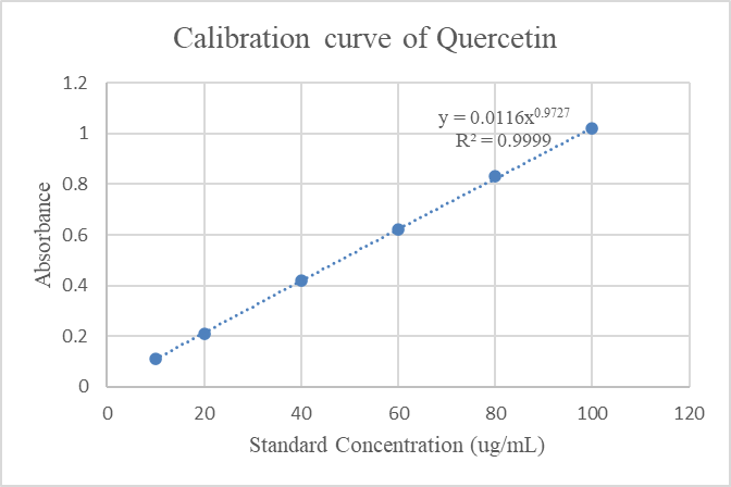
Fig.1: Standard graph of quercetin for estimation of total flavonoid content
3.3.2 Determination of total phenolic content (TPC)
The total phenolic content (TPC) of EANAT was estimated using the modified Folin–Ciocalteu method.(17) Gallic acid (10–50 µg/mL) was used as standard, and 1 mg/mL extract was reacted with diluted Folin–Ciocalteu reagent and sodium carbonate. After 10 minutes, absorbance was measured at 765 nm. The TPC of EANAT was found to be 19.58 µg GAE/mg extract. Standard calibration curve for gallic acid is shown in Fig 2.
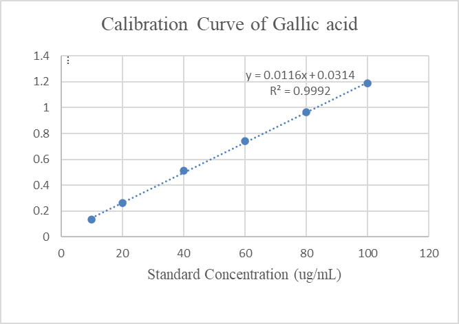
Fig.2: Standard graph of gallic acid for estimation of total phenolic content
3.4 Effect of EANAT on body weight & liver weight
The graph represents the changes in body weight across different treatment groups over a 60-day period in a study evaluating the effect of the ethyl acetate extract of Nyctanthes arbor-tristis (EANAT) on high cholesterol high fat (HCHF) diet-induced non-alcoholic fatty liver disease (NAFLD) in Wistar rats.
At Day 0, all groups exhibited comparable baseline body weights, indicating proper randomization. By Day 30 and Day 60, the disease control (DC) group showed a drastic rise in body weight (p < 0.05, denoted by #) compared to the normal control (NC), reflecting the obesogenic impact of the HCHF diet. However, treatment with EANAT at doses of 50, 100, and 200 mg/kg significantly prevented excessive weight gain in contrast to the DC group (p < 0.05 to p < 0.001), with the 200 mg/kg dose showing a near-normalization of body weight. The standard drug group treated with pioglitazone (3 mg/kg) also demonstrated a significant reduction in body weight gain compared to the DC group.
Liver weight is a crucial parameter for assessing hepatic steatosis and disease progression in NAFLD models. In the present study, the liver weight of animals in the normal control (NC) group was 9.4 ± 40.83 g, representing baseline physiological conditions. A drastic rise in liver weight was observed in the disease control (DC) group (40.4 ± 7.269 g), indicating hepatomegaly due to excessive lipid accumulation and inflammation resulting from HCHFD administration.
Treatment with pioglitazone (HCHFD+PGZ), used as the standard reference, led to a substantial reduction in liver weight (15.9 ± 41.52 g; p<0.001 vs. DC), suggesting reversal of hepatic fat deposition and inflammatory changes. Similarly, animals treated with the ethyl acetate extract of Nyctanthes arbor-tristis (EANAT) showed a dose-dependent decrease in liver weight. At 50 mg/kg, liver weight reduced to 27 ± 2.568 g (p<0.01); at 100 mg/kg, it was 23 ± 31.09 g (p<0.01); and the 200 mg/kg group showed a further reduction to 20 ± 21.09 g (p<0.01), all significantly lower than the DC group. Table 2 indicates liver weight of rats.
Table 2: Effect of administration of EANAT on liver weight in HCHF diet induced NAFLD in Wistar rats
|
Experimental Groups |
Liver Weight (g) |
|
Normal Control (NC) |
9 ± 0.50 |
|
Disease Control (DC) |
13 ± 0.71 |
|
HCHFD + Pioglitazone (3 mg/kg) |
6 ± 0.53 |
|
HCHFD + EANAT (50 mg/kg) |
7 ± 0.34 |
|
HCHFD + EANAT (100 mg/kg) |
12 ± 0.51 |
|
HCHFD + EANAT (200 mg/kg) |
10 ± 0.41 |
3.5 EANAT improved food & water intake:
NAFLD induction via HCHFD resulted in a significant decline in food intake in the DC group, especially noticeable by Day 60 (17.5 ± 0.05 g, ## p<0.01 vs NC), likely due to liver dysfunction, altered metabolism, and satiety regulation disruption. Pioglitazone (PGZ) and all EANAT-treated groups demonstrated a reversal of this decline, restoring food intake closer to normal levels by Day 60. Notably, the EANAT 200 mg/kg group showed the highest recovery (22.4 ± 0.79 g), even surpassing the NC group, indicating a strong restorative and appetite-normalizing effect. The dose-dependent increase in food intake suggests improved hepatic function, metabolic balance, and possibly anti-inflammatory or orexigenic (appetite-stimulating) actions of the extract. In HCHFD-induced NAFLD rats, a marked decline in water intake was observed in the disease control (DC) group by Day 60 (25.7 ± 0.22 mL), indicating metabolic disruption and liver-related dysfunction. In contrast, treatment with pioglitazone and EANAT at all tested doses (50, 100, and 200 mg/kg) led to a dose-dependent restoration of water intake. The EANAT 200 mg/kg group showed the most significant recovery (30.35 ± 0.17 mL), approaching normal levels. This suggests that EANAT effectively mitigates hepatic stress and improves systemic homeostasis, likely due to its hepatoprotective and antioxidant properties. The changes in food & water intake across different experimental groups during the study period are presented in Table 3.
Table:3 Effect of EANAT on Food and Water Intake in Experimental Groups
|
|
||||||
|
Experimental Groups (n=6) |
Food Intake (g/day) – Day 0 |
Food Intake – Day 30 |
Food Intake – Day 60 |
Water Intake (mL/day) – Day 0 |
Water Intake – Day 30 |
Water Intake – Day 60 |
|
Normal Control (NC) |
22.5 ± 0.11 |
22.1 ± 0.10 |
22.0 ± 0.04 |
31.2 ± 0.55 |
31.4 ± 0.42 |
31.3 ± 0.44 |
|
Disease Control (DC) |
20.4 ± 0.13 |
18.3 ± 0.12 |
17.5 ± 0.05 |
31.0 ± 0.28 |
25.9 ± 0.29 |
25.7 ± 0.22 |
|
HCHFD + Pioglitazone (3 mg/kg) |
22.3 ± 1.04 |
18.7 ± 0.99 |
21.1 ± 0.91 |
27.0 ± 0.28 |
26.6 ± 0.21 |
29.6 ± 0.26 |
|
HCHFD + EANAT (50 mg/kg) |
21.4 ± 1.05 |
17.2 ± 0.99 |
20.3 ± 0.91 |
30.9 ± 0.20 |
26.3 ± 0.17 |
28.6 ± 0.18 |
|
HCHFD + EANAT (100 mg/kg) |
19.5 ± 1.10 |
16.5 ± 0.96 |
20.0 ± 0.89 |
30.9 ± 0.29 |
26.8 ± 0.30 |
29.5 ± 0.18 |
|
HCHFD + EANAT (200 mg/kg) |
19.9 ± 1.30 |
16.1 ± 0.86 |
22.4 ± 0.79 |
30.95 ± 0.18 |
27.15 ± 0.26 |
30.35 ± 0.17 |
|
|
|
|||||
3.6 Effect of EANAT on serum lipid profile:
Administration of a high cholesterol high fat (HCHF) diet significantly altered serum lipid parameters in Wistar rats, as observed in the disease control (DC) group. TG, CHO, LDL, and VLDL levels were markedly elevated (p < 0.001), while There was a substantial reduction in the HDL values. (p < 0.001) compared to the normal control (NC) group, indicating the development of dyslipidemia associated with NAFLD. Treatment with ethyl acetate extract of Nyctanthes arbor-tristis (EANAT) at 50, 100, and 200 mg/kg resulted in a dose-dependent normalization of these lipid parameters.
EANAT significantly decreased TG and CHO levels at all doses, with the 200 mg/kg group showing the most pronounced effect (p < 0.01), comparable to the standard pioglitazone-treated group (3 mg/kg). Similarly, LDL and VLDL levels were significantly reduced in all EANAT-treated groups (p < 0.01 to p < 0.001), and the 200 mg/kg dose showed the highest efficacy. HDL levels, which were suppressed in the DC group, improved significantly following EANAT administration, particularly at higher doses (p < 0.05). Pioglitazone demonstrated the greatest improvement across all parameters.
3.7 Effect of EANAT on liver function enzymes:
The HCHF diet-induced NAFLD model resulted in a significant elevation of serum liver enzymes-alanine aminotransferase (ALT), aspartate aminotransferase (AST), and alkaline phosphatase (ALP) in the disease control (DC) group compared to the normal control (NC), indicating hepatic injury and impaired liver function (p < 0.001). As shown in Figure 3, treatment with ethyl acetate extract of Nyctanthes arbor-tristis (EANAT) at 50, 100, and 200 mg/kg demonstrated a dose-dependent normalization of these hepatic biomarkers.
ALT levels were significantly reduced in all EANAT-treated groups, with the highest reduction observed at 200 mg/kg (p < 0.01), closely approaching the values seen in the standard drug (pioglitazone)-treated group (p < 0.001). A comparable pattern was noticed for AST, where EANAT significantly lowered enzyme activity at all doses (p < 0.05 to p < 0.01), with the 200 mg/kg dose showing the most favorable outcome. ALP levels, which were markedly elevated in the DC group, were significantly reduced by EANAT in a dose-responsive manner (p < 0.05 to p < 0.01), with the 200 mg/kg dose again exhibiting comparable efficacy to the standard treatment.
3.8 EANAT attenuated oxidative stress biomarkers
The antioxidant parameters catalase (CAT), glutathione (GSH), and superoxide dismutase (SOD) were significantly altered in the high cholesterol high fat (HCHF) diet-induced non-alcoholic fatty liver disease (NAFLD) model. The disease control (DC) group exhibited a marked decrease in CAT, GSH, and SOD levels compared to the normal control (NC) group, indicating substantial oxidative stress. Treatment with the ethyl acetate extract of Nyctanthes arbor-tristis (EANAT) at doses of 50, 100, and 200 mg/kg significantly restored antioxidant enzyme activities in a dose-dependent manner. Notably, EANAT at 200 mg/kg showed a comparable effect to the standard drug Pioglitazone (3 mg/kg), with significant improvements in CAT and GSH levels (p < 0.01 and p < 0.001, respectively), and enhanced SOD activity (p < 0.01). These findings suggest that EANAT confers hepatoprotective effects by mitigating oxidative stress and enhancing endogenous antioxidant defense mechanisms in NAFLD.
3.9 Effect of EANAT on inflammatory biomarker
The pro-inflammatory cytokine tumor necrosis factor-alpha (TNF-α) was markedly elevated in the disease control (DC) group, indicating enhanced inflammatory signaling in high cholesterol high fat (HCHF) diet-induced non-alcoholic fatty liver disease (NAFLD). The TNF-α concentration significantly increased to approximately 45 pg/ml in the DC group compared to the normal control (NC), which remained near baseline levels (~10 pg/ml) (p < 0.001). Administration of ethyl acetate extract of Nyctanthes arbor-tristis (EANAT) resulted in a dose-dependent reduction in hepatic TNF-α levels. The 50 mg/kg dose showed a moderate yet significant reduction (p < 0.05), while the 100 and 200 mg/kg doses produced more pronounced decreases (p < 0.01) in TNF-α concentration, approaching levels observed in the standard treatment group (Pioglitazone, 3 mg/kg). These results suggest that EANAT effectively attenuates hepatic inflammation, likely by downregulating TNF-α-mediated inflammatory pathways, thereby contributing to its protective effect in NAFLD pathology.
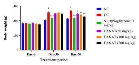
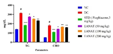
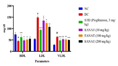
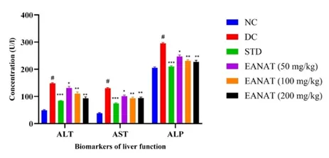
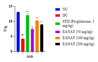
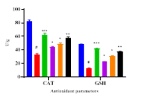
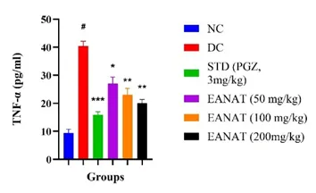
Fig 3. Effect of EANAT on biochemical parameters in HCHF diet induced NAFLD in rats. a) Body weight b) Triglycerides & Cholesterol c) HDL, LDL & VLDL d) Liver function biomarkers e) & f) Oxidative stress biomarkers g) Inflammatory biomarker- TNF alpha. EANAT ethyl acetate extract of Nyctanthes arbor-tristis NC, normal control; DC, HCHF diet control group; Data are expressed as mean ± S.E.M. (n = 6), #p < 0.01, as compared to NC; *p <0.05, **p < 0.01, ***p<0.001 as compared to DC. Data was analysed by one-way Analysis of Variance (ANOVA) followed by Dunnet's multiple tests for comparison.
3.10 Effect of EANAT on OGTT
According to the results of the Oral Glucose Tolerance Test (OGTT), (table 4) ,the disease control (DC) group's blood glucose levels were noticeable higher than those of the normal control (NC), suggesting that their glucose tolerance was compromised. Treatment with pioglitazone (PGZ) and the ethyl acetate extract of Nyctanthes arbor-tristis at doses of 50, 100, and 200 mg/kg (T1, T2, T3) demonstrated a dose-dependent improvement in glucose clearance. Among the extract-treated groups, T3 (200 mg/kg) exhibited the most notable effect, closely approaching the efficacy of PGZ, as reflected by lower glucose levels at all time points and a reduced area under the curve (AUC). These findings suggest that the extract, particularly at higher doses, improves glucose tolerance and mitigates insulin resistance in high-fat high-cholesterol diet-induced NAFLD.
Table 4: Effect of EANAT on OGTT
|
Group |
0 min |
30 min |
60 min |
90 min |
|
120 min |
AUC (mg min/dL) |
|
NC |
89.2 ± 2.4 |
123.5 ± 3.1 |
110.8 ± 2.7 |
99.6 ± 2.2 |
90.3 ± 2.1 |
12200 ± 300 |
|
DC |
108.5 ± 3.0 |
182.7 ± 4.4 |
169.2 ± 3.8 |
155.8 ± 3.5 |
140.6 ± 3.2 |
18780 ± 410 |
|
PGZ |
102.3 ± 2.8 |
145.6 ± 3.7 |
125.2 ± 3.0 |
108.9 ± 2.6 |
95.5 ± 2.4 |
14950 ± 320 |
|
T1 (50 mg/kg) |
106.8 ± 3.1 |
168.9 ± 3.9 |
148.2 ± 3.5 |
131.0 ± 3.1 |
118.6 ± 3.0 |
17240 ± 390 |
|
T2 (100 mg/kg) |
104.1 ± 2.9 |
158.2 ± 3.6 |
135.7 ± 3.1 |
117.5 ± 2.7 |
101.4 ± 2.5 |
15870 ± 360 |
|
T3 (200 mg/kg) |
100.4 ± 2.7 |
148.5 ± 3.3 |
122.8 ± 2.9 |
105.3 ± 2.6 |
92.2 ± 2.3 |
14620 ± 340 |
3.11 Effect of EANAT on histopathology of liver
Histological analysis confirmed NAFLD induction in the disease control (DC) group, showing marked vascular congestion, hepatocyte vacuolation, fatty degeneration, and mild ballooning. In contrast, the normal control (NC) group displayed preserved liver architecture without signs of steatosis or inflammation. Standard drug treatment led to near-normal hepatic structure with minimal fatty changes. NAT extract-treated groups showed dose-dependent improvement. T-1 exhibited moderate vacuolation and fatty changes, T-2 showed reduced degeneration and minimal inflammation, while T-3 demonstrated the greatest protective effect with mild hepatocellular swelling and minimal steatosis. The histopathological changes are shown in fig. 4.
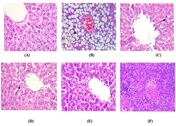
Fig.4.Effect of EANAT and PGZ treatment group on the histology of liver. Typical liver tissue sections (40 X) stained with haematoxylin and eosin (H&E). (A) normal control group (B) HCHF Diet Control group (C) HCHF Diet + Pioglitazone- (3mg/kg), (D) HCHF Diet +EANAT (50 mg/kg), (E) HCHF Diet + EANAT (100 mg/kg), (F) HCHF Diet + EANAT (200 mg/kg),
DISCUSSION
A sedentary lifestyle and the consumption of calorie-dense, high-fat diets are major contributors to visceral adiposity and the onset of non-alcoholic fatty liver disease (NAFLD) (18)In the present study, NAFLD was successfully induced in Wistar rats through the administration of a high-cholesterol high-fat diet (HCHFD), characterized by increased liver weight, hepatic lipid accumulation, elevated serum liver enzymes, dyslipidemia, impaired glucose metabolism, and oxidative stress. This model closely mimicked the pathophysiological hallmarks of human NAFLD, providing a reliable platform for evaluating therapeutic interventions (19)
NAFLD is a progressive metabolic disorder characterized by hepatic steatosis that can advance to non-alcoholic steatohepatitis (NASH), fibrosis, and cirrhosis in the absence of significant alcohol intake. In this study, the ethyl acetate extract of Nyctanthes arbor-tristis (EANAT) was evaluated for its protective potential against HCHF diet-induced NAFLD in Wistar rats.
The HCHF diet, comprising cholesterol, cholic acid, and corn oil, has been reported to induce hepatic steatosis by disrupting lipid homeostasis, promoting oxidative stress, and triggering inflammatory responses(20) Corn oil, rich in energy, promotes excessive fat deposition, while cholesterol and cholic acid enhance lipid absorption, leading to hepatic lipid overload. These effects were evident through a significant increase in body and liver weight in HCHFD-fed rats. Treatment with EANAT significantly attenuated these increases, suggesting modulation of lipid absorption and storage. These gross anatomical findings were confirmed by liver histology, which revealed a marked reduction in fat vacuole accumulation and restoration of normal lobular architecture in EANAT-treated rats compared to the distorted histoarchitecture observed in the disease control group.
Rats fed HCHFD showed conspicuous signs of dyslipidemia, which is defined by increased levels of total cholesterol (TC), triglycerides (TG), and LDL-C with lower HDL-C. This lipid imbalance reflects hepatic lipid accumulation and systemic metabolic dysfunction. EANAT significantly improved the lipid profile in a dose-dependent manner. The hypolipidemic effect may be attributed to its phytoconstituents such as flavonoids and iridoid glycosides, which are known to activate PPAR-α and suppress SREBP-1c, thereby enhancing lipid oxidation and reducing synthesis .(21,22) These biochemical improvements were further supported by histopathological evidence showing a decline in macrovesicular steatosis and preservation of hepatic sinusoidal architecture in the EANAT-treated groups. NAFLD is frequently linked to insulin resistance and glucose intolerance. Following oral glucose tolerance tests, the rats administered HCHFD showed increased fasting blood glucose levels and decreased glucose clearance, (OGTT), indicating systemic insulin resistance.(23) (24)EANAT administration significantly improved glucose tolerance and reduced fasting glucose levels. This antidiabetic effect may be mediated by enhanced insulin signaling and glucose uptake, potentially via modulation of IRS/PI3K/Akt pathways. Histologically, this metabolic correction was paralleled by reduced hepatocyte ballooning and decreased inflammatory infiltration in the liver, suggesting improved hepatocellular integrity and insulin sensitivity.
Serum transaminases-ALT, AST, and ALP serve as sensitive indicators of hepatocellular damage. In the disease control group, these enzymes were significantly elevated, reflecting liver injury. EANAT treated animals showed substantial normalization of these enzyme levels, suggesting hepatoprotection. These biochemical changes correlated with histopathological findings where the EANAT treated livers demonstrated a marked reduction in cytoplasmic vacuolations and inflammatory cell infiltration, further confirming the extract’s membrane-stabilizing and cytoprotective effects.(25)
Oxidative stress, characterized by elevated ROS and depleted antioxidant defenses, plays a central role in NAFLD progression. In HCHFD-fed rats, antioxidant enzyme levels including SOD, CAT, and GSH were significantly reduced.(26) EANAT administration restored these antioxidant parameters, suggesting enhanced redox homeostasis. This is possibly mediated by activation of the Nrf2 signaling pathway and the presence of polyphenolic antioxidants in the extract(27).(28) These antioxidative effects were confirmed histologically by reduced lipid peroxidation-associated damage in hepatocytes and preserved structural integrity of hepatic cords.
Inflammation, primarily mediated by TNF-α and IL-6, exacerbates hepatocyte injury and steatosis. The HCHFD group exhibited significantly elevated TNF-α levels, indicative of hepatic inflammation and immune cell activation. EANAT treatment significantly reduced TNF-α concentrations, implying an anti-inflammatory action possibly mediated by inhibition of NF-κB signaling (29).(30) Liver sections of EANAT-treated rats showed decreased mononuclear cell infiltration and a return to normal hepatic morphology, supporting its anti-inflammatory effect at the tissue level.
CONCLUSION
The current research provides compelling evidence that the ethyl acetate extract of Nyctanthes arbor-tristis leaves confers significant protection against HCHF diet-induced NAFLD in Wistar rats. EANAT improved serum lipid and glucose profiles, normalized liver enzyme levels, enhanced antioxidant defense, and reduced pro-inflammatory cytokines. All these effects were substantiated by histopathological findings, demonstrating restoration of hepatic architecture and reduction in fat accumulation and inflammation. These results corroborate the therapeutic potential of EANAT as a natural remedy for NAFLD through its multifactorial mechanism of action.
REFERENCES


Shital Doifode, Deepti Bandawane, Dnyaneshwar Bomnale, Nyctanthes Arbor-Tristis Leaves Extract Improves the Hepatic Damage in Non-Alcoholic Fatty Liver Disease by Alleviating Oxidative Stress & Inflammation in Fatty Wistar Rats, Int. J. of Pharm. Sci., 2026, Vol 4, Issue 8, 1662-1677, https://doi.org/10.5281/zenodo.21870598
 10.5281/zenodo.21870598