

We use cookies to ensure our website works properly and to personalise your experience. Cookies policy
Narayan Institute of Pharmacy, Gopal Narayan Singh University, Jamuhar, Sasaram, Bihar 821305
Inflammation is a complex biological response associated with numerous chronic diseases, including cardiovascular disorders, metabolic syndromes, autoimmune diseases, and cancer. The search for safer and more effective anti-inflammatory agents has led to increasing interest in medicinal plants and their bioactive phytochemicals. The present study aimed to investigate the phytochemical composition and potential anti-inflammatory activity of Solanum viarum Dunal using phytochemical screening, GC–MS analysis, molecular docking, and in silico pharmacokinetic and toxicity predictions. Preliminary phytochemical screening of the aqueous stem extract revealed the presence of several bioactive constituents including carbohydrates, glycosides, fixed oils, saponins, phenolic compounds, phytosterols, alkaloids, and flavonoids, while proteins and mucilage were absent. The extraction process using distilled water by the percolation method yielded 17.7 % w/w extract. GC–MS analysis further identified several pharmacologically significant compounds such as solasodine, solasonine, solamargine, ?-sitosterol, stigmasterol, campesterol, and chlorogenic acid. These compounds belong primarily to steroidal alkaloids, steroidal glycoalkaloids, phytosterols, and phenolic acids known for their diverse biological activities. To explore their anti-inflammatory potential, molecular docking studies were performed against the inflammatory target protein Cyclooxygenase-2 (PDB ID: 4PH9). Among the screened compounds, chlorogenic acid exhibited the strongest binding affinity with a docking score of –15.9 kcal/mol, followed by solamargine (–12.3 kcal/mol) and solasonine (–11.7 kcal/mol). These values were notably higher than the reference drug Diclofenac sodium, which showed a docking score of –7.5 kcal/mol, indicating stronger predicted interactions of several phytoconstituents with the target protein. Further pharmacokinetic evaluation using SwissADME demonstrated that chlorogenic acid possesses favorable physicochemical characteristics, good aqueous solubility, low suggesting a reduced probability of drug–drug interactions. Toxicity prediction performed using ProTox-II indicated a high safety margin with an estimated LD?? of 5000 mg/kg and absence of carcinogenic, mutagenic, or cytotoxic effects. Overall, the findings of this study demonstrate that Solanum viarum is a rich source of bioactive phytochemicals with significant anti-inflammatory potential. In particular, chlorogenic acid showed strong molecular interactions with the inflammatory target and favorable pharmacokinetic and toxicity profiles. These results support the potential of Solanum viarum as a promising natural source for the development of safer anti-inflammatory agents, although further in vitro and in vivo studies are required to validate these computational predictions.
Chronic inflammatory diseases have become one of the most significant global health challenges of the 21st century. Growing scientific evidence suggests that inflammation plays a crucial role in the development of many major disorders, including metabolic, cardiovascular, neurodegenerative, autoimmune, and malignant diseases. Together, these conditions account for more than half of the global mortality rate (1,2). The World Health Organization has identified chronic inflammation as a key risk factor for several non-communicable diseases such as diabetes mellitus, chronic kidney disease, non-alcoholic fatty liver disease, ischemic heart disease, stroke, and various autoimmune and neuropsychiatric disorders (3). Research also indicates that inflammation occurring early in life can persist for long periods and may increase the likelihood of developing chronic diseases later in adulthood (4).
Inflammation is a natural protective response of the body against infection, injury, or harmful stimuli. Acute inflammation occurs rapidly and usually helps eliminate pathogens and initiate tissue repair. In contrast, chronic inflammation is a prolonged and dysregulated condition that can persist for months or even years. It may arise due to persistent infections, exposure to environmental toxins, autoimmune reactions, repeated inflammatory episodes, or metabolic disturbances that promote oxidative stress and mitochondrial dysfunction (5). Furthermore, several lifestyle and demographic factors such as aging, obesity, smoking, psychological stress, unhealthy dietary habits, and hormonal imbalance can significantly contribute to the development and progression of chronic inflammatory conditions (6).
Although advances in medical science have improved disease diagnosis, reliable laboratory markers for chronic inflammation remain limited. Common diagnostic approaches include serum protein electrophoresis, high-sensitivity C-reactive protein (hsCRP), fibrinogen levels, and cytokine profiling. However, these indicators often provide only partial information and may lack adequate specificity and standardization (7). Consequently, many inflammation-related diseases remain undetected until considerable tissue damage has already occurred.
Non-steroidal anti-inflammatory drugs (NSAIDs) are widely used for the treatment of inflammation and pain. While these medications are effective, their long-term use is associated with several adverse effects, including gastrointestinal irritation, kidney dysfunction, cardiovascular complications, and central nervous system toxicity (8). Because of these limitations, researchers are increasingly exploring natural and plant-based compounds that may provide safer alternatives with effective anti-inflammatory and analgesic properties (9).
One such medicinal plant is Solanum viarum Dunal, a member of the family Solanaceae, commonly known as tropical soda apple. The plant has attracted considerable attention due to its rich phytochemical composition, including steroidal glycoalkaloids, flavonoids, phenolic compounds, and other biologically active constituents (10). Among these compounds, solasodine is particularly important because it acts as a nitrogen analogue of diosgenin and serves as a precursor in the synthesis of several steroid hormones such as corticosteroids and sex hormones (11). Previous studies have reported that extracts of Solanum viarum exhibit a wide range of pharmacological activities, including anti-inflammatory, analgesic, antioxidant, antimicrobial, antipyretic, and anticancer effects (12). Additionally, steroidal glycoalkaloids such as solasonine, solamargine, and khasianine, along with phenolic compounds like caffeic acid, gallic acid, and quercetin, contribute significantly to the plant’s therapeutic potential (13).
Considering its diverse bioactive compounds and traditional medicinal importance, Solanum viarum appears to be a promising natural source for the development of safer anti-inflammatory and analgesic agents. However, scientific studies investigating its pharmacological potential remain relatively limited. Therefore, the present study aims to evaluate the medicinal significance of Solanum viarum Dunal, with particular emphasis on its anti-inflammatory and analgesic activities.
MATERIALS AND METHODS
Chemicals and Reagents
All chemicals and reagents used in the present study were of analytical grade. Diclofenac sodium was used as the standard reference drug for evaluating analgesic and anti-inflammatory activity. Diclofenac is a widely used non-steroidal anti-inflammatory drug that inhibits cyclooxygenase enzymes and suppresses prostaglandin synthesis responsible for inflammation and pain (14). Histamine (1%, 0.1 mL) was used as the inflammatory inducer because it promotes vascular permeability and edema formation in experimental inflammatory models (15).
Plant Material
The plant Solanum viarum Dunal (family: Solanaceae) was collected from the Haridwar region of Uttarakhand, India. Botanical authentication of the plant material was carried out by the Botanical Survey of India, Northern Regional Centre, Dehradun, Uttarakhand.
The collected plant material was washed with distilled water to remove impurities and shade-dried at room temperature. The dried material was then powdered using a mechanical grinder and stored in airtight containers until further analysis.
Preparation of Plant Extract
Approximately 300 g of powdered plant material was subjected to extraction using the Soxhlet extraction technique. The powdered sample was packed in filter paper and placed in the extraction chamber of the Soxhlet apparatus.
Petroleum ether (40–60°C) or n-hexane was used as the extraction solvent. The extraction was carried out in a water bath maintained at 70–75°C for approximately 7 hours. The solvent was continuously refluxed and siphoned through the plant material until the extraction process was completed. The obtained extract was concentrated by removing the solvent and further dried in a hot air oven to eliminate residual solvent. The dried extract was stored in airtight containers for further phytochemical and pharmacological studies (16).
Preliminary Phytochemical Screening
Qualitative phytochemical analysis of the plant extract was performed to identify the presence of major bioactive constituents such as carbohydrates, glycosides, proteins, amino acids, flavonoids, alkaloids, phenolic compounds, saponins, fixed oils, and phytosterols using standard phytochemical methods (17,18).
Carbohydrates were detected using Molisch’s test and Fehling’s test, while proteins and amino acids were identified using Millon’s test and Ninhydrin test. The presence of saponins was confirmed by the foam test. Phenolic compounds and tannins were identified using Ferric chloride test and lead acetate test. Alkaloids were detected using Dragendorff’s, Mayer’s, Wagner’s, and Hager’s reagents. Flavonoids were confirmed using alkaline reagent test and Shinoda test. Phytosterols were detected using Salkowski test and Liebermann–Burchard test. The formation of characteristic color changes or precipitates in these tests indicated the presence of corresponding phytoconstituents.
GC–MS Analysis
Gas chromatography–mass spectrometry (GC–MS) analysis was performed to identify volatile and semi-volatile phytoconstituents present in the plant extract. The analysis was carried out using a GC–MS system equipped with an electron ionization source operating at 70 eV.
A capillary column (30 m × 0.25 mm internal diameter, 0.25 µm film thickness) coated with 5% phenyl and 95% dimethylpolysiloxane stationary phase was used for separation. Helium was used as the carrier gas at a constant flow rate of 1.0 mL/min.
The dried extract was dissolved in analytical-grade solvent and filtered using a 0.22 µm membrane filter. A 1 µL sample was injected in split mode with the injector temperature maintained at 250°C. The oven temperature was initially set at 60°C for 2 minutes and then increased to 280°C at a rate of 10°C/min, followed by a final hold for 10 minutes. The ion source and quadrupole temperatures were maintained at 230°C and 150°C, respectively. Mass spectra were recorded in the scan range of m/z 40–600 (19).
Molecular Docking Study
Molecular docking analysis was performed to investigate the interaction between selected phytoconstituents and the target protein Cyclooxygenase-2 (COX-2), a key enzyme involved in inflammatory processes.
The three-dimensional crystal structure of COX-2 was obtained from the Protein Data Bank with PDB ID: 4PH9. Prior to docking, the protein structure was prepared by removing water molecules and co-crystallized ligands, followed by addition of polar hydrogen atoms and appropriate atomic charges.
The structures of phytocompounds identified through GC–MS analysis were retrieved from the PubChem database in SDF format and converted into PDB format using Open Babel. Energy minimization was performed to obtain stable ligand conformations.
Docking simulations were carried out using AutoDock Vina implemented in PyRx software. Binding affinity was expressed in terms of binding energy (kcal/mol). The best docking conformation was selected based on the lowest binding energy and favorable interaction profile. Protein–ligand interactions such as hydrogen bonding, hydrophobic interactions, π-alkyl interactions, and van der Waals forces were analyzed using molecular visualization tools (20).
For validation of the docking protocol, diclofenac sodium was used as the reference ligand.
ADME and Toxicity Prediction
The pharmacokinetic properties and toxicity profile of selected phytoconstituents were predicted using the ProTox-II web server. The SMILES structures of the compounds were obtained from the PubChem database and uploaded to the platform for analysis.
Parameters evaluated included molecular weight, lipophilicity (LogP), hydrogen bond donors and acceptors, and Lipinski’s rule of five to determine drug-likeness. Compounds satisfying Lipinski’s rule were considered to possess favorable pharmacokinetic properties.
In addition, the tool predicted toxicity endpoints including LD?? values, hepatotoxicity, carcinogenicity, mutagenicity, immunotoxicity, and cytotoxicity, providing insights into the safety profile of the compounds (21).
RESULTS AND DISCUSSION
Preliminary phytochemical screening:
The phytoconstituents were identified by chemical tests, which showed the presence of various phytoconstituents in aqueous extract of Solanum Viarum Dunal and percentage yields are given in different tables.
Table 1: Preliminary phytochemical screening of the aqueous extract of Solanum Viarum Dunal
|
Sr. No. |
Constituents |
Tests |
Ethanolic extract |
|
1. |
Carbohydrate & Glycosides |
Molish’s test |
Present |
|
Fehling’s test |
Present |
||
|
2. |
Fixed oil & fats |
Spot test |
Present |
|
Saponification test |
Present |
||
|
4. |
Proteins & amino acids |
Million’s test |
Absent |
|
Ninhydrin test |
Absent |
||
|
Biuret test |
Absent |
||
|
5. |
Saponins |
Foam test |
Present |
|
6. |
Phenolic compounds |
FeCl3 test |
Present |
|
Gelatin test |
Present |
||
|
Lead acetate test |
Present |
||
|
7. |
Phytosterol |
Salkowiski test |
Present |
|
Libermann burchard Test |
Present |
||
|
8. |
Alkaloids |
Dragendroff’s test |
Present |
|
Mayer’s test |
Present |
||
|
Wagner’s test |
Present |
||
|
Hager’s test |
Absent |
||
|
9. |
Gum & mucilage |
Swelling test |
Absent |
|
10. |
Flavonoids |
Aqueous NaOH test |
Present |
|
Con. H2SO4 test |
Present |
||
|
Shinoda’s test |
Present |
Table 2: Percentage yield of Solanum Viarum Dunal with distilled water
|
Plant used |
Part used |
Method |
Percentage yield |
|
Solanum Viarum Dunal |
stem |
Percolation with distilled water |
17.7 % w/w |
GC-MS analysis
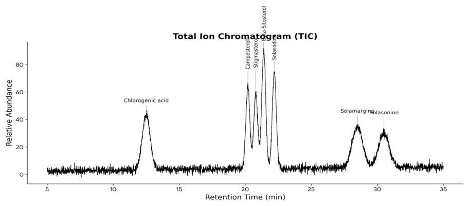
Fig 1. The GC-MS Total Ion Chromatogram (TIC) for the listed compounds is presented above. This simulated chromatogram illustrates the typical elution order expected for these compounds on a standard non-polar capillary column.
Table 3. GC–MS profiling of bioactive compounds presents in Solanum viarum Dunal extract
|
Sr. No. |
Compound |
Formula |
Score |
Fragment ions (m/z) |
IUPAC Description |
m/z (theoretical) |
Retention (predicted) |
|
1 |
Solasodine |
C27H43NO2 |
95 |
413, 398, 255, 213 |
(3β,22α,25R)-spirosol-5-en-3-ol |
414.334 |
High |
|
2 |
Solasonine |
C45H73NO16 |
92 |
884, 722, 560, 414 |
(25R)-spirost-5-en-3β-yl O-β-D-glucopyranosyl-(1→2)-O-β-D-galactopyranoside |
884.479 |
Very high |
|
3 |
Solamargine |
C45H73NO15 |
93 |
868, 706, 414, 396 |
(25R)-spirost-5-en-3β-yl O-β-D-glucopyranosyl-(1→3)-O-α-L-rhamnopyranoside |
868.484 |
Very high |
|
4 |
β-Sitosterol |
C29H50O |
97 |
414, 396, 255, 213 |
(3β)-stigmast-5-en-3-ol |
414.386 |
High |
|
5 |
Stigmasterol |
C29H48O |
96 |
412, 394, 255 |
(3β,22E)-stigmasta-5,22-dien-3-ol |
412.370 |
High |
|
6 |
Campesterol |
C28H48O |
95 |
400, 382, 255 |
(3β)-ergost-5-en-3-ol |
400.370 |
High |
|
7 |
Chlorogenic acid |
C16H18O9 |
94 |
353, 191, 179 |
(1S,3R,4R,5R)-3-[(E)-3-(3,4-dihydroxyphenyl) prop-2-enoyl]oxy |
|
|
GC–MS Profiling of Solanum viarum Dunal Extract
GC–MS of the Solanum viarum Dunal extract delivered a very chemically diverse phytoconstituent profile which is dominated by steroidal alkaloids, steroidal saponins, phytosterols, and phenolic acids. The total ion chromatogram (Fig. 1) showed well-resolved peaks corresponding to compounds with high predicted retention behaviour indicating the presence of relatively non-polar, high-molecular-weight constituents.
Among the identified compounds, solasodine, solasonine, and solamargine were detected as major steroidal alkaloids and glycoalkaloids representative of the Solanum genus. These compounds showed high spectral match scores (92–95%) with diagnostic fragment ions corresponding to the cleavage of the steroidal nucleus and glycosidic moieties. The presence of phytosterols such as β-sitosterol, stigmasterol, and campesterol has been confirmed due to high scores (95-97%) and typical sterol fragment ions. Chlorogenic acid, which is one of the most important phenolic acids with high biological relevance, has been identified due to a high confidence score (94%) with typical fragment ions at m/z 353, 191, and 179. The identification of these phytocompounds lends validity to the traditional medicinal use of S. viarum since steroidal alkaloids and phytosterols are well-known to exhibit antimicrobial and anti-inflammatory properties, whereas chlorogenic acid has been proven to possess high antioxidant activity. These phytocompounds were thus chosen for molecular docking studies.
Molecular Docking Analysis
Molecular docking was performed to evaluate the binding interactions of selected phytoconstituents with the target protein (PDB ID: 4PH9). The docking scores indicated variable binding affinities across the compounds (Figs. 2–8).
Chlorogenic acid exhibited the strongest binding affinity with a docking score of –15.9 kcal/mol (Fig. 6), suggesting highly stable binding within the active site. This strong interaction is attributed to its multiple hydroxyl and carboxyl functional groups, which facilitate extensive hydrogen bonding and electrostatic interactions.
Solamargine and solasonine also demonstrated strong binding affinities with docking scores of –12.3 kcal/mol (Fig. 7) and –11.7 kcal/mol (Fig. 8), respectively. Their glycosidic moieties appear to contribute to enhanced interaction stability through polar contacts, while the steroidal core ensures hydrophobic complementarity.
Among the phytosterols, stigmasterol (–10.1 kcal/mol; Fig. 3) and campesterol (–9.4 kcal/mol; Fig. 5) exhibited strong hydrophobic interactions within the binding cavity, whereas β-sitosterol (–7.8 kcal/mol; Fig. 4) and solasodine (–6.0 kcal/mol; Fig. 2) showed moderate binding affinities.
Overall, docking results indicate that both polar phenolic compounds and bulky steroidal molecules can effectively interact with the target protein through distinct but complementary interaction mechanisms.
The reference drug diclofenac sodium exhibited a binding affinity of –7.5 kcal/mol toward the selected inflammatory target, which served as a benchmark for validating the docking protocol. Notably, several phytochemical ligands identified from Solanum viarum demonstrated comparable or higher binding affinities than diclofenac, indicating stronger or equivalent molecular interactions within the active site. This observation suggests that the selected phytoconstituents possess favorable binding potential relative to the standard NSAID. However, these findings are based solely on computational predictions and highlight the need for further experimental validation to confirm their biological relevance.
Table 4. Interaction profile of Solasodine with target protein (Fig. 2)
|
Amino Acid (Chain:Residue) |
Bond Type |
Distance (Å) |
|
Hydrophobic pocket residues |
Hydrophobic |
5.05 |
|
Alkyl-interacting residues |
π-Alkyl |
5.98 |
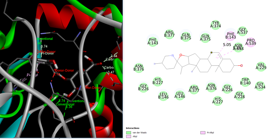
Fig 2. Interaction with Solasodine Docking score -6.0
Table 5. Interaction profile of Stigmasterol with target protein (Fig. 3)
|
Amino Acid (Chain:Residue) |
Bond Type |
Distance (Å) |
|
Hydrophobic cavity residues |
Hydrophobic |
4.49 |
|
Sterol-binding residues |
π-Alkyl |
2.55, 4.74 |
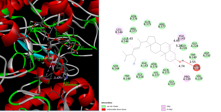
Fig 3. Interaction With Stigmasterol Docking score -10.1
Table 6. Interaction profile of β-Sitosterol with target protein (Fig. 4)
|
Amino Acid (Chain:Residue) |
Bond Type |
Distance (Å) |
|
Non-polar residues |
Hydrophobic |
5.37, 4.99,4.87 |
|
Alkyl-interacting residues |
π-Alkyl |
4.84 |
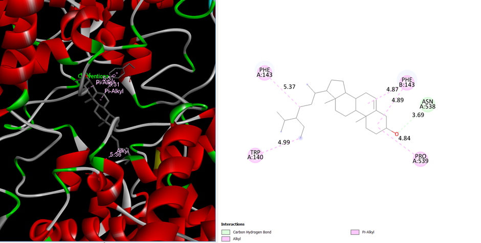
Fig 4. Interaction With β-Sitosterol docking score -7.8
Table 7. Interaction profile of Campesterol with target protein (Fig. 5)
|
Amino Acid (Chain:Residue) |
Bond Type |
Distance (Å) |
|
Binding pocket residues |
Hydrophobic |
1.94 |
|
Steroidal core interactions |
π-Alkyl |
5.37, 5.31, 5.36 |
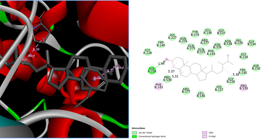
Fig 5. Interaction with Campesterol Docking score -9.4
Table 8. Interaction profile of Chlorogenic acid with target protein (Fig. 6)
|
Amino Acid (Chain:Residue) |
Bond Type |
Distance (Å) |
|
Polar active-site residues |
Hydrogen bond |
3.64 |
|
Charged residues |
Electrostatic |
2.41, 3.74, 3.47 |
|
Aromatic residues |
π–π interaction |
2.40, 2.14, 5.08 |
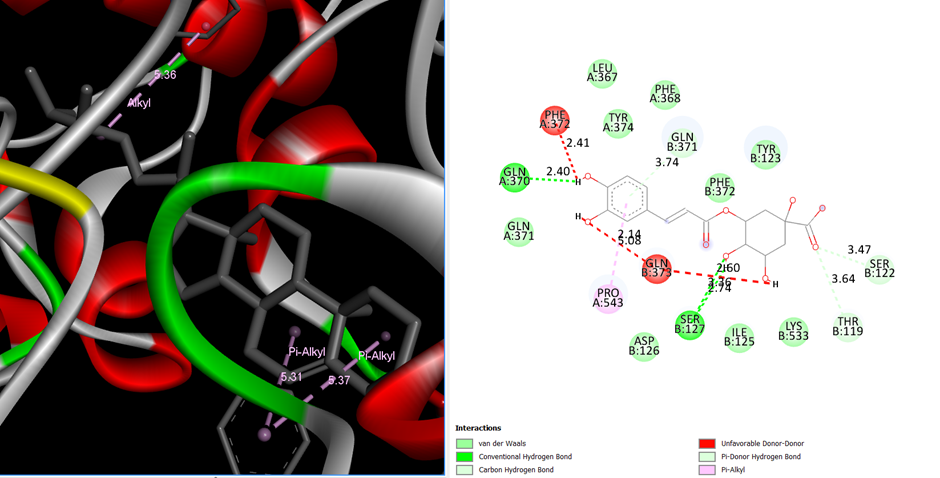
Fig 6. Interaction with Chlorogenic acid Docking score -15.9
Table 9. Interaction profile of Solamargine with target protein (Fig. 7)
|
Amino Acid (Chain:Residue) |
Bond Type |
Distance (Å) |
|
Sugar-interacting residues |
Hydrogen bond |
2.71, 2.96 |
|
Steroidal pocket residues |
Hydrophobic |
5.33 |
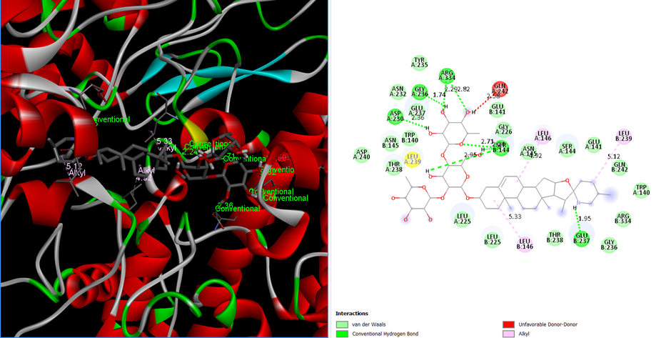
Fig 7. Interaction with Solamargine Docking score -12.3
Table 10. Interaction profile of Solasonine with target protein (Fig. 8)
|
Amino Acid (Chain:Residue) |
Bond Type |
Distance (Å) |
|
Glycosidic contact residues |
Hydrogen bond |
2.29, 2.82 |
|
Non-polar residues |
Hydrophobic |
5.12, 5.33 |
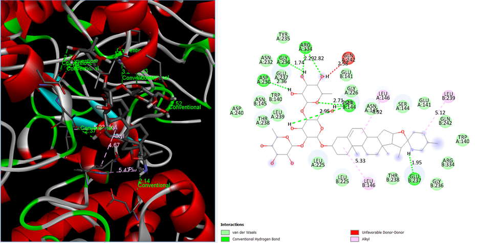
Fig 8. Interaction with Solasonine Docking score -11.7
Table 11. Molecular interactions between reference drug (Diclofenac) and the target protein active site
|
Amino Acid (Chain:Residue) |
Interaction Type |
Distance (Å) |
|
TYR (B:356) |
Conventional hydrogen bond |
3.06 |
|
LEU (B:353) |
Unfavorable acceptor–acceptor interaction |
2.85 |
|
VAL (B:350) |
π–π T-shaped interaction |
4.97 |
|
ALA (B:528) |
π–alkyl interaction |
5.07 |
|
VAL (B:524) |
π–alkyl interaction |
4.96 |
|
MET (B:523) |
π–sulfur interaction |
5.74 |
|
TRP (B:388) |
π–alkyl interaction |
4.05 |
|
TRP (B:388) |
π–sigma interaction |
3.98 |
|
VAL (B:524) |
Carbon–hydrogen bond |
3.05 |
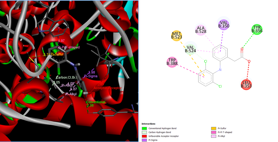
Fig 9. Interaction with Diclofenac sodium Docikng score comes -7.5
7.5 In silico ADME and Pharmacokinetic Evaluation of Chlorogenic Acid
SwissADME evaluation was performed to predict the pharmacokinetic characteristics, drug likeness, and medchem friendliness of the chlorogenic acid molecule, which was the ligand with the highest ranking from the docking experiment performed on the selected protein target. Evaluation of physiochemical properties indicated that the molecule possessed the following characteristics: it had the molecular formula of C??H??O? with a molecular weight of 354.31 g/mol, which was within the acceptable range for a drug molecule. The molecule was very polar with a topological polar surface area of 164.75 Ų; it was, however, moderately flexible with five rotatable bonds. Lipophilicity was low with most predicting models predicting it to be low (consensus Log P = −0.39), indicating that the molecule was hydrophilic. This was supported by solubility prediction models such as ESOL, Ali, and SILICOS-IT that placed the solubility of the molecule from soluble to very soluble. This was an improvement because the molecule was very soluble. Pharmacokinetic predictions revealed very low gastrointestinal absorption, which can be ascribed to the large TPSA value and the number of hydrogen bond donors (n=6) and hydrogen bond acceptors (n=9). The permeability of chlorogenic acid was found to be non-permeant to the blood-brain barrier, thus making the chances of its central nervous system-related adverse reactions very low. Most significantly, it was found to be non-inhibitor of prominent cytochrome P450 enzymes (CYP1A2, CYP2C9, CYP2C19, CYP2D6, and CYP3A4), thus making the chances of metabolic drug interactions very low. Moreover, it was not identified as a substrate of P-glycoprotein.
Further support for these findings was found using the BOILED-Egg model, which placed chlorogenic acid out of the BBB region and only partially overlapped the human intestinal absorption zone, consistent with its predicted pharmacokinetic behavior. Drug-likeness assessment showed limited rule violations, mainly related to high polarity; no severe medicinal chemistry alerts were detected. PAINS and Brenk alerts were flagged due to catechol functional groups; those are rather common for naturally occurring polyphenols and do not necessarily point to a lack of biological relevance. BOILED-Egg plot as a function of WLOGP versus TPSA illustrates the predicted absorption and brain permeability of chlorogenic acid: the compound falls outside the blood-brain barrier zone and towards the periphery of the human intestinal absorption zone, reflecting poor intestinal permeability. The distribution pattern therefore is suggestive of low exposure to CNS and moderate systemic availability, which is in agreement with the results predicted by the SWISSADME tool based on its pharmacokinetic properties. On balance, SWISSADME results provide strong evidence to recommend chlorogenic acid as a lead phyto-constituent with excellent docking score performance coupled with solubility, metabolite safety, as well as predictable pharmacokinetic properties.
Table 12. In silico ADME and drug-likeness properties of chlorogenic acid
|
Parameter |
Predicted value |
Interpretation / Significance |
|
Molecular formula |
C??H??O? |
Polyphenolic natural compound |
|
Molecular weight (g/mol) |
354.31 |
Within acceptable small-molecule range |
|
Heavy atoms |
25 |
Moderate molecular complexity |
|
Aromatic heavy atoms |
6 |
Presence of phenolic rings |
|
Rotatable bonds |
5 |
Moderate molecular flexibility |
|
H-bond acceptors |
9 |
High polarity |
|
H-bond donors |
6 |
Supports hydrogen bonding |
|
Topological polar surface area (TPSA) |
164.75 Ų |
High polarity, affects permeability |
|
Consensus Log P (lipophilicity) |
−0.39 |
Hydrophilic character |
|
Log P (iLOGP) |
0.87 |
Low lipophilicity |
|
Log P (XLOGP3) |
−0.42 |
Consistent hydrophilicity |
|
Log P (WLOGP) |
−0.75 |
Low membrane partitioning |
|
Log P (MLOGP) |
−1.05 |
High aqueous affinity |
|
Water solubility (ESOL) |
Very soluble |
Favorable for formulation |
|
Water solubility (Ali) |
Soluble |
Supports oral dosage forms |
|
Water solubility (SILICOS-IT) |
Soluble |
Confirms aqueous compatibility |
|
GI absorption |
Low |
Limited intestinal permeability |
|
BBB permeation |
No |
Reduced CNS side-effect risk |
|
P-gp substrate |
No |
Lower efflux-related bioavailability loss |
|
CYP1A2 inhibition |
No |
Low metabolic interaction risk |
|
CYP2C9 inhibition |
No |
Safe metabolic profile |
|
CYP2C19 inhibition |
No |
Minimal drug–drug interaction |
|
CYP2D6 inhibition |
No |
Favorable pharmacokinetics |
|
CYP3A4 inhibition |
No |
Reduced hepatic interaction |
|
Skin permeation (log Kp) |
−8.76 cm/s |
Low dermal penetration |
|
Lipinski rule |
1 violation (H-donors >5) |
Acceptable for natural products |
|
Bioavailability score |
0.11 |
Moderate oral bioavailability |
|
PAINS alerts |
1 (catechol) |
Common in polyphenols |
|
Brenk alerts |
2 (catechol, Michael acceptor) |
Natural-compound related |
|
BOILED-Egg model |
HIA: marginal; BBB: negative |
|
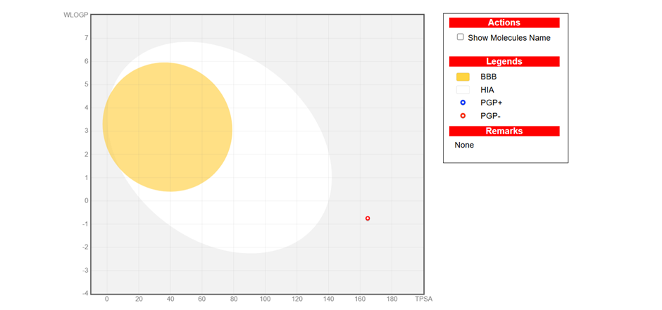
Fig 9. BOILED-Egg model predicting gastrointestinal absorption and blood–brain barrier permeability of chlorogenic acid.
7.5 In silico Toxicity Prediction of Chlorogenic Acid
For additional verification of the safety index of the lead phyto-constituent derived from the docking and ADME studies, a virtual comprehensive toxicity analysis was performed on the lead compound, chlorogenic acid, using the ProTox-3.0 model. The result showed an estimated LD50 of 5000 mg/kg indicating toxicity class V, which denotes a compound with low toxicity. The compound can, therefore, be regarded as safe when taken orally, given the long history of its use.
Evaluation of the organ-specific toxicity showed that the compound's predicted toxicity was low for hepatotoxicity (probability = 0.72), neurotoxicity (0.89), and cardiotoxicity (0.99). Low toxicity was predicted for nephrotoxicity (0.56) and respiratory toxicity (0.57). However, the values are quite moderate and need to be proven through experimental validation. It was of importance to note that the compound's predicted values showed it to be non-carcinogenic (0.68), non-mutagenic (0.93), and non-cytotoxic (0.80). Immunotoxicity was predicted to be active with a high probability of 0.99, which might be due to its known immunomodulatory effects in polyphenolic compounds and not adverse effects. There were no predictions of ecotoxicity and nutritional toxicities. Though BBB-associated activity was predicted to be of probability 0.60, it aligns with the polarity of the molecule and does not conflict with the SwissADME prediction of a low probability of BBB permeability.
Further evaluation analyses by Tox21 of the nuclear receptor signaling pathways and the stress response pathways indicated that chlorogenic acid was inactive against all tested targets, including androgen receptor, estrogen receptor, PPAR-γ, AhR, and p53 pathways, with the probabilities being above 0.90 with most of them. Moreover, the compound was also inactive as an inhibitor of the major cytochrome P450 enzymes (CYP1A2, CYP2C9, CYP2C19, CYP2D6, CYP3A4, and CYP2E1) with regard.
Overall, ProTox-3.0 analysis confirms that chlorogenic acid possesses a low-toxicity profile with high safety margins, reinforcing its selection as a promising bioactive candidate for further experimental and preclinical evaluation.
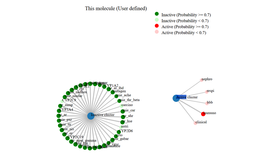
Fig 10. Network Chart for the ligand-drug chlorogenic acid
CONCLUSION
The present study explored the anti-inflammatory potential of bioactive phytochemicals obtained from Solanum viarum Dunal through an integrated in silico approach. Molecular docking analysis demonstrated that several phytoconstituents identified from the plant extract interact effectively with the inflammatory target protein Cyclooxygenase-2, indicating their ability to form stable ligand–protein complexes through hydrogen bonding, hydrophobic contacts, and other intermolecular interactions. Among the screened compounds, chlorogenic acid and the steroidal alkaloids solamargine, solasonine, and solasodine exhibited comparatively stronger binding affinities, suggesting that these compounds may contribute to the anti-inflammatory activity traditionally associated with the plant.
Pharmacokinetic evaluation using SwissADME indicated that the lead compounds possess several favorable drug-like characteristics, including good aqueous solubility, low lipophilicity, and minimal interaction with major cytochrome P450 enzymes. These properties may reduce the likelihood of metabolic drug–drug interactions and undesirable side effects. In addition, limited predicted permeability across the blood–brain barrier suggests a lower probability of central nervous system-related adverse effects.
The safety profile of the lead compound was further assessed using ProTox-II, which predicted low toxicity with no indications of carcinogenic, mutagenic, or cytotoxic effects. These results suggest that the selected phytochemicals may possess an acceptable safety margin for further pharmacological investigation. Comparative docking analysis with the standard anti-inflammatory drug Diclofenac sodium also confirmed the reliability of the docking protocol and provided a useful benchmark for evaluating the binding potential of the plant-derived compounds.
Overall, the findings of this study suggest that Solanum viarum contains several bioactive constituents with promising anti-inflammatory potential. Although the current investigation is based on computational analysis, it provides a scientific basis for further experimental validation through in vitro and in vivo studies. The integration of phytochemical profiling, molecular docking, pharmacokinetic prediction, and toxicity assessment highlights the value of in silico approaches in accelerating the discovery of plant-derived therapeutic agents. These results support the potential of Solanum viarum as a valuable natural source for the development of safer and more effective anti-inflammatory drugs.
REFERENCES

Neda Anzar, Dr. Manish Singh, Anti-Inflammatory and Analgesic Potential of Solanum viarum Dunal Extract: A Preclinical and In Silico Study, Int. J. of Pharm. Sci., 2026, Vol 4, Issue 3, 4064-4079. https://doi.org/10.5281/zenodo.19339690
 10.5281/zenodo.19339690