We use cookies to ensure our website works properly and to personalise your experience. Cookies policy
1,2,3,4 Government College of Pharmacy, Kathora Naka, Amravati, Maharashtra, India 444604
5 Government College of Pharmacy, Ratnagiri, Maharashtra, India 415612.
Background: Alcoholic Liver Disease (ALD) remains a major cause of chronic liver morbidity and mortality worldwide, particularly in developing regions where alcohol misuse is prevalent. Early detection and assessment of disease severity are essential to prevent progression to advanced fibrosis and cirrhosis. Objective: To assess the extent of liver damage in patients diagnosed with Alcoholic Liver Disease at District General Hospital, Amravati, using demographic, clinical, laboratory, radiological, and non-invasive fibrosis markers. Methods: A hospital-based prospective observational study was conducted among 210 patients with confirmed ALD. Patients were categorized into alcoholic fatty liver, alcoholic hepatitis, and alcoholic cirrhosis. Demographic data, alcohol consumption patterns, hematological parameters, liver function tests, ultrasonographic findings, and non-invasive fibrosis indices (De Ritis ratio, APRI, and FIB-4) were analyzed. Results: The study population showed a strong male predominance (97%), with most patients belonging to the 31–50 years age group. Daily alcohol intake was the predominant pattern, and higher consumption (?180 ml/day) over prolonged duration (5–20 years) was strongly associated with advanced disease. Laboratory findings revealed anemia, thrombocytopenia, elevated AST and ALT levels, hyperbilirubinemia, and hypoalbuminemia. Non-invasive fibrosis markers indicated significant fibrosis in a substantial proportion of patients, particularly in the cirrhosis subgroup. Ultrasonography commonly demonstrated hepatomegaly, fatty infiltration, ascites, and splenomegaly. Conclusion: Chronic heavy alcohol consumption, especially in rural and socioeconomically vulnerable populations, is strongly associated with progressive liver damage. Integrated assessment using clinical evaluation, laboratory parameters, imaging, and non-invasive fibrosis scores is essential for early detection and severity stratification of Alcoholic Liver Disease.
Alcoholic liver disease (ALD) represents a major cause of chronic liver morbidity and mortality worldwide and constitutes a significant public health burden, particularly in regions with high per capita alcohol consumption. ALD encompasses a wide pathological spectrum ranging from simple hepatic steatosis (fatty liver) to alcoholic hepatitis and ultimately cirrhosis, which may progress to hepatic failure and hepatocellular carcinoma. The disease develops as a consequence of sustained and excessive alcohol intake, leading to progressive structural and functional deterioration of hepatic tissue.1-5
The earliest and potentially reversible stage of ALD is alcoholic fatty liver, characterized by the accumulation of lipid droplets within hepatocytes, particularly around the portal tracts. Continued alcohol exposure leads to hepatocellular injury, necrosis, inflammation, and steatosis, collectively referred to as alcoholic hepatitis. Persistent inflammatory insult activates hepatic stellate cells, resulting in extracellular matrix deposition, fibrosis, and architectural distortion of the liver parenchyma. Over time, this culminates in cirrhosis, an irreversible stage marked by nodular regeneration and progressive hepatic insufficiency.6-8
Globally, chronic alcohol consumption accounts for millions of deaths annually and contributes substantially to liver-related mortality. A significant proportion of individuals with chronic alcohol use develop varying degrees of hepatic fibrosis, and a subset progresses to cirrhosis and hepatocellular carcinoma. Epidemiological data indicate that the risk of ALD correlates with both the quantity and duration of alcohol intake. Regular consumption exceeding 30–50 g of alcohol per day for several years markedly increases the likelihood of hepatic steatosis, while higher intake levels significantly elevate the risk of cirrhosis. Women exhibit increased susceptibility to alcohol-induced liver injury at comparatively lower consumption levels, possibly due to differences in alcohol metabolism and hormonal influences.9-11
The pathogenesis of ALD is multifactorial and involves complex metabolic, immunological, and oxidative mechanisms. Ethanol metabolism occurs primarily via alcohol dehydrogenase and the microsomal ethanol-oxidizing system, generating acetaldehyde and reactive oxygen species. These metabolites alter the intracellular redox state, impair mitochondrial β-oxidation, enhance lipid synthesis, and induce oxidative stress. In addition, increased gut permeability leads to endotoxin translocation into the portal circulation, activating Kupffer cells and triggering pro-inflammatory cytokine release, including tumor necrosis factor-α. These events collectively contribute to hepatocyte apoptosis, necrosis, and fibrogenesis.12-13
Clinically, ALD may remain asymptomatic in its early stages, particularly in fatty liver disease. As the disease advances, patients may present with jaundice, anorexia, nausea, vomiting, ascites, fatigue, muscle weakness, and neuropsychiatric manifestations. Decompensated cirrhosis is associated with life-threatening complications such as variceal hemorrhage, hepatic encephalopathy, spontaneous bacterial peritonitis, hepatorenal syndrome, and hepatopulmonary syndrome.14-15
Accurate and timely diagnosis is crucial for preventing disease progression. Assessment typically includes a detailed history of alcohol consumption, screening tools such as the CAGE questionnaire and AUDIT, and laboratory evaluation including complete blood count, liver function tests (AST, ALT, bilirubin, GGT), serum albumin, coagulation profile (PT, INR), and lipid profile. Imaging modalities such as abdominal ultrasonography assist in evaluating structural abnormalities, while liver biopsy remains the gold standard for definitive staging when diagnosis is uncertain. Non-invasive fibrosis assessment tools, including composite scoring systems based on biochemical parameters, are increasingly utilized to estimate the degree of hepatic fibrosis.
Management of ALD primarily centers on complete alcohol abstinence, which can reverse early-stage disease and significantly improve prognosis. Nutritional therapy plays a critical supportive role, as malnutrition is common in ALD patients. Pharmacological interventions such as corticosteroids, pentoxifylline, N-acetylcysteine, and antioxidants may be indicated in selected cases of severe alcoholic hepatitis. In advanced disease with liver failure, orthotopic liver transplantation remains the definitive therapeutic option.16-17
Despite advancements in diagnostic and therapeutic strategies, ALD continues to pose a substantial clinical challenge, particularly in district-level healthcare settings where late presentation and limited resources may affect outcomes. Therefore, systematic assessment of liver damage among patients with alcoholic liver disease at District General Hospital, Amravati, is essential to evaluate disease severity, biochemical alterations, and clinical patterns in the regional population. Such data may contribute to improved early detection, optimized management strategies, and reduction in ALD-related morbidity and mortality at the community level.18
MATERIALS AND METHODS:19-32
Study Design and Setting
The present investigation was conducted as a hospital-based prospective observational study with a case-control approach at District General Hospital, Amravati. The study was carried out in the Department of General Medicine, including the inpatient wards, Intensive Care Unit (ICU), and Outpatient Department (OPD).
The study aimed to assess the extent of liver damage in patients diagnosed with Alcoholic Liver Disease (ALD) using clinical, biochemical, hematological, radiological, and non-invasive fibrosis assessment parameters.
Source of Data
Data were collected from patient case records, laboratory investigation reports, radiological findings, and direct patient interviews. All relevant information was documented using a structured Patient Data Collection Form designed specifically for the study.
Study Population
Selection of Subjects
Patients diagnosed with Alcoholic Liver Disease and attending the General Medicine OPD, admitted to wards, or treated in ICU during the study period were screened for eligibility.
Inclusion Criteria
Patients were enrolled in the study if they were aged above 18 years and had a confirmed diagnosis of Alcoholic Liver Disease (ALD) based on clinical, laboratory, and radiological findings. Only individuals with a documented history of chronic alcohol consumption were considered eligible. In addition, participation was strictly voluntary, and written informed consent was obtained from all subjects prior to inclusion in the study.
Exclusion Criteria
Patients younger than 18 years of age were excluded from the study. Individuals who declined to provide consent or were unwilling to participate were also excluded. Pregnant and lactating women were not considered for inclusion due to physiological variations that could influence study parameters. Furthermore, patients lacking essential baseline investigations, including complete blood count (CBC), liver function tests (LFT), C-reactive protein (CRP), and abdominal ultrasonography, were excluded to ensure uniformity and completeness of data for analysis.
Study Variables and Parameters Assessed
The assessment of liver damage in patients with Alcoholic Liver Disease was performed using a comprehensive set of demographic, social, and clinical variables. These parameters were selected to evaluate disease severity, identify associated risk factors, and establish correlations between alcohol exposure and hepatic injury.Demographic Parameters
Demographic information was collected to understand the distribution of ALD and its association with biological and socio-environmental determinants. The recorded variables included age, gender, locality (urban or rural residence), educational status, body mass index (BMI), and associated comorbid conditions such as hypertension, diabetes mellitus, chronic obstructive pulmonary disease (COPD), asthma, and anemia.
Age and gender were analyzed as biological factors influencing alcohol metabolism and susceptibility to hepatic injury. Locality and educational status were evaluated as indicators of socioeconomic background, lifestyle patterns, and accessibility to healthcare services, which may impact alcohol consumption behavior and disease progression. Body mass index was assessed to determine the contribution of metabolic factors, particularly obesity, in exacerbating liver damage. The presence of comorbid conditions was documented, as these may accelerate hepatic injury and influence the clinical course of ALD.
Social History Parameters
A detailed alcohol consumption history was obtained from each participant to assess exposure patterns and their relationship with liver damage. Information collected included the type of alcohol consumed (such as locally brewed spirits, beer, wine, whisky, or others), average daily quantity intake measured in milliliters, frequency of consumption (daily, weekly, monthly, or occasional), duration of alcohol use in years, and the time elapsed since last alcohol intake.
These variables were analyzed to evaluate the cumulative alcohol burden and its dose-dependent impact on hepatic injury. Particular emphasis was placed on correlating the duration and quantity of alcohol intake with biochemical abnormalities and markers of liver dysfunction to better understand the progression and severity of Alcoholic Liver Disease.
Laboratory Parameters
(A) Hematological Parameters
All enrolled patients underwent complete blood count (CBC) analysis as part of routine laboratory evaluation. The hematological parameters assessed included hemoglobin concentration (Hb), red blood cell (RBC) count, white blood cell (WBC) count, mean corpuscular volume (MCV), mean corpuscular hemoglobin (MCH), and platelet count.
These indices were evaluated to identify hematological abnormalities commonly associated with Alcoholic Liver Disease. Particular attention was given to macrocytosis, reflected by elevated MCV values, which is frequently observed in chronic alcohol users. The presence of anemia, leukocytosis, and thrombocytopenia was also documented. Thrombocytopenia was considered a potential indicator of portal hypertension and hypersplenism, whereas leukocytosis was interpreted as a marker of inflammation or infection. Collectively, these hematological findings provided supportive evidence regarding the severity and progression of chronic liver disease.
(B) Biochemical Parameters
Biochemical investigations primarily focused on liver function and renal status. Liver function tests (LFTs) included estimation of total bilirubin, direct bilirubin, indirect bilirubin, serum glutamic-oxaloacetic transaminase (SGOT/AST), serum glutamate pyruvate transaminase (SGPT/ALT), and serum albumin. These markers were used to evaluate hepatocellular injury, cholestasis, and hepatic synthetic capacity.
Renal function was assessed through measurement of serum urea, blood urea nitrogen (BUN), and serum creatinine to detect any associated renal impairment, including hepatorenal dysfunction. In addition, electrolyte levels and inflammatory markers such as C-reactive protein (CRP) were recorded. Elevated transaminases (AST and ALT), increased bilirubin levels, and reduced serum albumin were considered indicative of hepatocellular damage and compromised synthetic function of the liver.
Radiological Assessment
All eligible patients underwent ultrasonography (USG) of the abdomen as a non-invasive imaging modality to evaluate structural liver changes. The ultrasound examination assessed liver size (hepatomegaly), fatty infiltration, surface nodularity suggestive of cirrhosis, presence of ascites, and features consistent with portal hypertension.
Radiological findings were correlated with laboratory parameters to establish the relationship between structural alterations and biochemical evidence of hepatic dysfunction. This integrated assessment improved the accuracy of disease staging and severity evaluation.
Non-Invasive Fibrosis Assessment Scores
To estimate the degree of hepatic fibrosis without invasive procedures, validated non-invasive scoring systems were calculated using laboratory parameters.
De Ritis Ratio
A value greater than 2 was considered suggestive of alcoholic liver injury, as alcohol-related hepatocellular damage typically results in a disproportionate elevation of AST compared to ALT.
AST to Platelet Ratio Index (APRI)
An APRI score less than 0.5 was interpreted as minimal fibrosis, whereas a value greater than 1.5 indicated significant fibrosis or cirrhosis. This index was used as a simple, cost-effective tool for fibrosis assessment, particularly in resource-limited settings.
Fibrosis-4 (FIB-4) Score
A score below 1.3 suggested minimal fibrosis, values between 1.3 and 2.67 were considered indeterminate, and scores above 2.67 were indicative of advanced fibrosis or cirrhosis. The FIB-4 score was utilized to enhance non-invasive staging of liver disease.
Patient Data Collection Form
|
Name : |
Age |
Gender |
||||
|
Region : urban/rural |
Marital status: |
Blood Group |
||||
|
Weight : |
Education: |
Occupation |
||||
|
Caste: |
Eating Habit: Veg / Non-Veg |
|||||
|
Nutritional Status: |
||||||
|
Vaccinated for hepatitis: Yes/No |
||||||
|
Chief Complaints: |
||||||
|
Sign and Symptoms: |
||||||
|
Social History : Smoker/Tobacco chewer/Alcoholics/Allergy |
Types of Alcohol: Desi/Wine/Beer/Whisky/Others |
|||||
|
Frequency Of alcohol: Daily/ weekly/Monthly/Occasional |
Daily intake of Alcohol(ml): 90/180/360/720/more |
|||||
|
Duration of consumption(years): |
Last Consumption: |
|||||
|
Family History : |
||||||
|
Other Comorbid Conditions |
||||||
|
Past Medical History |
||||||
|
Past Medication History |
||||||
|
Laboratory investigations: |
||||||
|
Haematological parameters |
Liver Function Test |
Coagulation Profile |
||||
|
WBC |
Total Bilirubin |
Prothrombin Time |
||||
|
Lym% |
Direct Bilirubin |
Prothrombin ratio |
||||
|
Gran% |
Indirect Bilirubin |
INR |
||||
|
RBC |
SGOT |
CRP |
||||
|
Hb |
SGPT |
|
||||
|
HCT |
Dirits Ratio |
|
||||
|
MCV |
|
|
||||
|
MCH |
Kidney Function Test |
Serum Electrolyte: |
||||
|
MCHC |
Sr. Creatinine |
Potassium: |
||||
|
RDWCV |
Sr. Uric acid |
Sodium: |
||||
|
RDWSD |
|
Magnesium: |
||||
|
PLT |
USG Report: |
Calcium: |
||||
|
MPV |
Chloride: |
|||||
|
PDW |
|
|||||
|
PCT |
|
|||||
|
PLCR |
|
|||||
|
PLCC |
|
|||||
|
Other Investigation: |
||||||
|
Diagnosis: |
||||||
|
Treatment: |
||||||
|
Sr. No. |
Drugs |
Dose |
Route |
Freq. |
||
|
1 |
|
|
|
|
||
|
2 |
|
|
|
|
||
Statistical Analysis
All collected data were systematically entered into Microsoft Excel and subsequently analyzed using appropriate statistical software. Descriptive statistical methods were applied to summarize demographic, clinical, laboratory, and radiological findings. Continuous variables were expressed as mean ± standard deviation, whereas categorical variables were presented as frequencies and percentages.
Correlation analyses were performed to evaluate the association between alcohol consumption patterns (type, quantity, duration, and frequency) and markers of liver injury, including transaminase levels and fibrosis indices. Statistical significance was determined using appropriate tests, and results were interpreted to identify meaningful relationships between alcohol exposure and severity of hepatic damage.
RESULTS:
Amongst the sample size of 210, all the patients with alcoholic liver disease were categorized into 3 subgroups as Alcoholic fatty liver (75), Alcoholic hepatitis (44) and Alcoholic cirrhosis (91).
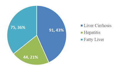
Figure 1: Distribution of sample size into categories
Demographics Parameters
The demographic details analyzed in our study were gender, age, region, educational status and eating habit, occupation, BMI and co-morbidities.
|
|
Categories |
No of cases |
Frequency |
|
Gender |
Male |
204 |
97 % |
|
Female |
6 |
3 % |
|
|
Age |
20-30 |
34 |
16 % |
|
31-40 |
64 |
31 % |
|
|
41-50 |
60 |
29 % |
|
|
51-60 |
39 |
19 % |
|
|
Region |
Rural |
163 |
69 % |
|
Urban |
74 |
31 % |
|
|
Education |
Below SSC |
100 |
48 % |
|
HSC |
87 |
41 % |
|
|
Graduate |
15 |
7 % |
|
|
Illiterate |
8 |
4 % |
|
|
Eating habit |
Vegetarian |
187 |
89 % |
|
Non vegetarian |
23 |
11 % |
|
|
Occupation |
Privet job |
65 |
31 % |
|
Labour |
111 |
53 % |
|
|
Business |
9 |
4 % |
|
|
Not working |
25 |
12 % |
|
|
BMI |
Healthy weight |
97 |
46 % |
|
Underweight |
51 |
24 % |
|
|
Overweight |
48 |
23 % |
|
|
Obese |
14 |
7 % |
|
|
Co morbidities |
HTN |
28 |
43 % |
|
DM |
13 |
20 % |
|
|
Anemia |
6 |
9 % |
|
|
Asthma |
6 |
9 % |
|
|
COPD |
4 |
6 % |
|
|
HTN+DM |
8 |
12 % |
Gender (n=210)
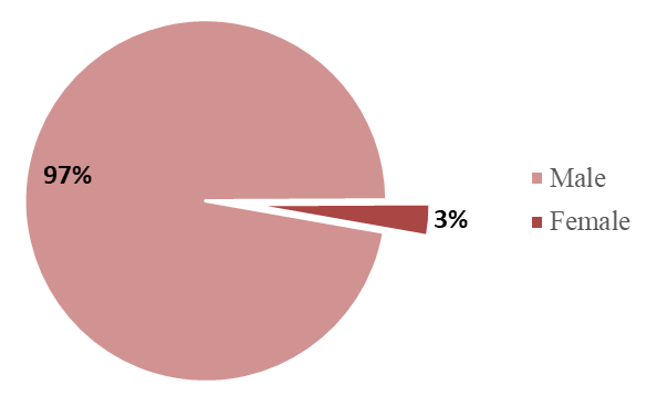
The study showed predominance of males 204 (97%). The females enrolled in our study were 6 (3%). The ratio being 1:34 for females is to males.
Further, cross categorizing gender with subtype of alcoholic liver disease showed male predominance across liver cirrhosis (89 cases), fatty liver (71 cases), and alcoholic hepatitis (44 cases).
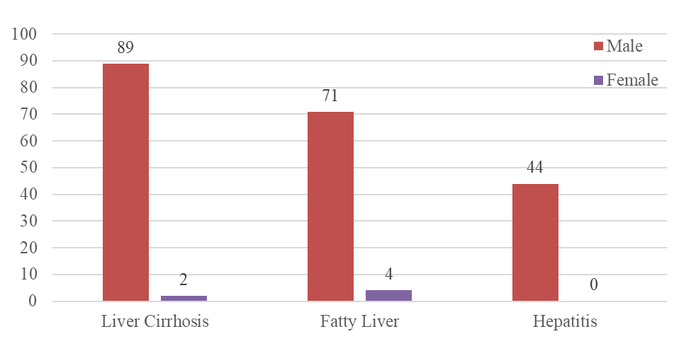
Figure 3: Distribution of patients according to gender-category
Age (n=210)
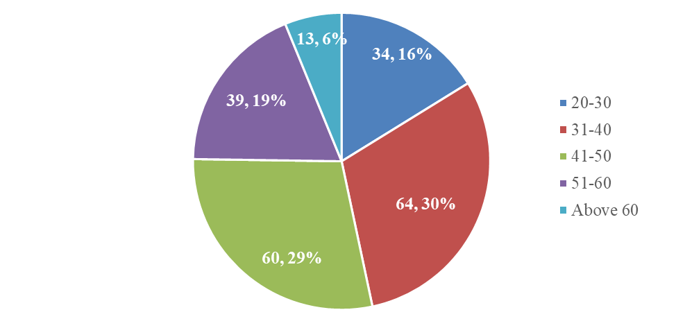
ALD predominantly affected the 31–40 year age group (30%, 64 cases), followed by 41-50 years (29%, 60 cases), indicating a higher incidence in the productive age groups across all three categories.
Region (n=210)
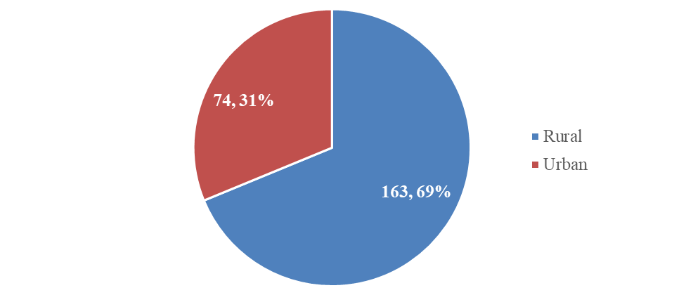
Our study depicted the maximum patients comes from the rural area 163 (69%) and remaining from urban area 74 (31%). In general, the patients coming to the District General Hospital are from low socio-economic background.
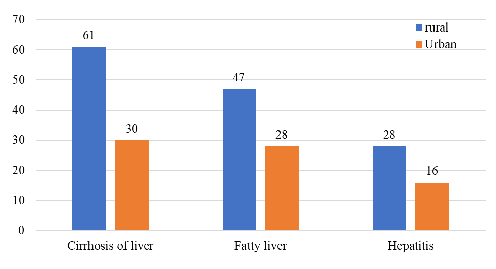
Also, the maximum number of patient in each subcategory of ALD are from rural areas. cirrhosis (67%), fatty liver (63%), hepatitis (64%).
Education (n=210)
Out of an entire sample size of 210, maximum people had education status below matriculation100 (48%). This parameter was studied to analyze the impact of literacy on the development of ALD and whether the lack of higher education leads to higher prevalence of ALD.
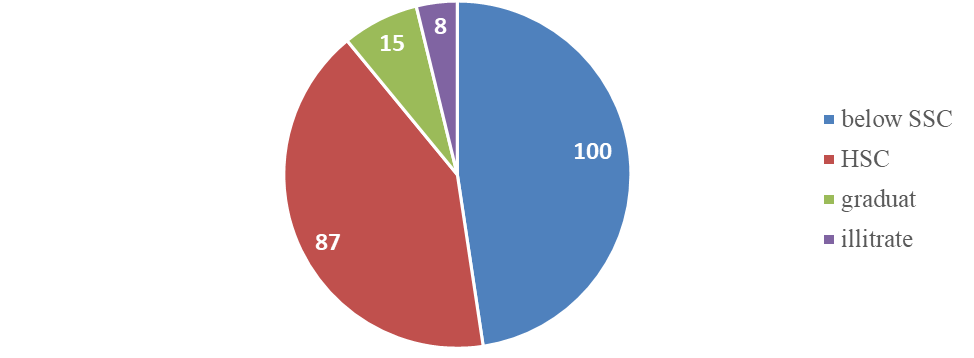
Figure 7: Distribution of patients according to educational status
Eating Habit (n=210)
Non-vegetarian diet was more prevalent in ALD patients, seen in 86% cirrhosis, 93% fatty liver, and 89% hepatitis cases.
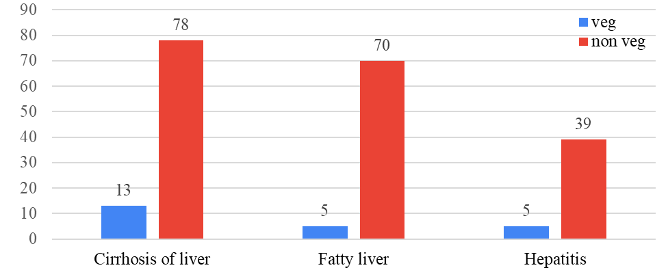
Figure 8: Distribution of patients according to eating habit
Blood Group (n=210)
Blood group distributions are shown for each ALD category, with B+ being the most common across all groups.
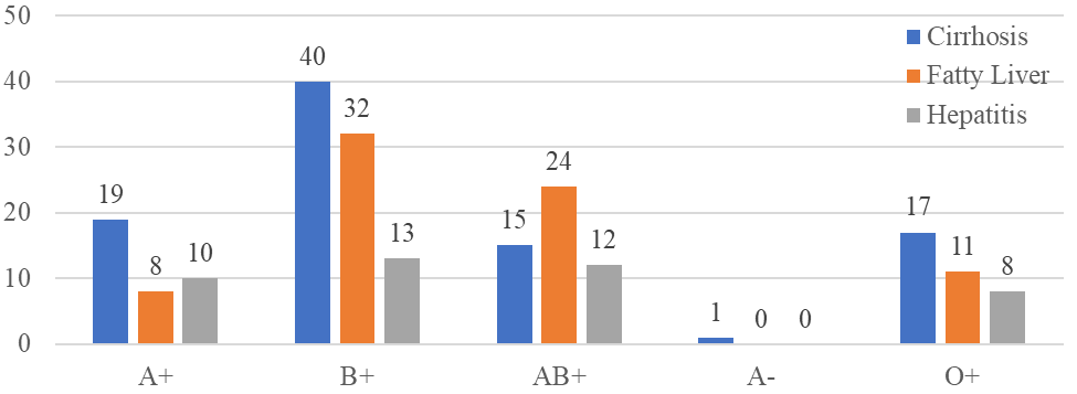
Figure 9: Distribution of patients according to blood group
Occupation (n=210)
Private jobs and labour were the most common occupations among cirrhosis, fatty liver and hepatitis cases.
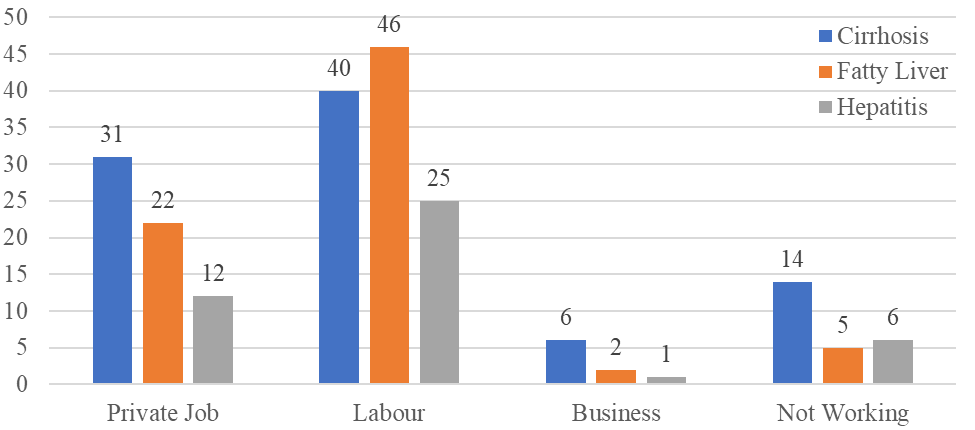
Figure 10: Distribution of patients according to occupation
Body Mass Index (BMI) (n=210)
A high proportion of cirrhosis (48%, 44 cases) and fatty liver (44%, 33 cases) cases had a healthy BMI range, while alcoholic hepatitis cases were more evenly distributed across BMI categories.
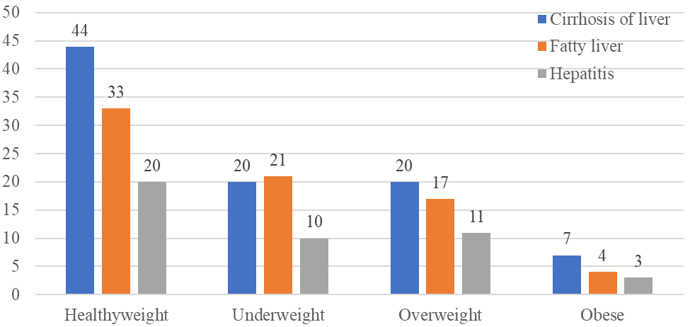
Figure 11: Distribution of patients according to BMI
Social history (n=210)
Alcohol consumption, with or without smoking/tobacco, was the primary risk factor across all ALD categories.
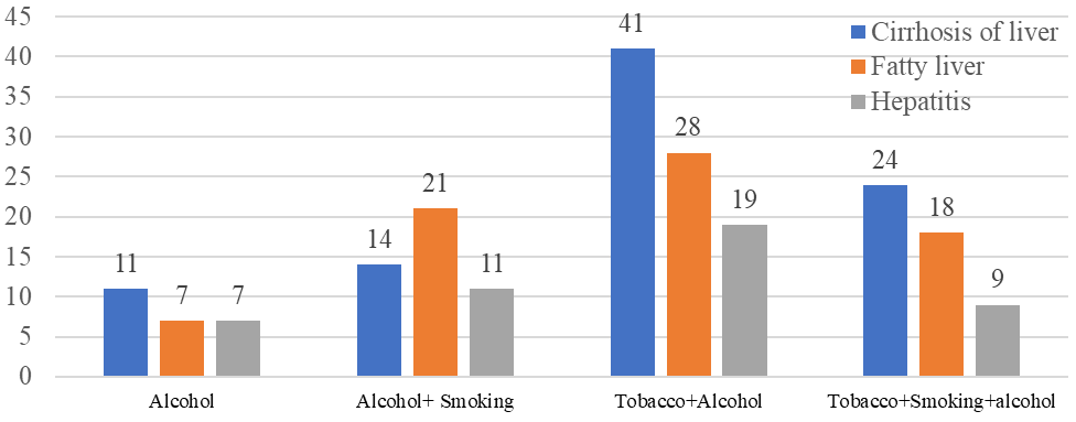
Figure 12: Distribution of patients according to social history
Frequency of alcohol consumed (n=210)
Daily alcohol consumption was the most common pattern among cirrhosis, fatty liver and hepatitis cases.
81% of people consumed alcohol on a regular basis and the most prominent consumed alcohol was local alcohol. Cirrhosis of liver shows a strong correlation with the frequency of alcohol consumption, being most prevalent among daily drinkers and significantly less common among those who drink weekly or less. Fatty liver, with the highest incidence among daily drinkers. However, it shows a slightly more gradual decline in cases among weekly and occasional drinkers compared to cirrhosis. While the number of hepatitis cases is highest among daily drinkers, it is interesting to note that it is slightly higher among weekly drinkers compared to fatty liver The daily intake of patients who developed cirrhosis of the liver consumed>180ml of alcohol. In our study, maximum no. of patients consumed alcohol for greater than 5 years.
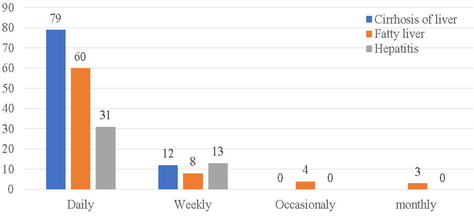
Figure 13: Distribution of patients according to frequency of alcohol consumed
Daily Intake (n=210)
The majority of cirrhosis (60%, 55 cases) and fatty liver (55%, 41 cases) cases consumed 180ml of alcohol per day, while alcoholic hepatitis cases had a more varied pattern
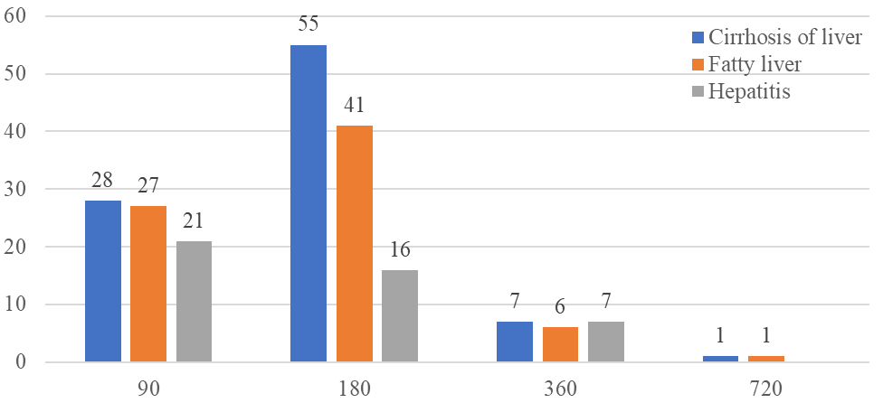
Figure 14: Distribution of patients according to daily intake of alcohol
Duration of consumption (n=210)
Most cirrhosis cases were 10-20 years, and fatty liver cases had 5-15 years of alcohol consumption duration. While hepatitis spread between 5-20 years having wider range.
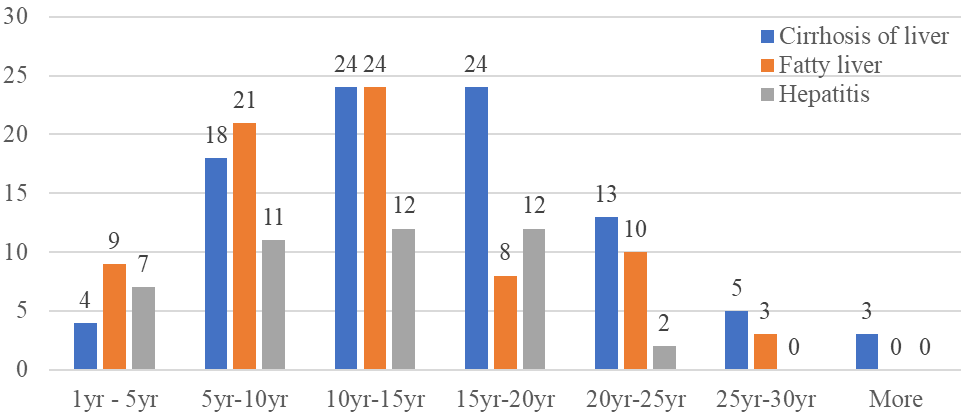
Figure 15: Distribution of patients according to duration of consumption
Co-morbidities (n=210)
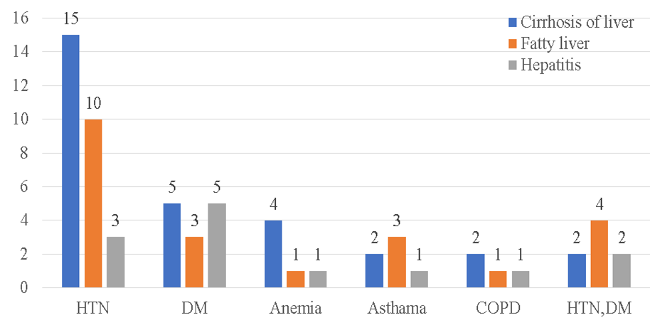
Hypertension and diabetes were notable comorbidities present in ALD patients across all categories. From the entire population of 210, 69% people were without any comorbidity. From the remaining 31%, observed comorbidities were hypertension, diabetes, anemia, COPD and asthma.
Haematological parameters
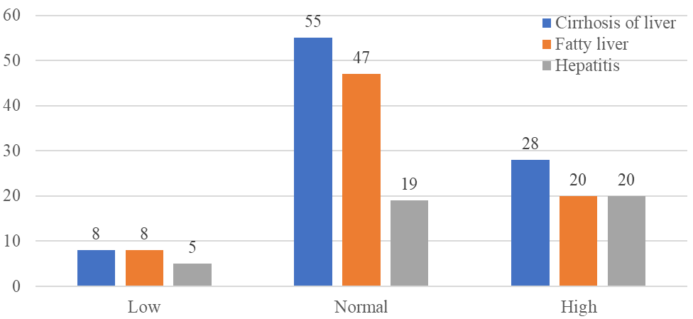
Figure 17: Distribution of patients according to variation in WBC
White Blood Cell (WBC) (n=210)
Most cirrhosis (60%, 55 cases) and fatty liver (63%, 47 cases) cases had normal WBC counts, while alcoholic hepatitis showed a higher proportion (46%, 20 cases) of elevated WBC, indicating underlying inflammation.
Red Blood Cell (RBC) (n=210)
A significant proportion of cirrhosis (72%), fatty liver (54%), and hepatitis (58%) cases showed low RBC counts, reflecting anemia associated with chronic liver disease.
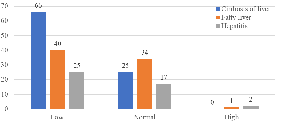
Figure 18: Distribution of patients according to variation in RBC
Hemoglobin (HGB) (n=210)
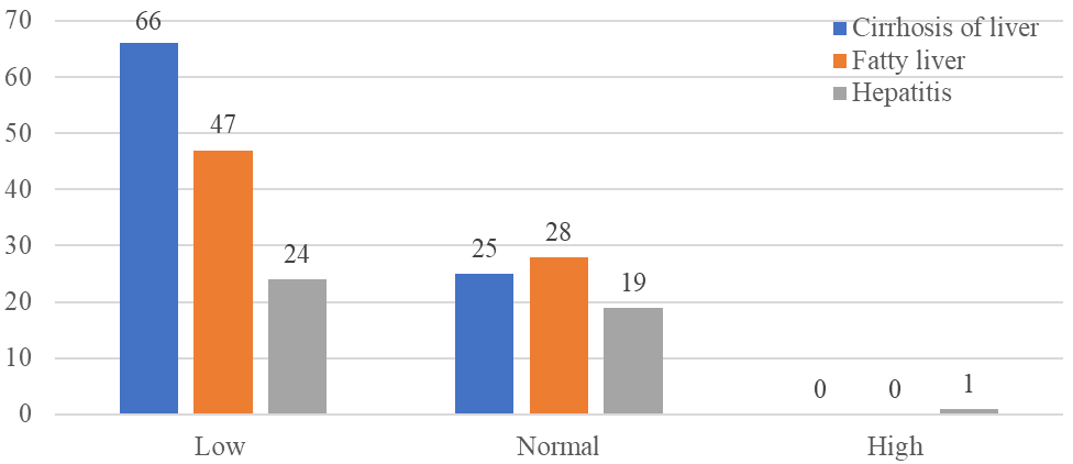
Figure 19: Distribution of patients according to variation in HGB
Low hemoglobin levels were seen in 72% of cirrhosis, 53% of fatty liver, and 46% of hepatitis cases, further confirming the presence of anemia in ALD patients.
Mean Corpuscular Volume (MCV) (n=210)
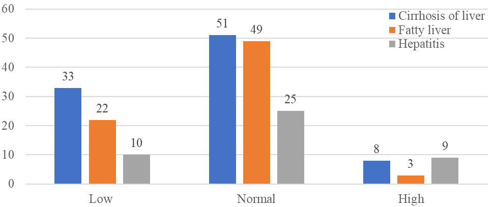
While most cases had normal MCV, microcytosis (Low MCV) was observed in 36% (33 cases) of cirrhosis, 29% (22 cases) of fatty liver, and 22% (10 cases) of alcoholic hepatitis, potentially due to alcohol-related effects.
The majority of cases across cirrhosis, fatty liver, and hepatitis had normal MCH levels, with some showing microcytic and macrocytic patterns.
Platelet count (PLT) (n=210)
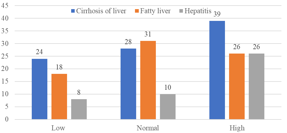
While most cases had normal PLT, Thrombocytopenia (low platelets) was prevalent in 29% (23 cases) of cirrhosis, 23% (17 cases) of fatty liver, and 19% (11 cases) of alcoholic hepatitis cases, indicating hypersplenism or decreased thrombopoietin production.
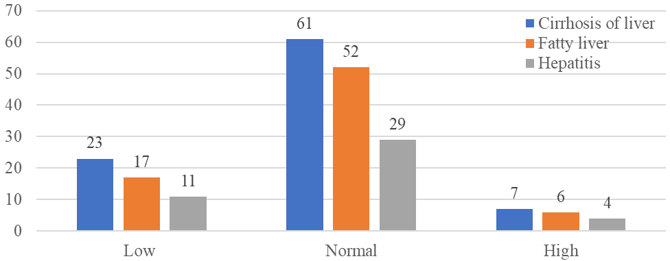
Figure 22: Distribution of patients according to variation in PLT
Biochemical Parameters
Serum Urea (n=210)
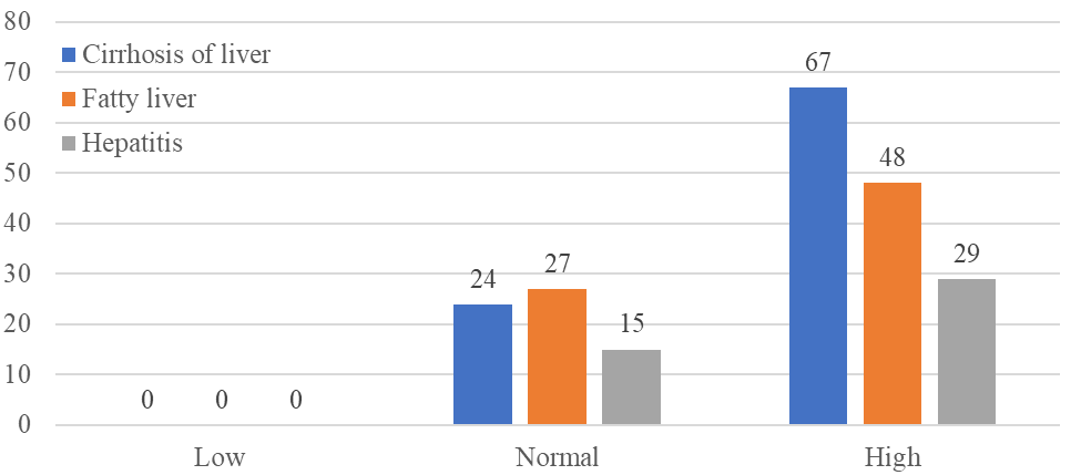
Most cases across all three categories had normal Sr. urea, but some showed elevated levels, indicating impaired kidney function.
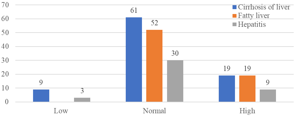
Figure 23: Distribution of patients according to variation in Sr. Urea
Serum Creatinine (n=210)
Most cases across all three categories had normal creatinine levels, but some showed elevated levels, indicating impaired kidney function.
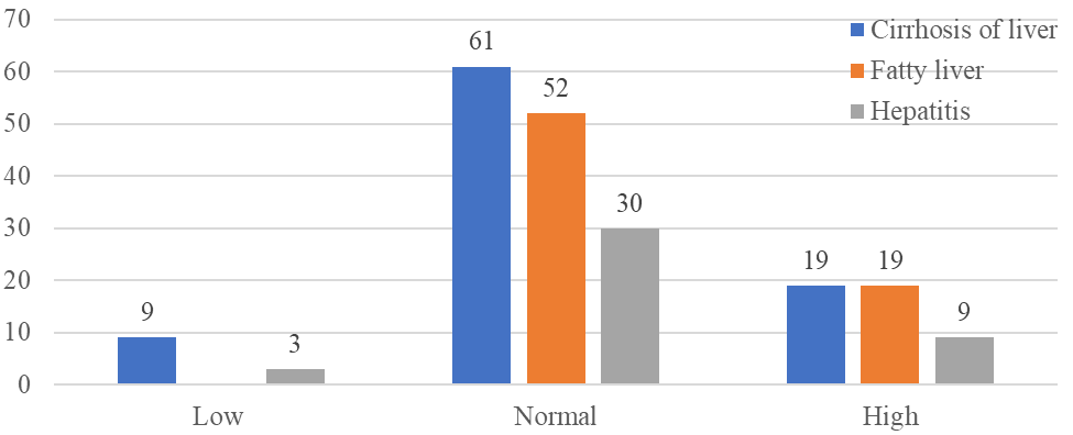
Figure 24: Distribution of patients according to variation in Sr. Creatinine
Serum Bilirubin Total (n=202)
Elevated total bilirubin levels were common across cirrhosis (mean 4.05 mg/dL), alcoholic hepatitis (mean 4.19 mg/dL), and fatty liver (mean 2.19 mg/dL), suggesting varying degrees of hepatocellular injury.
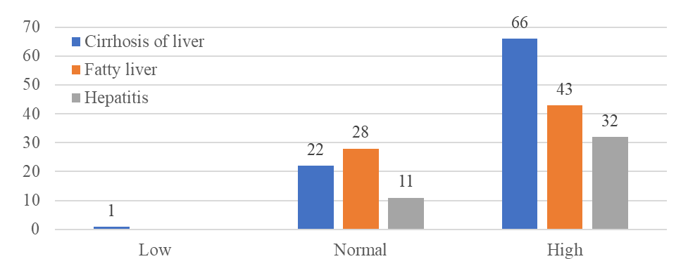
Figure 25: Distribution of patients according to variation in Sr. Bilirubin Total (n=202)
Serum Albumin (n=143)
Hypoalbuminemia (low albumin) was observed in cirrhosis (mean 3.04 g/dL), alcoholic hepatitis (mean 3.22 g/dL), and fatty liver (mean 3.51 g/dL) cases, indicating impaired synthetic liver function.
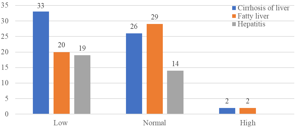
Figure 26: Distribution of patients according to variation in Sr. Albumin (n=143)
Serum Glutamic-Oxaloacetic Transaminase (SGOT) (n=210)
Serum Glutamic-Pyruvic Transaminase (SGPT) (n=210)
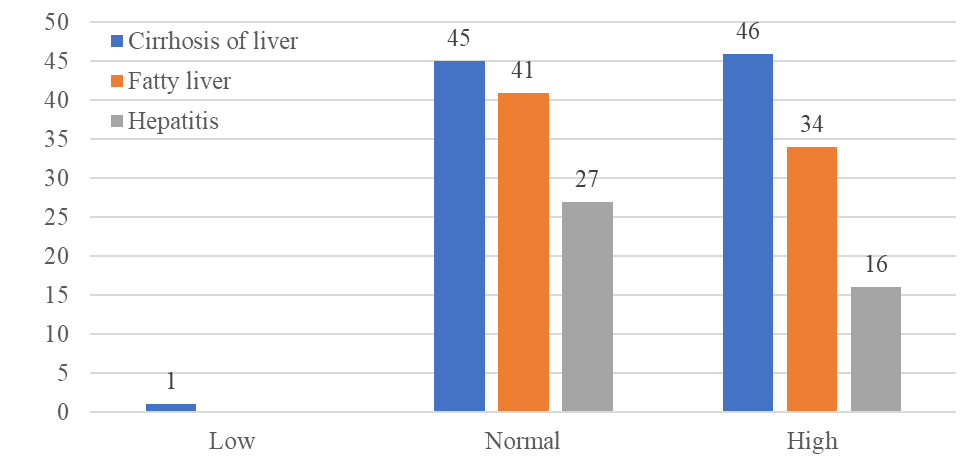
Figure 28: Distribution of patients according to variation in SGPT
Elevated SGOT and SGPT levels, markers of hepatocellular injury, were seen across cirrhosis, alcoholic hepatitis, and fatty liver categories, with the highest mean values in cirrhosis.
De rities ratio (n=210)
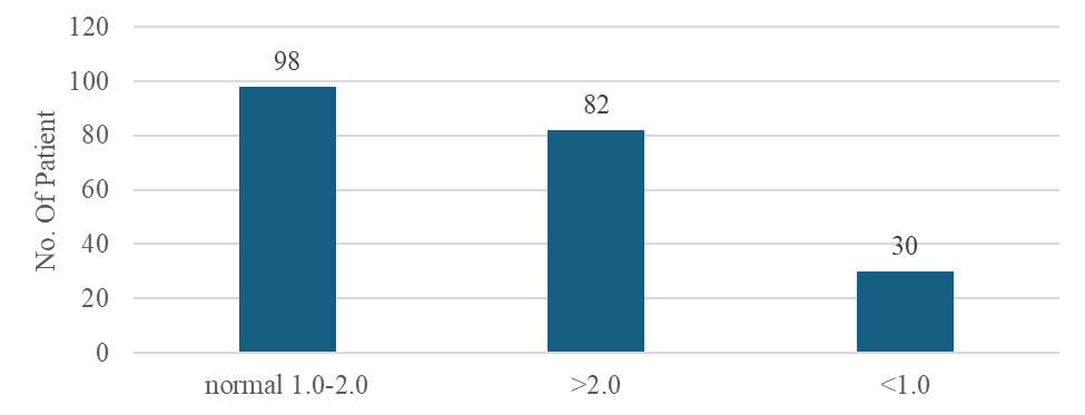
This chart shows the distribution of the De Ritis ratio (AST/ALT) across ALD categories, with mean values around 2.0 for fatty liver (2.04) and alcoholic hepatitis (2.29), but slightly higher for cirrhosis (2.45). Only 14.3% had a ratio >2.0, the traditional marker for alcoholic liver disease.
AST to Platelet Ratio Index (APRI) (n=210)
The APRI (AST to Platelet Ratio Index) is a non-invasive marker of hepatic fibrosis. 25.2% of patients had APRI >1.5, indicating significant fibrosis/cirrhosis, with the highest mean APRI of 2.08 in the cirrhosis category.
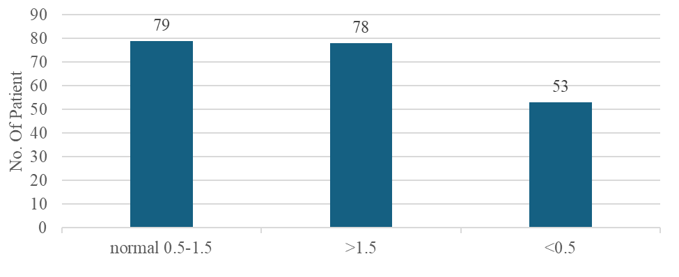
Figure 30: Distribution of patients according to variation in APRI
Fibrosis-4 (FIB-4) (n=210)
The FIB-4 (Fibrosis-4 Score) data is provided only for the cirrhosis category. 63.7% had FIB-4 >2.67, suggesting advanced fibrosis/cirrhosis, while only 14.3% had FIB-4 <1.3, indicating minimal fibrosis. The high mean FIB-4 of 6.84 reflects its utility in identifying cirrhosis.
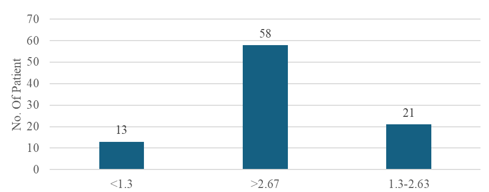
Figure 31: Distribution of patients according to variation in FIB-4
Alkaline Phosphatase (ALP) (n=210)
While most cases had normal ALP levels, some showed elevated ALP across cirrhosis, fatty liver, and alcoholic hepatitis categories, potentially indicating biliary obstruction or infiltrative liver disease.
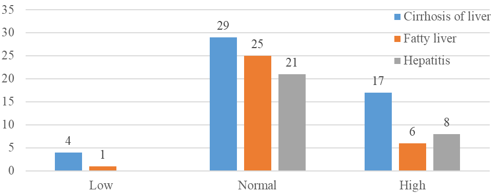
Figure 32: Distribution of patients according to variation in API
Serum Total Protein (n=210)
The majority of cases across cirrhosis, fatty liver, and alcoholic hepatitis categories had normal total protein levels.
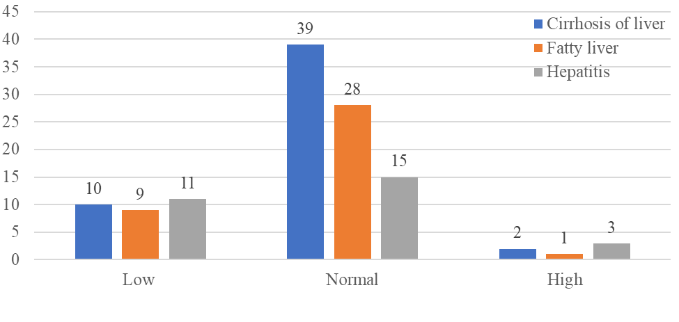
Figure 33: Distribution of patients according to variation in Sr. Total Protein
Ultrasound Sonography (USG) (n=210)
Common imaging findings included ascites, splenomegaly, hepatomegaly, and fatty liver, indicating advanced liver disease across all three categories.
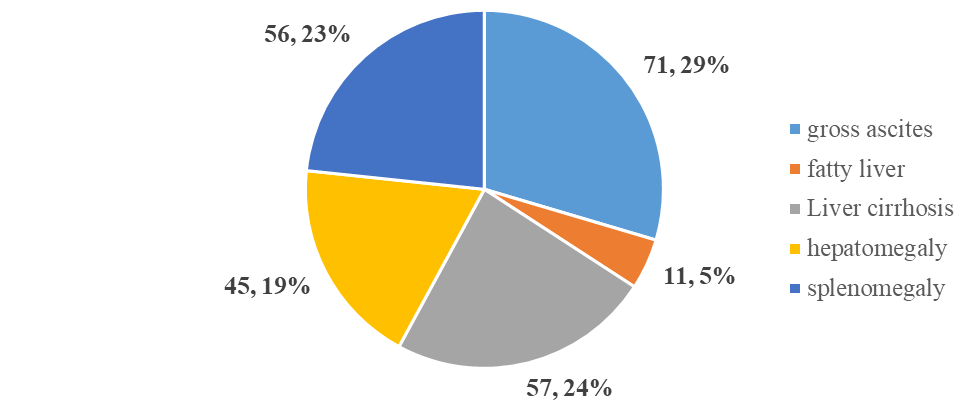
Figure 34: Distribution of patients according to variation in USG
DISCUSSSION:
The present study evaluated demographic, clinical, laboratory, and radiological characteristics of patients with Alcoholic Liver Disease (ALD). A marked male predominance (97%) was observed, consistent with earlier reports indicating higher alcohol consumption among men. The majority of patients belonged to the economically productive age groups of 31–50 years, highlighting the substantial social and occupational impact of ALD. A greater proportion of cases were from rural areas and had lower educational status, suggesting a strong association between socioeconomic factors and disease occurrence.
Daily alcohol intake was the most frequent pattern identified, and higher consumption (≥180 ml/day) was commonly observed among patients with cirrhosis and fatty liver. A clear dose–response relationship between alcohol quantity, duration of use, and disease severity was evident, supporting the established role of chronic heavy drinking in hepatic injury progression.
Hematological abnormalities such as anemia and thrombocytopenia were frequently documented, reflecting chronic liver dysfunction and portal hypertension. Elevated transaminases (AST and ALT) and increased total bilirubin levels were observed across disease categories, indicating hepatocellular injury. Serum albumin levels were reduced, particularly in advanced stages, demonstrating impaired synthetic function. While most patients had normal MCV and MCH values, a subset showed variations, partially aligning with previous studies.
Non-invasive fibrosis markers, including the De Ritis ratio, APRI, and FIB-4 score, were useful in identifying advanced liver damage. The mean AST/ALT (De Ritis) ratio exceeded 2 in most categories and was highest in cirrhosis, suggesting progressive hepatocellular injury with prolonged alcohol exposure. Approximately one-fourth of patients had APRI values above 1.5, indicating significant fibrosis, with the highest scores seen in cirrhosis cases.
Correlated well with biochemical derangements and clinical severity.
Overall, the findings reinforce existing evidence that demographic profile, alcohol consumption pattern, laboratory abnormalities, and imaging features collectively play a critical role in assessing the severity and progression of Alcoholic Liver Disease.
CONCLUSION
The present study evaluated the extent of hepatic injury in 210 patients diagnosed with Alcoholic Liver Disease, classified into alcoholic fatty liver, alcoholic hepatitis, and alcoholic cirrhosis. A pronounced male predominance was observed, with most patients belonging to the economically active age group of 31–50 years. A higher proportion of cases originated from rural areas and individuals with lower educational status, indicating the influence of socioeconomic determinants on disease occurrence.
Chronic daily alcohol intake emerged as the predominant consumption pattern and showed a strong association with disease severity. A significant number of patients with cirrhosis and fatty liver reported consuming 180 ml or more of alcohol per day over duration of 5–20 years, supporting the cumulative dose-dependent effect of alcohol on liver damage. Laboratory evaluation demonstrated frequent occurrence of anemia, thrombocytopenia, and elevated hepatic transaminases, reflecting ongoing hepatocellular injury and compromised liver function. Non-invasive fibrosis indices, including APRI and FIB-4, indicated a considerable proportion of patients with advanced fibrosis or cirrhosis. Radiological findings such as hepatomegaly, fatty infiltration, ascites, and splenomegaly further substantiated the presence of progressive liver disease.
Overall, the study highlights the critical role of alcohol consumption patterns, demographic factors, and integrated laboratory and imaging assessment in determining the severity and progression of Alcoholic Liver Disease.
CONFLICTS OF INTERESTS:
All authors have declared no conflict of interest.
REFERENCES

Om Khandarkar, Vedant Kale, Kallyani Solanke, Pratiksha Wanjari, Dr. Hrishikesh Gupta, Assessment of Liver Damage in Patient with Alcoholic Liver Disease at District General Hospital, Amravati, Int. J. of Pharm. Sci., 2026, Vol 4, Issue 3, 602-623. https://doi.org/10.5281/zenodo.18894755
 10.5281/zenodo.18894755