

We use cookies to ensure our website works properly and to personalise your experience. Cookies policy
College of Pharmaceutical Sciences, Government Medical College, Thiruvanandhapuram, Kerala, India-695011.
Uterine leiomyoma (UL), the most common benign tumor of the female reproductive tract, imposes a substantial economic and clinical burden, yet current management options—including GnRH agonists, selective progesterone receptor modulators, and hysterectomy—remain limited by short-term use , side effects, or invasiveness. UL arises from a somatic driver mutation in a single myometrial progenitor cell, most commonly in MED12, followed by HMGA2 overexpression or FH inactivation, with these alterations occurring in a mutually exclusive manner across molecularly distinct tumor subtypes. Clonal expansion is further promoted by ovarian steroid hormones, particularly progesterone acting through PR-A/PR-B to activate Wnt/?-catenin, MAPK, and PI3K/AKT/mTOR signaling, alongside estrogen-mediated upregulation of progesterone receptor expression. A convergent feature across genetic subtypes is aberrant activation of the Wnt/?-catenin pathway, which links driver mutations and hormonal signaling to proliferation, stem-cell maintenance, and excessive extracellular matrix (ECM) deposition, the latter compounded by TGF-?, activin-A, PDGF, and specific microRNAs. Epigenetic dysregulation, marked by elevated DNMT1 expression, further reinforces these pathways. This review evaluates non-hormonal druggable targets emerging from this biology, including nodes within the Wnt/?-catenin cascade (PORCN, tankyrase, ?-catenin/TCF and ?-catenin/CBP complexes, RSPO3, WNT9A), the TDO2–kynurenine–AHR axis downstream of MED12 mutation, the ITGA11–collagen–ECM fibrosis pathway, EIF2AK3/PERK-mediated stress signaling, PARP1-associated DNA-repair vulnerability, and the miR-148a-3p–TXNIP/NRP1 regulatory axis. Among these, the TDO2–kynurenine–AHR axis currently holds the strongest preclinical validation, particularly in MED12-mutant fibroids. Collectively, these findings point toward a shift from hormone-dependent therapy to molecularly stratified, non-hormonal treatment strategies for UL, though most candidate targets still require further mechanistic and translational validation
Uterine leiomyoma (UL) places a substantial financial strain on the U.S. healthcare system, with estimated costs ranging from USD 5.9 billion to USD 34.4 billion when both direct medical expenses and indirect losses are considered1. This cost burden mirrors the disease's clinical impact: although roughly half of those with UL have no symptoms, affected individuals can experience irregular or heavy menstrual bleeding, significant anemia, pelvic pain and pressure, difficulty conceiving, and complications during pregnancy 1,2. Treatment options vary based on symptom presentation and include the levonorgestrel-releasing IUD, GnRH agonists, selective progesterone receptor modulators (SPRMs), and oral contraceptive pills 3,4. Of these, GnRH agonists are considered the most effective at shrinking fibroid volume and easing symptoms5. Their use, however, is capped at six months because prolonged treatment is linked to adverse effects such as bone density loss and diabetes risk5. Given these limitations, hysterectomy remains the only therapy that offers a definitive cure for symptomatic UL an option that is unappealing for those hoping to avoid surgery or preserve their uterus. Even though UL is the most frequently diagnosed benign tumor of the female reproductive system, the medical armamentarium for managing its symptoms and complications is still notably limited6.
A central obstacle to developing better UL treatments is that its underlying cause remains poorly understood. ULs are known to be benign, clonal, hormone-responsive tumors, but the biological pathways that drive their formation are still largely unclear 7. This gap has pushed researchers to investigate the genetic and epigenetic changes responsible for UL tumor development across different patient populations. The expectation is that clarifying these mechanisms will pave the way toward individualized treatment approaches for UL patients7. This review surveys the current state of knowledge on the genetic and epigenetic underpinnings of UL, its genetic epidemiology, and emerging gene-based therapeutic strategies, with the goal of identifying where UL treatment development still has room to advance.
Genetic initiation and clonal origin
Uterine leiomyoma (UL) pathogenesis begins with a somatic driver mutation in a single myometrial smooth muscle stem/progenitor cell, which then undergoes clonal expansion to form the tumor mass10. The most common initiating event is a mutation in mediator complex subunit 12 (MED12), typically a missense or small in-frame insertion-deletion affecting exons 1–2 of the gene; this is found in approximately 70–80% of fibroids across multiple ethnic populations, South African, and U.S. cohorts 8,10. MED12 is a component of the Mediator complex, a 26-subunit transcriptional regulator that bridges DNA regulatory sequences to RNA polymerase II, so its disruption has broad downstream effects on gene transcription10. A second major subtype involves overexpression of high mobility group AT-hook 2 (HMGA2), most often driven by a t(12;14)(q14-15;q24) chromosomal translocation, accounting for roughly 10% of fibroids and occurring almost exclusively in tumors that lack MED12 mutations (2,3); HMGA2, a chromatin architectural protein, has been shown to promote tumorigenesis partly through downstream activation of the proto-oncogene PLAG1 and through dysregulation of G1/S cell-cycle checkpoint control9,19. A smaller subset of fibroids arises from biallelic inactivation of fumarate hydratase (FH), which can occur sporadically or in the context of hereditary leiomyomatosis and renal cell cancer (HLRCC) syndrome; FH loss causes fumarate accumulation, altering cellular metabolism and pseudohypoxic signaling 10.Other recurrent but less common chromosomal changes include interstitial 7q deletions, 6p21 rearrangements, trisomy 12, and COL4A5-COL4A6 deletions 10,20. Critically, large tumor-profiling studies have confirmed that MED12, HMGA2, and FH alterations are mutually exclusive within a given tumor, meaning no single fibroid carries more than one of these driver events supporting a model in which UL is not one disease but several molecularly distinct entities converging on a similar clinical phenotype, and explaining why fibroids within the same uterus are frequently genetically unrelated to one another3.
Hormonal promotion of growth
Following initiation, expansion of the mutant clone is strongly dependent on ovarian steroid hormones. Progesterone is now regarded as the principal growth-promoting hormone in UL: acting through progesterone receptors PR-A and PR-B, it activates the Wnt/β-catenin, MAPK, and PI3K/AKT/mTOR signaling cascades, which collectively drive cell proliferation, inhibit apoptosis, stimulate cytokine release, promote angiogenesis, and increase extracellular matrix (ECM) accumulation11. This progesterone-driven model underlies the mechanism of selective progesterone receptor modulators (SPRMs) such as ulipristal acetate, which interfere with this signaling axis and can induce tumor regression11. Estrogen contributes both directly, by promoting proliferation, and indirectly, by upregulating progesterone receptor expression in fibroid tissue, so that estrogen and progesterone act synergistically rather than independently 11,21; prolonged estrogen dominance has similarly been implicated in disrupting normal uterine physiology in ways that favour fibroid formation11.
Downstream signalling and extracellular matrix dysregulation
A convergent feature across genetically distinct UL subtypes is activation of the Wnt/β-catenin pathway, which appears to link diverse initiating mutations (MED12, HMGA2) and hormonal signaling to a shared downstream growth program11,21. Fibroids are also distinguished histologically by excessive, disorganized ECM — a fibrotic phenotype that in some respects is a more defining feature of the tumor than smooth muscle cell number alone17,18. This ECM consists predominantly of collagen types I and III, along with fibronectin, laminins, and proteoglycans, produced largely by myofibroblasts within the tumor18,19. ECM accumulation is regulated by growth factors — particularly TGF-β1 and TGF-β3, activin-A, and PDGF — as well as by cytokines such as TNF-α, steroid hormones, and specific microRNAs including the miR-29 family, miR-200c, and miR-93/106b17. TGF-β and activin-A in particular have been identified as key drivers of this fibrotic ECM accumulation17. The ECM is not merely a passive structural byproduct: elevated ECM components can activate mechanotransduction signaling, including the integrin–Rho/p38 MAPK/ERK pathway, creating bidirectional signaling between leiomyoma cells and their surrounding matrix that further reinforces the pathogenic phenotype 17. Additional cytokines and growth factors implicated in this process include epidermal growth factor, insulin-like growth factors 1 and 2, basic fibroblast growth factor, VEGF, and monocyte chemotactic protein-1, based on cellular-biology models of leiomyoma developed from the broader literature18.
Epigenetic contributions
Epigenetic dysregulation adds a further layer to UL pathogenesis. Leiomyoma tissue shows elevated expression of DNA methyltransferase 1 (DNMT1) relative to adjacent normal myometrium, and pharmacologic reversal of DNA methylation using agents such as 5-aza-2′-deoxycytidine has been shown to reduce cell proliferation, ECM formation, and Wnt/β-catenin pathway activity in primary leiomyoma cells — directly linking altered methylation to the same downstream pathways implicated in hormonal and genetic drivers of the disease19.
Wnt/β-catenin Signaling as a Druggable Target in Uterine Leiomyoma
The canonical Wnt/β-catenin cascade is a fundamental regulator of cell division, tissue differentiation, stem-cell renewal, and matrix remodeling. Under resting conditions, β-catenin is kept in check through continuous phosphorylation by a destruction complex composed of APC, AXIN, CK1, and GSK-3β, targeting it for ubiquitin-mediated proteasomal breakdown. Once a Wnt ligand engages Frizzled receptors together with the LRP5/6 co-receptor, Dishevelled becomes active and disables this destruction complex, permitting β-catenin to build up in the cytoplasm before translocating into the nucleus, where it partners predominantly with TCF/LEF transcription factors to switch on proliferative genes such as MYC and cyclin D1 (CCND1).
In uterine leiomyoma (UL), this cascade becomes abnormally active and drives both proliferation of tumor cells and expansion of the fibroid stem-cell compartment. Elevated levels of several Wnt ligands — WNT4, WNT5A, WNT11, and WNT16 — have been documented in fibroid tissue and in leiomyoma stem-cell fractions specifically22. Notably, WNT4 can trigger β-catenin activity through an AKT-dependent route, which in turn boosts MYC and cyclin D1 expression23. Supporting a causal role for this pathway, forced β-catenin activation in mouse uterine tissue was sufficient to generate myometrial overgrowth and mesenchymal tumors closely resembling leiomyoma24. Because β-catenin signaling also appears to sustain leiomyoma stem/progenitor cells, blocking β-catenin activity in experimental tumor models suppressed leiomyoma-like tumor growth, pointing to Wnt/β-catenin as a mechanism that helps maintain — not just initiate — these tumors 22,23.
This pathway also intersects with MED12 mutation, the most prevalent molecular alteration in UL, present in roughly 70% of cases. Mutant MED12 has been shown experimentally to amplify WNT4 and downstream β-catenin activity, suggesting that even though MED12 itself may be difficult to target directly, intervening further downstream in the Wnt/β-catenin cascade could still suppress growth in this large MED12-mutant patient subgroup24.
Several nodes within this cascade offer distinct opportunities for pharmacological intervention, including WNT4/FZD engagement, PORCN, tankyrase, β-catenin itself, and the β-catenin/TCF and β-catenin/CBP transcriptional complexes. PORCN stands out as an attractive upstream target because Wnt ligands require PORCN-mediated palmitoylation before they can be secreted; blocking this enzyme therefore chokes off Wnt ligand output before signaling even begins. Compounds demonstrating this mechanism include LGK-974, Wnt-C59, ETC-159, and IWP-221. A second strategy works by reinforcing the destruction complex rather than blocking ligand release: tankyrase 1/2 inhibitors such as XAV939 stabilize AXIN, thereby accelerating β-catenin degradation22. In primary human leiomyoma cells, both XAV939 and niclosamide were shown to suppress canonical Wnt/β-catenin activity and slow cell growth, with ICAT producing comparable inhibitory effects23.
A separate approach targets the transcriptional output of nuclear β-catenin rather than upstream ligand production or destruction-complex stability. Since β-catenin must partner with CBP to drive transcription of Wnt-responsive genes, small molecules like ICG-001 that disrupt this specific protein-protein interaction can shut down pathway output at the transcriptional level29. Other compounds — JW67, JW74, and KYA1797K — instead promote β-catenin breakdown by acting on the destruction complex through distinct mechanisms23. Because these strategies intervene at different points along the pathway, they offer multiple independent avenues for developing more selective inhibitors.
Wnt/β-catenin signaling may hold particular relevance to the fibrotic features of UL, given documented crosstalk between β-catenin and TGF-β-driven extracellular matrix regulation. Constitutive β-catenin activity in experimental uterine tissue coincided with elevated TGF-β3 and leiomyoma-like lesion formation, while blocking TGF-β3-driven β-catenin signaling reduced leiomyoma cell growth — indicating that Wnt/β-catenin inhibition might simultaneously curb both cell proliferation and the excess matrix deposition characteristic of these tumors24.
Taken together, Wnt/β-catenin signaling represents a compelling non-hormonal drug target for UL because it links multiple core aspects of fibroid biology — stem/progenitor cell expansion, MYC/cyclin D1-driven proliferation, MED12-associated signaling, AKT crosstalk, mechanotransduction, and matrix accumulation — into a single pathway. Inhibiting canonical Wnt signaling has already shown antiproliferative activity in primary human leiomyoma models22,23. That said, because Wnt signaling is also indispensable for normal tissue maintenance and reproductive physiology, systemic pathway blockade carries a real risk of off-target effects. Future drug development in this space will likely need to prioritize UL-selective downstream nodes or molecularly stratified patient subgroups rather than blanket inhibition of the entire pathway.
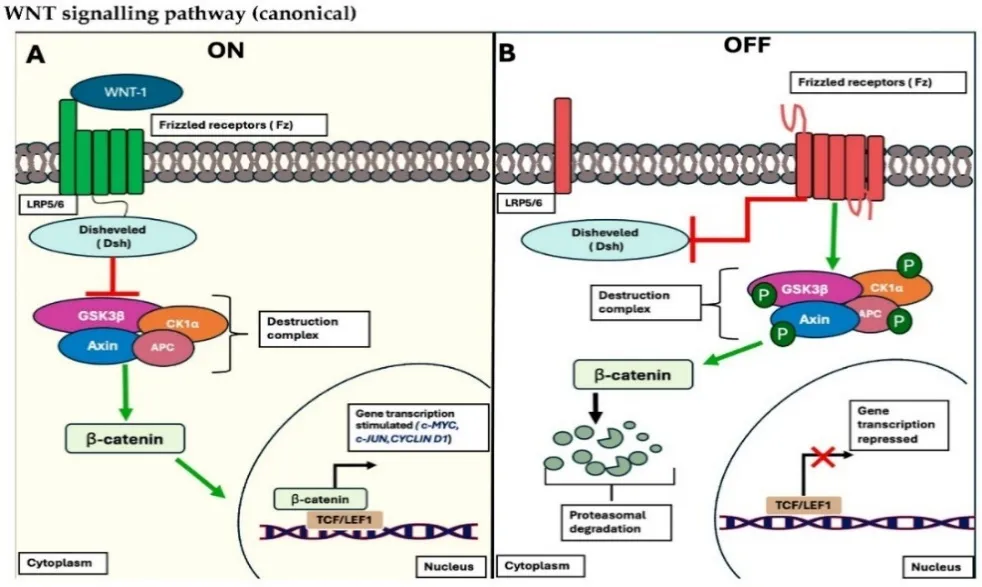
Figure 1: WNT signalling pathway (canonical)
Emerging Non-Hormonal Druggable Molecular Targets in Uterine Leiomyoma
1. The TDO2–Kynurenine–AHR Axis
Among the candidate non-hormonal targets, the tryptophan 2,3-dioxygenase (TDO2)–kynurenine–aryl hydrocarbon receptor (AHR) axis currently has the strongest mechanistic backing, especially in leiomyomas driven by MED12 mutations. Combined chromatin and transcriptomic analyses have singled out TDO2 as a key downstream effector of mutant MED1225,26. TDO2 converts tryptophan into kynurenine, a metabolite that acts as an endogenous AHR ligand. Rising kynurenine levels drive AHR from the cytoplasm into the nucleus, switching on AHR-dependent gene programs25,26. In fibroid cells, this signaling cascade boosts proliferation, blunts apoptosis, and raises expression of the AHR target genes CYP1A1 and CYP1B1. Silencing or pharmacologically blocking TDO2/AHR reverses these effects, indicating a causal rather than incidental role in disease biology25,26.
The enzymatic nature of TDO2 makes it a classic small-molecule target, while AHR offers a parallel receptor-based route to intervention. Experimental agents — the TDO2 inhibitor 680C91 and the AHR antagonist CH223191 — have each dampened pathway activity in preclinical fibroid models, and apigenin has been explored more recently as a way to selectively suppress this axis in MED12-mutant cells27. This positions the TDO2–kynurenine–AHR pathway as a promising precision-medicine strategy for genetically defined fibroid subtypes, though clinical safety and efficacy remain to be established.
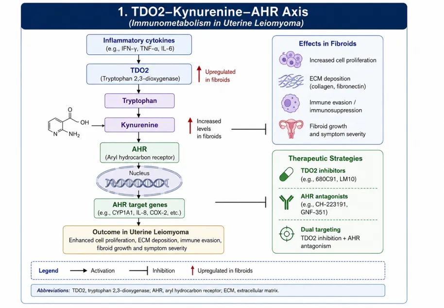
Figure 2:TDO2–Kynurenine–AHR Signalling Axis in Uterine Leiomyoma Progression
2. ITGA11 and the Collagen–ECM–Fibrosis Pathway
Integrin α11 (ITGA11) pairs with β1 to form the α11β1 collagen receptor, which governs cell–matrix adhesion and collagen remodeling — processes central to the fibrotic character of uterine fibroids. Fibroid tissue is known to express this collagen-binding integrin, and transcriptomic studies show ITGA11 is upregulated relative to normal myometrium28.
A recent proteome-wide Mendelian randomization study strengthened the case for ITGA11, identifying it as one of six proteins with genetic evidence replicated across independent datasets, and linking it specifically to the ECM/fibrosis dimension of fibroid pathology25. This supports a model in which dysregulated integrin–collagen signaling contributes to disease susceptibility, and suggests that disrupting ITGA11–collagen binding could, in principle, limit the abnormal matrix architecture that sustains tumor growth. Unlike PARP inhibitors, however, no ITGA11-targeted therapy yet exists for fibroids — it remains a target for future antibody, blocking-peptide, or small-molecule development rather than a validated treatment25.
3. RSPO3 and Wnt/β-Catenin Signaling
R-spondin 3 (RSPO3) is a secreted amplifier of canonical Wnt signaling that could offer a more targeted alternative to inhibiting β-catenin directly. It acts through LGR receptors together with the RNF43/ZNRF3 ubiquitin ligase system, increasing surface availability of Wnt receptors and thereby enhancing stem-cell maintenance, proliferation, and tissue remodeling. Wnt/β-catenin signaling is already well established in fibroid biology: WNT ligands are dysregulated in leiomyoma tissue, WNT4 drives AKT-mediated β-catenin activation, and downstream β-catenin signaling induces proliferative genes such as MYC and cyclin D1.
What makes RSPO3 novel is its recent emergence as a genetically supported, potentially druggable node within this pathway. Proteome-wide Mendelian randomization analyses found replicated colocalization between RSPO3 and fibroid risk, and elevated RSPO3 levels have been reported in fibroid tissue25. Because RSPO3 is a secreted extracellular protein, it may be more tractable for neutralizing antibodies or ligand-blocking approaches than intracellular β-catenin itself. Even so, this evidence currently identifies RSPO3 as a candidate requiring functional validation in fibroid-specific models rather than a proven therapeutic target25.
4. WNT9A and β-Catenin Signaling
WNT9A represents another Wnt-pathway component recently flagged by genetic studies of uterine fibroids. Wnt signaling already has an established role in fibroid stem-cell biology, and experimental activation of β-catenin can generate leiomyoma-like mesenchymal tumors in animal models. Multiple Wnt ligands are dysregulated in human fibroid tissue, feeding proliferative transcriptional programs.
The same proteome-wide Mendelian randomization analysis that highlighted RSPO3 also identified WNT9A among six proteins with replicated, colocalized genetic links to fibroid risk25. This places WNT9A alongside RSPO3 as a genetically supported, ligand-level entry point into Wnt signaling — potentially allowing more selective modulation than pathway-wide β-catenin suppression. A plausible drug-development approach would target WNT9A secretion or receptor engagement, but direct experimental evidence that inhibiting WNT9A slows fibroid growth is still needed before it can be considered validated25.
5. EIF2AK3/PERK and the Integrated Stress Response
EIF2AK3 encodes PERK (protein kinase R-like endoplasmic reticulum kinase), a core sensor of the integrated stress response that activates when misfolded proteins accumulate in the endoplasmic reticulum. Once triggered, PERK phosphorylates eIF2α, reshaping protein translation and stress-gene expression to help cells cope with metabolic and proteotoxic strain.
EIF2AK3's relevance to fibroids comes from the same proteome-wide Mendelian randomization work, which found replicated genetic colocalization between EIF2AK3 and fibroid risk, situating it within a PERK-driven stress-response pathway relevant to disease biology [1]. The appeal of this target lies in the possibility that fibroid cells rely on adaptive stress signaling to sustain growth and survival. However, because PERK also maintains normal cellular homeostasis, therapeutic strategies would likely need to achieve selective rather than broad inhibition. As with RSPO3 and WNT9A, EIF2AK3 remains an emerging candidate awaiting fibroid-specific functional studies25.
6. PARP1 and DNA-Repair Vulnerability
PARP1 (poly[ADP-ribose] polymerase-1) senses DNA strand breaks and coordinates their repair. Unlike hormonal or growth-factor-based strategies, targeting PARP1 exploits a DNA-repair weakness in genetically distinct fibroid subtypes rather than directly suppressing proliferative signaling.
PARP1 was likewise identified in the proteome-wide Mendelian randomization study as one of six proteins with replicated genetic evidence linking it to fibroid risk, and its expression is elevated in fibroid tissue relative to myometrium25. PARP1 is especially attractive from a translational standpoint because PARP inhibitors are already an established oncology drug class, offering a wealth of existing data on target engagement and medicinal chemistry. That said, findings from cancer cannot be assumed to transfer directly to a benign condition like fibroids — PARP inhibitors carry known adverse-effect profiles, and any therapeutic window for fibroids would need to be considerably wider than what is tolerated in oncology. PARP1 is therefore best framed as a genetically supported repurposing candidate for molecularly selected patients, not an established fibroid therapy25.
7. The miR-148a-3p–TXNIP/NRP1 Axis
miR-148a-3p represents a fundamentally different class of target: a regulatory microRNA rather than a protein. Experimental work has shown that miR-148a-3p is expressed in fibroid tissue, and that silencing it with a locked-nucleic-acid (LNA) anti-miRNA reduces proliferation of primary fibroid cells, while artificially raising its levels increases proliferation30. These findings point to miR-148a-3p as an active driver of fibroid cell behavior rather than a passive marker of disease.
The therapeutic rationale rests on anti-miRNA technology: an antisense oligonucleotide or LNA construct can sequester miR-148a-3p and block its regulatory effects on downstream genes, including TXNIP and NRP1, which may mechanistically connect this miRNA to fibroid proliferation30. This approach is notable for targeting a regulatory RNA rather than a conventional receptor or enzyme, but practical hurdles — tissue-specific delivery, oligonucleotide stability, and off-target activity — remain significant barriers to clinical translation30.
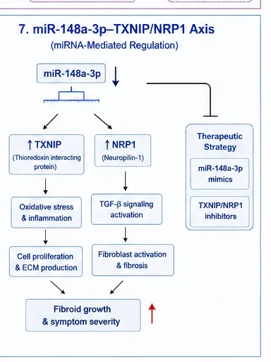
Figure 3: miR-148a-3p–TXNIP/NRP1 Regulatory Axis in Uterine Leiomyoma Pathogenesis.
8. Wnt/β-Catenin Signaling via Tankyrase Inhibition
The Wnt/β-catenin pathway offers several potential points of pharmacological intervention beyond RSPO3 and WNT9A. Under resting conditions, the APC–AXIN–GSK-3β destruction complex marks β-catenin for degradation; Wnt pathway activation suppresses this complex, allowing β-catenin to accumulate and enter the nucleus to drive proliferative gene expression. In fibroids specifically, WNT4 has been shown to activate AKT-dependent β-catenin signaling, and blocking canonical Wnt signaling slows human fibroid cell growth31.
Tankyrase 1/2 is a particularly attractive node because it regulates AXIN stability; inhibiting tankyrase stabilizes AXIN and promotes β-catenin degradation. Compounds such as XAV939 and JW55 have been developed on this principle as experimental Wnt-pathway inhibitors.
Summary: Priorities for Non-Hormonal Drug Development
TDO2 — a conventional medicinal-chemistry enzyme target
ITGA11 — an antibody or small-molecule ECM target
RSPO3 — an antibody or ligand-blocking Wnt target
WNT9A — a ligand-specific Wnt inhibition strategy
PARP1 — a drug-repurposing or novel selective-inhibitor opportunity
Of these, the TDO2–kynurenine–AHR axis currently stands out as the most mechanistically validated, since its inhibition has already demonstrated measurable effects on proliferation and apoptosis in fibroid models, including those driven by MED12 mutations 25,26.
REFERENCES


Shabna Rasheed, Dr. Rajalakshmi, Molecular Targets for Drug Design and Development of Uterine Leiomyoma, Int. J. of Pharm. Sci., 2026, Vol 4, Issue 9, 860-870, https://doi.org/10.5281/zenodo.22655493
 10.5281/zenodo.22655493