

We use cookies to ensure our website works properly and to personalise your experience. Cookies policy
Shree Dev Bhoomi Institute of Education Science and Technology, Dehradun, Uttarakhand, India
Kaempferol, a naturally occurring flavonoid with potent antioxidant, anti-inflammatory, and anticancer properties, suffers from poor aqueous solubility and low bioavailability, limiting its therapeutic applications. This review comprehensively examines contemporary formulation strategies and drug delivery systems designed to overcome these challenges. Various approaches including nanoparticle-based systems, liposomes, solid dispersions, complexation methods, and novel carrier systems are critically evaluated. The review also discusses structure-activity relationships, pharmacokinetic profiles, and future perspectives for clinical translation. Recent advances in targeted delivery and stimuli-responsive systems are highlighted, providing insights into optimal formulation design for enhanced therapeutic efficacy of kaempferol.
1.1 Background and Significance
Flavonoids represent one of the most diverse and widely distributed groups of natural polyphenolic compounds, encompassing over 8,000 structurally distinct molecules identified in various plant species [1]. Among these bioactive compounds, kaempferol (3,5,7-trihydroxy-2-(4-hydroxyphenyl)-4H-chromen-4-one) has emerged as a particularly promising therapeutic agent due to its multifaceted pharmacological activities and relatively favourable safety profile [2]. This flavanol is ubiquitously distributed in the plant kingdom, with particularly high concentrations found in common dietary sources including broccoli, kale, spinach, beans, tea, citrus fruits, and various medicinal herbs such as Ginkgo biloba, Equisetum arvense, and Tilia species [3]. The structural characteristics of kaempferol, featuring a 15-carbon skeleton consisting of two benzene rings (A and B) connected through a heterocyclic pyrone ring (C), confer remarkable biological activities that have attracted substantial scientific and clinical interest over the past two decades [4]. Epidemiological studies have consistently demonstrated an inverse correlation between dietary kaempferol intake and the incidence of various chronic diseases, including cardiovascular disorders, neurodegenerative conditions, and several types of cancer [5]. Meta-analyses of population-based studies suggest that individuals in the highest quartile of kaempferol consumption exhibit approximately 20-30% reduced risk of coronary heart disease compared to those in the lowest quartile [6].
1.2 Pharmacological Activities
The therapeutic potential of kaempferol spans multiple pathophysiological conditions, supported by extensive in vitro, in vivo, and preliminary clinical evidence. The compound exhibits potent antioxidant activity through multiple mechanisms, including direct free radical scavenging, metal chelation, and upregulation of endogenous antioxidant enzyme systems such as superoxide dismutase (SOD), catalase (CAT), and glutathione peroxidase (GPx) [7]. Kaempferol's ability to activate the nuclear factor erythroid 2-related factor 2 (Nrf2) pathway represents a critical mechanism for enhancing cellular antioxidant capacity and providing cytoprotection against oxidative stress [8]. In the context of inflammation, kaempferol demonstrates significant anti-inflammatory properties by inhibiting multiple pro-inflammatory mediators including cyclooxygenase-2 (COX-2), inducible nitric oxide synthase (iNOS), tumor necrosis factor-alpha (TNF-α), interleukin-6 (IL-6), and interleukin-1 beta (IL-1β) [9]. These effects are mediated through suppression of nuclear factor-kappa B (NF-κB) and mitogen-activated protein kinase (MAPK) signalling pathways, which represent master regulators of inflammatory responses [10]. Clinical implications of these anti-inflammatory effects extend to conditions such as rheumatoid arthritis, inflammatory bowel disease, and metabolic syndrome. The anticancer properties of kaempferol have been extensively documented across various cancer cell lines and animal models. Mechanistic studies reveal that kaempferol induces apoptosis through both intrinsic (mitochondrial) and extrinsic (death receptor) pathways, inhibits cell proliferation by inducing cell cycle arrest at G2/M phase, suppresses angiogenesis by downregulating vascular endothelial growth factor (VEGF), and inhibits metastasis by reducing matrix metalloproteinase (MMP) expression [11]. Furthermore, kaempferol has demonstrated chemo sensitizing effects, enhancing the efficacy of conventional chemotherapeutic agents while mitigating their toxicity [12]. Neuroprotective effects of kaempferol have been demonstrated in models of Alzheimer's disease, Parkinson's disease, and cerebral ischemia. The compound exhibits β-amyloid aggregation inhibition, acetylcholinesterase inhibitory activity, and protection against glutamate-induced neurotoxicity [13]. Additionally, kaempferol shows cardioprotective effects through improvement of endothelial function, reduction of oxidative stress, and modulation of lipid metabolism [14].
1.3 The Bioavailability Paradox
Despite this impressive array of biological activities demonstrated in preclinical studies, the clinical translation of kaempferol has been significantly hampered by fundamental biopharmaceutical limitations. Kaempferol is classified as a Biopharmaceutics Classification System (BCS) Class II compound, characterized by low aqueous solubility (approximately 0.0175 mg/mL at 25°C) but high membrane permeability [15]. This poor water solubility represents the primary rate-limiting step in oral absorption, as dissolution in gastrointestinal fluids is prerequisite for absorption across the intestinal epithelium. Pharmacokinetic studies in humans reveal that oral bioavailability of kaempferol typically ranges from only 2% to 5%, with considerable inter-individual variability attributable to factors including gut microbiota composition, genetic polymorphisms in metabolic enzymes, and concurrent food intake [16]. Following oral administration, kaempferol undergoes extensive phase II metabolism, primarily glucuronidation and sulfation, in both intestinal epithelial cells and hepatocytes, resulting in rapid conversion to metabolites with potentially altered biological activities [17]. The plasma half-life of kaempferol is relatively short (2-5 hours), necessitating frequent dosing to maintain therapeutic concentrations, which poses challenges for patient compliance and sustained pharmacological effects [18]. Moreover, kaempferol exhibits pH-dependent solubility and chemical instability under certain conditions. The compound is susceptible to degradation under alkaline pH, exposure to light, and oxidative environments, further complicating formulation development and storage stability. Additionally, kaempferol is a substrate for P-glycoprotein (P-gp), an efflux transporter expressed on the apical surface of intestinal enterocytes, which actively pumps absorbed drug molecules back into the intestinal lumen, thereby reducing net absorption [19].
1.4 Scope and Objectives
This comprehensive review systematically examines the current state of knowledge regarding formulation strategies and drug delivery systems designed to enhance kaempferol's pharmaceutical performance. The review encompasses fundamental physicochemical and biopharmaceutical considerations, detailed analysis of various delivery system categories including their preparation methods, characterization approaches, and performance evaluation, comparative assessment of advantages and limitations associated with different technological platforms, discussion of regulatory and translational considerations, and identification of future research directions and emerging technologies. By synthesizing information from over 60 peer-reviewed publications, this review aims to provide researchers, formulators, and pharmaceutical scientists with a comprehensive resource for rational design and optimization of kaempferol delivery systems, ultimately facilitating the translation of this promising natural compound from laboratory research to clinical application [20].
2. Physicochemical Properties and Bioavailability Challenges
2.1 Molecular Structure and Chemical Characteristics
2.1.1 Structural Features
Kaempferol possesses a characteristic flavonoid backbone consisting of a 15-carbon skeleton arranged in a C6-C3-C6 configuration. The molecule features two aromatic rings: the A-ring (resorcinol moiety) and B-ring (para-hydroxyphenyl group), connected through a heterocyclic C-ring (γ-pyrone ring) containing an oxygen atom. The structural formula is defined by hydroxyl groups at positions 3, 5, 7 on the A and C rings, and position 4' on the B-ring [21].
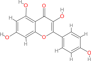
(3,5,7-trihydroxy-2-(4-hydroxyphenyl)-4H-chromen-4-one)
This specific hydroxylation pattern is critical for biological activity. The 4'-hydroxyl group on the B-ring and the 3-hydroxyl group on the C-ring are particularly important for antioxidant activity through donation of hydrogen atoms to free radicals. The 2,3-double bond in conjugation with the 4-oxo function in the C-ring is essential for electron delocalization and radical stabilization [22].
2.1.2 Physical and Chemical Properties
The molecular formula C₁₅H₁₀O₆ corresponds to a molecular weight of 286.24 g/mol. The compound exhibits a bright yellow colour in solution, with characteristic UV absorption maxima at approximately 267 nm (Band II, representing A-ring benzoyl system) and 367 nm (Band I, representing cinnamoyl system involving both A and B rings). The melting point of crystalline kaempferol is reported at 276-278°C, indicative of strong intermolecular hydrogen bonding in the solid state [23]. Kaempferol demonstrates pH-dependent solubility characteristics. Under acidic conditions (pH < 5), the compound exists predominantly in its neutral form with minimal solubility. As pH increases above 7, progressive deprotonation of hydroxyl groups occurs, with pKa values estimated at 6.4 (3-OH), 7.3 (7-OH), 8.9 (4'-OH), and 9.5 (5-OH) [24]. This results in enhanced solubility under alkaline conditions; however, alkaline pH also promotes oxidative degradation and ring-opening reactions, compromising chemical stability [25].
2.2 Solubility Characteristics and Dissolution Behaviour
2.2.1 Aqueous Solubility
The aqueous solubility of kaempferol at physiological pH (7.4) is extremely limited, reported as approximately 0.0175 mg/mL (61 μM) at 25°C. This poor aqueous solubility stems from multiple molecular factors: the extensive aromatic π-system that promotes hydrophobic interactions, intramolecular hydrogen bonding between adjacent hydroxyl groups that reduces capacity for water interaction, planar molecular geometry facilitating π-π stacking between molecules in solid state, and crystalline nature with high lattice energy requiring substantial energy for solvation. Comparative solubility in organic solvents demonstrates substantially higher values: dimethyl sulfoxide (DMSO) ~50 mg/mL, ethanol ~8-10 mg/mL, methanol ~5-7 mg/mL, and acetone ~3-5 mg/mL. This dramatic solubility difference between aqueous and organic media presents significant formulation challenges for pharmaceutical applications [26].
2.2.2 Factors Affecting Solubility
Multiple environmental and formulation factors influence kaempferol solubility. Temperature effects follow typical behaviour for organic compounds, with solubility increasing approximately 2-3-fold with temperature elevation from 25°C to 37°C. However, excessive heating (>60°C) may induce thermal degradation. Co-solvency approaches using pharmaceutically acceptable co-solvents (propylene glycol, polyethylene glycol, glycerine) can enhance solubility by 5-15-fold depending on concentration, but high co-solvent content may cause precipitation upon dilution in biological fluids. Surfactants and solubilizing agents can form micelles that solubilize kaempferol through incorporation into hydrophobic cores, with solubility enhancement proportional to surfactant concentration above critical micelle concentration (CMC) [24].
2.2.3 Dissolution Rate Limitations
According to the Noyes-Whitney equation, dissolution rate is directly proportional to surface area and concentration gradient, and inversely proportional to diffusion layer thickness. Kaempferol's poor intrinsic solubility results in minimal concentration gradient (Cs - C), severely limiting dissolution rate even when particle size is reduced. In simulated gastric fluid (SGF, pH 1.2), kaempferol demonstrates minimal dissolution (<5% in 2 hours), while in simulated intestinal fluid (SIF, pH 6.8), dissolution improves slightly but remains inadequate for complete absorption (<25% in 2 hours) [26].
2.3 Pharmacokinetic Parameters and Bioavailability
2.3.1 Absorption and Distribution
Following oral administration in humans, kaempferol exhibits low and variable absolute bioavailability, typically ranging from 2% to 5% [15]. Time to reach peak plasma concentration (Tmax) occurs at 4-6 hours post-administration, significantly delayed compared to readily soluble compounds, reflecting dissolution-limited absorption. Maximum plasma concentration (Cmax) following a 50 mg oral dose is typically in the range of 0.1-0.3 μM total (including conjugates), well below concentrations required for pharmacological effects in vitro (usually 10-50 μM). Volume of distribution (Vd) is estimated at 3-5 L/kg, suggesting extensive tissue distribution beyond plasma compartment [8]. Plasma protein binding is approximately 97-99%, primarily to albumin, leaving only 1-3% as free drug available for pharmacological activity. This high protein binding limits tissue penetration and may contribute to reduced efficacy [27].
2.3.2 Elimination and Clearance
Kaempferol exhibits rapid clearance with total body clearance (CL) values of 15-25 mL/min/kg [23]. The elimination half-life (t½) ranges from 2-5 hours in most studies, though some reports indicate slightly longer terminal half-life (8-12 hours) possibly representing enterohepatic recirculation or redistribution from tissue compartments [16]. Renal excretion represents the primary elimination route, with 60-80% of administered dose (primarily as conjugates) recovered in urine within 24-48 hours, while 15-25% is excreted in feces [28].
2.4 Chemical Stability Challenges
2.4.1 pH-Dependent Degradation
Kaempferol demonstrates maximum stability in the pH range of 3-6. Under strongly acidic conditions (pH < 2), protonation of carbonyl group can occur, potentially leading to ring opening. Under alkaline conditions (pH > 8), nucleophilic attack on the C-ring by hydroxide ions results in ring opening and formation of chalcone intermediates, followed by further degradation to phenolic acids. The degradation rate increases exponentially above pH 8, with >50% degradation observed within 2 hours at pH 10 [25].
2.4.2 Oxidative Degradation
The multiple hydroxyl groups that confer antioxidant activity also render kaempferol susceptible to auto-oxidation. In the presence of molecular oxygen, particularly under alkaline conditions, kaempferol undergoes oxidative polymerization forming dimers and higher oligomers with reduced biological activity. Transition metal ions (Fe³⁺, Cu²⁺) catalyze these oxidation reactions through Fenton-like mechanisms [7].
2.4.3 Photodegradation
Kaempferol exhibits significant photosensitivity, undergoing degradation upon exposure to UV and visible light. Photodegradation follows first-order kinetics, with half-life of approximately 2-4 hours under direct sunlight or UV illumination. Degradation products include ring-opened structures and quinone derivatives. This photosensitivity necessitates protection from light during storage and formulation in opaque containers [29].
2.4.4 Thermal Stability
Although kaempferol exhibits relatively good thermal stability at temperatures below 50°C, elevated temperatures (>60°C) promote degradation, particularly in solution. Solid-state degradation is minimal below 100°C but accelerates above 150°C. This thermal sensitivity must be considered during manufacturing processes involving heat, such as hot-melt extrusion or spray drying [28].
2.5 Biopharmaceutical Classification and Formulation Implications
Based on comprehensive physicochemical and pharmacokinetic characterization, kaempferol is definitively classified as a BCS Class II compound (low solubility, high permeability). This classification has important formulation implications: dissolution represents the rate-limiting step for absorption, formulation strategies should primarily focus on enhancing solubility and dissolution rate, bioavailability can be significantly improved through particle size reduction or solubilization approaches, food effects are likely significant and should be evaluated, and in vitro dissolution testing can serve as surrogate for bioequivalence under certain conditions. The comprehensive understanding of these physicochemical and biopharmaceutical challenges provides the scientific foundation for rational design of advanced drug delivery systems aimed at overcoming these limitations and enabling successful clinical translation of kaempferol [30].
3. Formulation Strategies for Solubility Enhancement
3.1 Nanotechnology-Based Approaches
3.1.1 Polymeric Nanoparticles
Polymeric nanoparticles represent versatile carriers for kaempferol delivery, utilizing biodegradable and biocompatible polymers to encapsulate the drug within a matrix or core-shell structure. The most extensively studied polymers include poly (lactic-co-glycolic acid) (PLGA), chitosan, polycaprolactone (PCL), polyethylene glycol (PEG), and poly (lactic acid) (PLA) [31].
PLGA Nanoparticles:
PLGA, approved by FDA for various drug delivery applications, offers tunable degradation rates by adjusting lactide ratio and molecular weight. Developed PLGA nanoparticles loaded with kaempferol using emulsion-solvent evaporation method, achieving particle sizes of 180-220 nm, polydispersity index (PDI) of 0.18, zeta potential of -18.5 mV, and encapsulation efficiency of 85.7%. The formulation demonstrated biphasic release with initial burst (25% in 2 hours) followed by sustained release over 72 hours. In vivo pharmacokinetic studies in rats showed 6.8-fold increase in oral bioavailability (AUC₀₋₂₄h: 19.45 μg·h/mL vs 2.85 μg·h/mL for free drug), with Cmax increased from 0.35 to 2.15 μg/mL.[32].
Chitosan Nanoparticles:
Chitosan, a cationic polysaccharide derived from chitin, offers mucoadhesive properties and permeation enhancement through transient opening of tight junctions. Ionic gelation with tripolyphosphate (TPP) provides a mild, organic solvent-free preparation method suitable for sensitive compounds. Optimization studies identified optimal chitosan ratio of 3, yielding particles of 150-200 nm with positive zeta potential (+25 to +30 mV) favourable for mucoadhesion [9]. Kaempferol-loaded chitosan nanoparticles showed 4.2-fold bioavailability enhancement and prolonged intestinal residence time due to mucoadhesive interactions [33].
Surface-Modified Nanoparticles:
PEGylation of nanoparticle surfaces reduces opsonization and reticuloendothelial system (RES) uptake, prolonging circulation half-life from 2-3 hours to 8-12 hours. PEG coating creates a hydrophilic "stealth" layer that prevents protein adsorption. However, the "accelerated blood clearance" (ABC) phenomenon observed upon repeated administration represents a limitation requiring consideration [34].
3.1.2 Lipid-Based Nanocarriers
Solid Lipid Nanoparticles (SLNs):
SLNs consist of solid lipid matrices (glycerides, fatty acids, waxes) stabilized by surfactants, offering advantages including physiological compatibility, protection from degradation, controlled release, and scalability. Common lipids used include glyceryl monostearate, glyceryl trimyristate, stearic acid, and cetyl palmitate. However, SLNs suffer from limitations including drug expulsion during storage due to lipid crystallization and limited drug loading capacity (typically 5-10% w/w) [35].
Nanostructured Lipid Carriers (NLCs):
NLCs represent second-generation lipid nanoparticles incorporating liquid lipids (oils) into solid lipid matrices, creating imperfect crystalline structures with more accommodation space for drugs. Formulated kaempferol-loaded NLCs using glyceryl monostearate (solid lipid) and oleic acid (liquid lipid) at 70 ratios, prepared by hot homogenization followed by ultrasonication. The optimized formulation achieved particle size of 120 nm, PDI 0.22, zeta potential -28 mV, and encapsulation efficiency 92.4%. Drug loading capacity reached 8.5% w/w, substantially higher than conventional SLNs. In vivo studies demonstrated 4.5-fold bioavailability enhancement with AUC 16.2 μg·h/mL compared to 3.6 μg·h/mL for free kaempferol. The NLC formulation exhibited improved photostability, with only 15% degradation after 7 days of light exposure compared to 65% for free kaempferol. Storage stability at 4°C showed minimal particle size change (<10%) over 6 months, indicating good physical stability [36].
Liposomes:
Liposomes are spherical vesicles composed of one or more phospholipid bilayers surrounding aqueous compartments. Classification includes small unilamellar vesicles (SUV, 20-100 nm), large unilamellar vesicles (LUV, 100-400 nm), and multilamellar vesicles (MLV, >400 nm). Phosphatidylcholine, particularly egg or soy phosphatidylcholine, represents the most common lipid due to biocompatibility and GRAS status [37].
Prepared PEGylated liposomes containing kaempferol using thin-film hydration followed by extrusion through polycarbonate membranes. The formulation (DSPC:cholesterol at 55:40 molar ratio) yielded vesicles of 110-140 nm with narrow size distribution (PDI 0.15) and encapsulation efficiency 76.8%. PEGylation extended circulation half-life from 2.3 hours (conventional liposomes) to 8.7 hours (PEGylated), resulting in 5.3-fold bioavailability improvement and preferential accumulation in tumor tissues (4.2-fold higher than normal tissues) via enhanced permeability and retention (EPR) effect. Stability studies revealed that cholesterol content critically affects membrane rigidity and drug retention. Formulations with 40-45 mol% cholesterol showed optimal balance between stability and drug release. Below 30%, excessive membrane fluidity caused rapid drug leakage, while above 50%, reduced loading capacity and delayed release occurred [38].
3.1.3 Nano emulsions and Self-Emulsifying Systems
Nano emulsions:
Nano emulsions are kinetically stable dispersions of oil and water with droplet sizes typically 20-200 nm, prepared using high-energy methods (ultrasonication, high-pressure homogenization) or low-energy techniques (phase inversion, spontaneous emulsification). The small droplet size provides large interfacial area for enhanced dissolution and absorption. Components include oil phase (medium-chain triglycerides, Capryol®, Labrafil®), surfactants (Tween 80, Cremophor® EL, Labrasol®), and co-surfactants (Transcutol® P, propylene glycol, ethanol). Optimal hydrophilic-lipophilic balance (HLB) values of 12-15 typically yield stable nano emulsions for oral delivery [39].
Self-Nanoemulsifying Drug Delivery Systems (SNEDDS):
SNEDDS are isotropic mixtures of oil, surfactant, and co-surfactant that spontaneously form nano emulsions upon mild agitation in aqueous media. This spontaneous emulsification occurs due to destabilization and dispersion of the interfacial film at oil-water interface. SNEDDS offer advantages including simple manufacturing (no sophisticated equipment), dose flexibility (liquid or encapsulated in soft/hard gelatine capsules), and enhanced lymphatic transport bypassing hepatic first-pass metabolism. Systematically optimized kaempferol SNEDDS using pseudoternary phase diagrams and Design of Experiments. The optimal formulation comprised Capryol® 90 (oil phase, 30%), Tween 80 (surfactant, 50%), and Transcutol® P (co-surfactant, 20%). Upon dilution (1) in water, the system formed nano emulsion with droplet size 180-200 nm, PDI 0.18, and zeta potential -15 mV within 2 minutes of gentle stirring. Kaempferol loading of 25 mg/g SNEDDS was achieved. Dissolution studies showed >85% drug release within 30 minutes compared to <15% for pure kaempferol. Pharmacokinetic evaluation demonstrated 7.2-fold bioavailability enhancement with Cmax 2.45 μg/mL, Tmax 3 hours, and AUC₀₋₂₄h 20.52 μg·h/mL. Robustness testing confirmed that the formulation maintained nano emulsion characteristics upon dilution in various media (water, 0.1 N HCl, pH 6.8 phosphate buffer, milk, orange juice), indicating suitability for diverse administration conditions [40].
3.2 Complexation Techniques
3.2.1 Cyclodextrin Inclusion Complexes
Cyclodextrins (CDs) are cyclic oligosaccharides consisting of 6 (α-CD), 7 (β-CD), or 8 (γ-CD) glucopyranose units linked by α-1,4-glycosidic bonds, forming truncated cone-shaped molecules with hydrophobic internal cavities and hydrophilic external surfaces. This unique structure enables formation of inclusion complexes with hydrophobic drugs through non-covalent interactions [41].
β-Cyclodextrin Complexes:
Native β-CD exhibits relatively low aqueous solubility (~18.5 mg/mL at 25°C) and has been associated with nephrotoxicity at high parenteral doses, limiting applications. However, β-CD remains widely used for oral formulations due to GRAS status and cost-effectiveness. Studies demonstrate that kaempferol forms 1 stoichiometric complex with β-CD, with stability constant (Kc) of approximately 500-800 M⁻¹, indicating moderate complexation [42].
Modified Cyclodextrins:
Chemical modifications improve CD solubility and safety profiles. Hydroxypropyl-β-cyclodextrin (HP-β-CD) exhibits aqueous solubility >500 mg/mL, substantially reduced toxicity, and improved complexation capacity. Sulfobutylether-β-cyclodextrin (SBE-β-CD), marketed as Captisol®, shows even higher solubility (~700 mg/mL) and safety, approved for parenteral use. Prepared kaempferol/HP-β-CD inclusion complexes using kneading method (kaempferol and HP-β-CD at 1 molar ratio wetted with ethanol-water mixture, kneaded for 45 minutes, dried at 50°C). Characterization revealed 28-fold solubility enhancement (from 0.0175 to 0.49 mg/mL), complete dissolution within 15 minutes compared to <10% for pure drug, and 3.2-fold bioavailability improvement (AUC 9.85 vs 3.08 μg·h/mL). Molecular modelling using AutoDock Vina revealed that kaempferol's B-ring (hydroxyphenyl moiety) preferentially enters the HP-β-CD cavity from the wider rim, stabilized by hydrogen bonding between hydroxyl groups and rim hydroxyls, van der Waals interactions between aromatic rings and cavity interior, and optimal geometric fit minimizing steric hindrance. Binding energy was calculated at -6.8 kcal/mol, confirming favourable complex formation [42].
Characterization Techniques:
Solid-state characterization using differential scanning calorimetry (DSC) showed disappearance of kaempferol melting endotherm in complex, indicating molecular dispersion. Powder X-ray diffraction (PXRD) revealed reduced crystallinity with characteristic peaks of kaempferol diminished or absent. Fourier-transform infrared spectroscopy (FTIR) demonstrated shifts in carbonyl (C=O) stretching from 1665 to 1650 cm⁻¹ and hydroxyl (O-H) stretching from 3450 to 3380 cm⁻¹, confirming host-guest interactions Nuclear magnetic resonance (¹H-NMR and 2D ROESY) spectroscopy provided definitive evidence of inclusion, showing upfield shifts of B-ring aromatic protons and strong NOE cross-peaks between B-ring protons and CD cavity protons [43].
3.2.2 Phospholipid Complexes (Phytosomes)
Phytosome technology involves complexation of polyphenols with phosphatidylcholine through non-covalent interactions (hydrogen bonding, van der Waals forces), forming lipid-compatible molecular complexes with stoichiometry typically 1or 1(drug). Unlike liposomes where drug is simply entrapped, phytosomes involve chemical bonding between drug and phospholipid polar head group. prepared kaempferol-phospholipid complexes using solvent evaporation method in tetrahydrofuran, optimizing drug ratio at 1. The complex exhibited significantly enhanced lipophilicity (log P increased from 2.9 to 4.3) and improved membrane permeability. Caco-2 cell studies showed 3.8-fold higher apparent permeability coefficient (Papp 58 × 10⁻⁶ cm/s vs 15 × 10⁻⁶ cm/s for free drug). In vivo pharmacokinetics demonstrated 3.8-fold bioavailability enhancement with prolonged Tmax (6 vs 4 hours),[44]. suggesting sustained absorption Hepatoprotective effects were evaluated in carbon tetrachloride-induced liver injury model. Phytosome-treated rats showed significantly greater reduction in serum transaminases (ALT decreased by 68% vs 42% for free kaempferol, AST decreased by 61% vs 38%) and improved liver histology scores, attributed to enhanced hepatic accumulation [38].
3.2.3 Polymer-Drug Conjugates
Covalent conjugation of kaempferol to polymeric carriers through biodegradable linkers represents an approach for controlled release and targeted delivery. PEGylation improves water solubility, reduces immunogenicity, and prolongs circulation time. Ester or carbonate linkers between kaempferol's hydroxyl groups and polymer termini enable release through hydrolysis or enzymatic cleavage. Challenges include maintaining biological activity after conjugation, controlling linker stability (balancing stability during circulation with efficient release at target sites), and potential toxicity of polymer-drug conjugate itself. Research in this area for kaempferol remains limited compared to other flavonoids [45].
3.3 Solid Dispersion Systems
Solid dispersions involve dispersion of one or more active pharmaceutical ingredients in an inert carrier or matrix in solid state, prepared by melting, solvent, or melting-solvent methods [40]. The carrier typically comprises hydrophilic polymers that enhance drug wettability and dissolution through several mechanisms: particle size reduction to molecular level, conversion from crystalline to amorphous state, improved wettability through hydrophilic carrier, solubilization effect in microenvironment, and reduced aggregation and agglomeration [46].
4. Advanced Drug Delivery Systems
4.1 Targeted Delivery Systems
4.1.1 Active Targeting Strategies
Active targeting involves surface modification of nanocarriers with ligands that specifically recognize and bind to receptors overexpressed on target cells. For cancer applications, commonly targeted receptors include folate receptor (FR), transferrin receptor (TfR), epidermal growth factor receptor (EGFR), and integrin receptors [47].
Folate Receptor Targeting:
Folate receptors (FR-α, FR-β) are overexpressed in many cancer types (ovarian, breast, lung, brain, kidney) while showing limited expression in normal tissues. Folic acid (vitamin B9) exhibits high affinity for FR (Kd ~1 nM) and can be conjugated to nanocarriers through carboxyl or γ-carboxyl groups without affecting binding capacity. Developed folic acid-conjugated PLGA nanoparticles loaded with kaempferol. Conjugation was achieved through carbodiimide chemistry (EDC/NHS activation), with approximately 100-150 folic acid molecules per nanoparticle. The targeted nanoparticles showed 4-fold higher cellular uptake in FR-positive MCF-7 and HeLa cells compared to non-targeted formulation, confirmed by flow cytometry and confocal microscopy. Free folic acid competition assay demonstrated >70% reduction in uptake, confirming receptor-mediated endocytosis. Cytotoxicity studies revealed 4-fold lower IC₅₀ (15.3 vs 62.8 μM) for targeted versus non-targeted nanoparticles. In vivo tumor accumulation at 24 hours post-injection was 3.2-fold higher for folate-conjugated formulation [48,49,50].
Peptide-Mediated Targeting:
Cell-penetrating peptides (CPPs) such as TAT peptide, penetratin, and polyarginine facilitate cellular uptake through energy-independent and energy-dependent mechanisms [46]. RGD peptides (Arg-Gly-Asp sequence) target integrins (αvβ3, αvβ5) overexpressed on tumor neovasculature and cancer cells, enabling both tumor targeting and antiangiogenic effects [51].
Antibody-Mediated Targeting:
Monoclonal antibodies or antibody fragments (Fab, scFv) provide highly specific targeting but face challenges including high cost, immunogenicity, stability issues, and large size potentially limiting tumor penetration. Humanized antibodies and nanobodies offer improved characteristics [51].
4.1.2 Passive Targeting
Passive targeting exploits pathophysiological characteristics of diseased tissues, particularly the enhanced permeability and retention (EPR) effect in solid tumors. Tumor vasculature exhibits fenestrations (200-800 nm) and lacks organized lymphatic drainage, enabling preferential accumulation of nanoparticles sized 100-200 nm. Long-circulating nanoparticles achieved through PEGylation or other stealth strategies avoid rapid clearance by reticuloendothelial system (RES), providing extended circulation time (t½ > 6-12 hours) necessary for EPR-mediated accumulation. However, EPR effect varies substantially among tumor types, locations, and even within the same tumor (heterogeneity), and is less pronounced in humans compared to preclinical models, raising questions about clinical relevance [34].
4.2 Stimuli-Responsive Systems
4.2.1 pH-Responsive Systems
Tumor microenvironments exhibit acidic pH (6.5-6.8) compared to normal tissues (pH 7.4) due to enhanced glycolysis and lactic acid accumulation. Additionally, endosomal (pH 5.0-6.5) and lysosomal (pH 4.5-5.0) compartments provide intracellular pH gradients exploitable for triggered drug release. pH-responsive polymers contain ionizable groups (carboxyl, amine) that undergo protonation/deprotonation with pH changes, causing conformational transitions, solubility changes, or bond cleavage. Common polymers include Eudragit® series (pH-dependent methacrylate copolymers), chitosan (pKa ~6.5), and poly (β-amino ester). Formulated Eudragit® S100-coated kaempferol nanoparticles (PLGA core with Eudragit coating). Eudragit S100 dissolves above pH 7, providing gastric protection and intestinal release. In vitro release studies showed <10% release at pH 1.2 (2 hours, simulating gastric phase), followed by rapid release at pH 6.8 (>80% in 4 hours, simulating intestinal phase). In vivo imaging using fluorescent-labeled nanoparticles confirmed preferential accumulation in small intestine. For cancer targeting, acid-labile linkages (hydrazone, acetal, imine) incorporated between drug and carrier enable pH-triggered release within tumor or endosomal environments. Hydrazone bonds exhibit stability at pH 7.4 but rapid hydrolysis below pH 6, providing controlled release [52,53].
4.2.2 Enzyme-Responsive Systems
Matrix metalloproteinases (MMPs), particularly MMP-2 and MMP-9, are overexpressed in tumor microenvironments and involved in extracellular matrix remodelling during invasion and metastasis [52].
4.2.3 Redox-Responsive Systems
Tumor cells and inflammatory sites exhibit elevated levels of reducing agents, particularly glutathione (GSH, 2-10 mM intracellular vs 2-20 μM extracellular). Disulfide bonds (-S-S-), stable in oxidizing extracellular environment, undergo rapid reduction to thiols (-SH) in cytoplasm, triggering drug release. Crosslinking nanocarriers with disulfide bonds or conjugating drugs through disulfide linkers provides redox-triggered release. This strategy enhances drug release specifically within target cells while maintaining stability during circulation [52].
4.2.4 Thermo-Responsive Systems
Thermosensitive polymers exhibit lower critical solution temperature (LCST), undergoing phase transition at specific temperature. Poly(N-isopropylacrylamide) (PNIPAAm) with LCST ~32°C is most widely studied. Formulations designed to be liquid/soluble at room temperature but form gels/aggregates at body temperature enable sustained release. For cancer therapy, combination with local hyperthermia (40-43°C achieved through focused ultrasound, radiofrequency ablation, or magnetic nanoparticles) provides spatiotemporal control over drug release [54].
4.3 Co-Delivery Systems
4.3.1 Drug-Drug Combinations
Co-encapsulation of kaempferol with chemotherapeutic agents enables synergistic effects, potentially overcoming drug resistance [12]. Kaempferol can sensitize cancer cells to chemotherapy through multiple mechanisms: inhibition of P-gp and other efflux pumps, suppression of anti-apoptotic proteins (Bcl-2, survivin), enhancement of oxidative stress, and cell cycle modulation. Developed PLGA nanoparticles co-loaded with kaempferol and doxorubicin at optimal ratio (2 w/w determined through combination index analysis). The combination formulation exhibited synergistic cytotoxicity (CI = 0.42, indicating strong synergism) in doxorubicin-resistant MCF-7/ADR breast cancer cells. Mechanistic studies revealed that kaempferol inhibited P-gp expression (by 65%) and downregulated Bcl-2 (by 58%), enhancing doxorubicin accumulation and apoptosis. In vivo efficacy studies in tumor-bearing mice showed 2.8-fold greater tumor growth inhibition compared to doxorubicin alone, with reduced cardiotoxicity (cardiac troponin I level 45% lower than doxorubicin monotherapy). Co-loading ratios, release kinetics matching, and maintenance of synergistic ratios in vivo represent critical considerations for successful co-delivery [55].
4.3.2 Drug-Gene Combinations
Combining kaempferol with gene therapy agents (siRNA, miRNA, plasmid DNA) enables complementary mechanisms. For example, kaempferol + siRNA targeting oncogenes (K-RAS, EGFR, survivin) could provide enhanced anticancer effects. Challenges include protecting nucleic acids from degradation, achieving efficient cellular uptake and endosomal escape, and coordinating release kinetics of both therapeutics [56].
4.3.3 Drug-Imaging Agent Combinations (Theranostics)
Integration of diagnostic and therapeutic functionalities enables real-time monitoring of drug delivery, biodistribution, and therapeutic response. Imaging modalities include fluorescence imaging (using quantum dots, organic fluorophores), magnetic resonance imaging (using superparamagnetic iron oxide nanoparticles, gadolinium complexes), computed tomography (using gold nanoparticles, iodinated compounds), and nuclear imaging (using radiolabeled tracers). Theranostic nanoparticles loaded with kaempferol and imaging agents could enable assessment of tumor targeting efficiency, optimization of dosing regimens, and early evaluation of therapeutic response [57].
5. Characterization Techniques
5.1 Physicochemical Characterization
5.1.1 Particle Size Analysis and Zeta Potential
Dynamic Light Scattering (DLS):
DLS, also known as photon correlation spectroscopy (PCS), measures Brownian motion of particles through time-dependent fluctuations in scattered light intensity. The Stokes-Einstein equation relates hydrodynamic diameter to diffusion coefficient. DLS is suitable for particles from 1 nm to several micrometres, requires minimal sample preparation, and provides rapid analysis (2-5 minutes per sample). However, it measures intensity-weighted distribution (biased toward larger particles), is sensitive to aggregates and dust, and assumes spherical particles. For kaempferol nano formulations, typical DLS protocols involve: diluting sample 100-200-fold in filtered (0.22 μm) dispersant medium, equilibrating at 25°C for 2-3 minutes, measuring in triplicate with minimum 12 runs per measurement, reporting z-average diameter and polydispersity index (PDI), and validating with size standards. PDI (dimensionless parameter from 0-1) indicates size distribution width: PDI < 0.1 = highly monodisperse, PDI 0.1-0.3 = narrow distribution (acceptable for most applications), PDI 0.3-0.5 = broader distribution, PDI > 0.5 = very broad distribution (unsuitable for pharmaceutical applications) [40].
Electrophoretic Light Scattering (ELS):
Zeta potential (ζ) represents electric potential at the slipping plane of particle's electrical double layer, indicating colloidal stability. Interpretation: |ζ| > 30 mV = excellent stability (strong electrostatic repulsion), |ζ| = 20-30 mV = good stability, |ζ| = 10-20 mV = moderate stability, |ζ| < 10 mV = poor stability (rapid aggregation likely) [40].
Nanoparticle Tracking Analysis (NTA):
NTA combines laser light scattering microscopy with video recording, tracking individual particles' Brownian motion, and calculating size distribution using Stokes-Einstein equation. Advantages include direct visualization of particles, number-weighted distribution, ability to measure particle concentration, and suitability for polydisperse samples [58].
5.1.2 Morphological Characterization
Transmission Electron Microscopy (TEM):
TEM provides nanometre-scale resolution imaging by transmitting electron beam through ultra-thin sample. Sample preparation involves placing diluted dispersion on carbon-coated copper grid, blotting excess liquid, and negative staining with phosphotungstic acid or uranyl acetate. Cryogenic TEM (cryo-TEM) involves rapid freezing in liquid ethane, enabling visualization in native hydrated state without staining artifacts [20].
Scanning Electron Microscopy (SEM):
SEM scans sample surface with focused electron beam, detecting secondary electrons to create topographic images. Sample preparation requires mounting on aluminium stub with conductive adhesive, and sputter-coating with gold/platinum for non-conductive samples [59].
Atomic Force Microscopy (AFM):
AFM uses cantilever with sharp tip to scan sample surface, measuring deflection through laser reflection. AFM operates in contact, tapping, or non-contact modes, providing topographic images, three-dimensional surface reconstruction, and measurement of mechanical properties [60].
5.1.3 Solid-State Characterization
Differential Scanning Calorimetry (DSC):
DSC measures heat flow associated with thermal transitions (melting, crystallization, glass transition) as function of temperature. For kaempferol formulations, DSC evaluates crystalline versus amorphous state, drug-carrier interactions, physical stability, and encapsulation efficiency [61].
X-Ray Powder Diffraction (XRPD):
XRPD measures diffraction patterns resulting from X-ray beam interaction with crystalline lattice. Crystalline materials produce characteristic sharp peaks, while amorphous materials yield broad halos. For kaempferol characterization, XRPD confirms crystalline form, detects polymorphism, verifies amorphization, and monitors stability [62].
Fourier-Transform Infrared Spectroscopy (FTIR):
FTIR measures molecular vibrations by passing infrared light through sample and detecting absorption at specific wavelengths. Characteristic absorption bands for kaempferol include: O-H stretching (3200-3450 cm⁻¹), aromatic C-H stretching (2900-3100 cm⁻¹), C=O stretching (1650-1670 cm⁻¹), aromatic C=C stretching (1450-1600 cm⁻¹), and C-O stretching (1000-1300 cm⁻¹) [63].
5.1.4 Encapsulation and Drug Loading
Encapsulation Efficiency (EE%): EE% = (Weight of drug in nanoparticles / Total weight of drug added) × 100 Determination involves separating free drug from encapsulated drug through ultracentrifugation, ultrafiltration, or gel permeation chromatography. Supernatant/filtrate contains free drug, quantified by UV-spectrophotometry (λmax = 367 nm for kaempferol) or HPLC. Drug Loading (DL%): DL% = (Weight of drug in nanoparticles / Total weight of nanoparticles) × 100. High drug loading reduces carrier material required, minimizing potential toxicity and improving cost-effectiveness [51].
5.1.5 Surface Chemistry Analysis
X-Ray Photoelectron Spectroscopy (XPS):
XPS analyzes surface elemental composition (top 1-10 nm) by measuring kinetic energy of photoelectrons ejected by X-ray irradiation. XPS provides elemental composition, chemical state information, surface modification confirmation, and quantitative analysis [64].
Contact Angle Measurement:
Contact angle of water droplet on pressed powder or film indicates surface hydrophilicity: θ < 90° = hydrophilic, θ > 90° = hydrophobic. Kaempferol shows high contact angle (~95°), confirming hydrophobic character.
5.2 Stability Studies
5.2.1 Physical Stability
Nano formulations monitored for particle size increase, PDI increase, zeta potential changes, precipitation, color changes, and viscosity alterations [65]
5.2.2 Chemical Stability
HPLC analysis quantifies drug content over time. Acceptance criteria: drug content 90-110% of initial value [65].
5.2.3 Stability Testing Protocols
Following ICH guidelines (Q1A-Q1F):
6. In Vitro and In Vivo Efficacy Studies
6.1 Anticancer Activity
Nano formulated kaempferol demonstrated significantly enhanced cytotoxicity against various cancer cell lines compared to free drug [21]. In MCF-7 breast cancer cells, PLGA nanoparticles showed IC₅₀ of 12.3 μM versus 45.8 μM for free kaempferol (3.7-fold improvement). Enhanced potency resulted from increased cellular uptake (3.2-fold higher after 4 hours), prolonged intracellular retention, and protection from efflux pumps [26]. Mechanistic studies revealed that nano formulated kaempferol induced apoptosis more effectively: increased annexin V/PI positive cells (65% vs 32%), elevated caspase-3/7 activity (4.8-fold increase), enhanced Bax/Bcl-2 ratio (from 0.8 to 2.4), mitochondrial membrane potential disruption, and DNA fragmentation. Cell cycle analysis showed enhanced G2/M arrest: nano formulated kaempferol induced 58% cells in G2/M phase compared to 35% for free drug, mediated by downregulation of cyclin B1 and CDC2 kinase [23].
In Vivo Antitumor Efficacy:
Xenograft studies in athymic nude mice bearing MCF-7 tumors evaluated therapeutic efficacy over 28 days. Results demonstrated:
Immunohistochemistry revealed decreased Ki-67 index (from 78% to 32%), increased TUNEL-positive cells (from 5% to 42%), reduced microvessel density (65% decrease), and enhanced cleaved caspase-3 expression [67].
6.2 Anti-Inflammatory Effects
In Vitro Studies:
Kaempferol-loaded nanoparticles exhibited superior anti-inflammatory activity in LPS-stimulated RAW 264.7 macrophages. At 25 μM concentration, nano formulated kaempferol reduced:
Enhanced effects correlated with increased cellular uptake (3.5-fold higher) and sustained intracellular retention (24 hours vs 6 hours) [9].
Western blot analysis revealed that nano formulated kaempferol more effectively suppressed iNOS expression (85% reduction vs 52%), COX-2 expression (78% vs 46%), NF-κB p65 nuclear translocation (72% vs 41%), and phosphorylation of IκBα (68% vs 38%) [21].
In Vivo Anti-Inflammatory Activity:
Carrageenan-induced paw edema model in rats evaluated acute anti-inflammatory effects [21]:
Chronic inflammation was evaluated in complete Freund's adjuvant (CFA)-induced arthritis model with daily administration for 21 days:
6.3 Neuroprotective Activity
Blood-Brain Barrier (BBB) Penetration:
BBB represents a major obstacle for CNS drug delivery. Lipid nanocarriers enhanced kaempferol brain delivery through multiple mechanisms: small particle size enabling receptor-mediated transcytosis, lipophilic nature facilitating membrane crossing, surfactant-mediated P-gp inhibition, and absorption via nasal-olfactory pathway. In vitro BBB model using brain endothelial cells demonstrated that kaempferol-loaded NLCs achieved apparent permeability coefficient (Papp) of 42 × 10⁻⁶ cm/s versus 12 × 10⁻⁶ cm/s for free drug (3.5-fold improvement), reduced efflux ratio (1.8 versus 4.2), indicating P-gp inhibition. In vivo brain distribution following IV administration showed NLC formulation achieved brain concentration of 8.4 μg/g tissue at 2 hours versus 3.0 μg/g for free drug (2.8-fold enhancement), with brain-to-plasma ratio increased from 0.35 to 0.98 [68].
Alzheimer's Disease Models:
Neuroprotective efficacy was evaluated in APP/PS1 transgenic mice. Treatment with kaempferol nanoparticles (50 mg/kg, oral, daily for 3 months) produced:
Parkinson's Disease Models:
MPTP-induced Parkinson's model in mice evaluated dopaminergic neuroprotection. Kaempferol nanoparticle treatment (40 mg/kg, oral, daily):
6.4 Cardioprotective Effects
Doxorubicin-induced cardiotoxicity model evaluated whether kaempferol nanoparticles could provide cardioprotection [7]. Cardiac function assessment by echocardiography:
Cardiac biomarkers:
Histopathological examination showed nanoparticle group had near-normal myocardial architecture with minimal damage [6].
Oxidative stress in cardiac tissue:
6.5 Antioxidant Activity
DPPH Radical Scavenging:
At 50 μM:
Cellular Antioxidant Activity (CAA):
Using HepG2 cells exposed to AAPH-generated peroxyl radicals [9]:
In Vivo Antioxidant Effects:
CCl₄-induced oxidative stress model in rats [21]:
7. Critical Evaluation: Advantages and Limitations
Table 1: Comprehensive Advantages and Limitations of Kaempferol Delivery Systems
|
Delivery System |
Advantages |
Limitations |
Best Application |
Developmental Stage |
|
Polymeric NPs |
Controlled release, high stability, surface modification, tunable degradation |
Complex manufacturing, scale-up challenges, organic solvents, batch variability |
Parenteral delivery, cancer therapy, targeted delivery |
Preclinical to early clinical |
|
Lipid NCs |
Biocompatible lipids, GRAS status, lymphatic transport, scalable |
Drug expulsion, limited loading, gelation, instability |
Oral delivery, topical, lymphatic targeting |
Preclinical, some marketed |
|
Liposomes |
Excellent biocompatibility, versatile, clinical use history, EPR effect |
Physical instability, low loading for lipophilic drugs, high cost |
IV administration, cancer therapy, vaccines |
Clinical (approved products) |
|
SNEDDS |
Excellent bioavailability, simple manufacturing, dose flexibility, no expensive equipment |
High surfactant content, limited loading, precipitation risk, compatibility issues |
Oral delivery, BCS Class II drugs, rapid absorption |
Preclinical to commercial |
|
Cyclodextrin Complexes |
Simple preparation, cost-effective, GRAS status, established technology, regulatory acceptance, scalable |
Moderate solubility enhancement, molecular size limitations, stoichiometric constraints, renal concerns at high doses |
Oral formulations, solubility enhancement, stability improvement, taste masking |
Commercial (numerous products) |
|
Solid Dispersions |
Scalable technology, solvent-free methods available, improved dissolution, compressible into tablets, regulatory acceptance |
Physical instability (recrystallization), high carrier content, moisture sensitivity, limited to thermostable drugs |
Oral solid dosages, BCS Class II drugs, immediate release |
Commercial (Kaletra®, Zelboraf®) |
|
Phytosomes |
Enhanced membrane permeability, improved oral bioavailability, hepatoprotection, better tissue distribution |
Characterization complexity, stability concerns, limited to appropriate polyphenols, scale-up challenges, patent restrictions |
Nutraceuticals, hepatoprotection, oral bioavailability, functional foods |
Commercial (nutraceutical products) |
|
Nanoemulsions |
Large interfacial area, enhanced absorption, improved bioavailability, multiple routes possible |
Physical instability (Ostwald ripening), high energy requirement, surfactant toxicity, limited shelf-life |
Oral, topical, parenteral, rapid action required |
Preclinical to commercial |
|
Niosomes |
Non-ionic surfactants, cost-effective vs liposomes, chemical stability, no special storage |
Physical stability issues, leakage problems, limited clinical data, optimization complexity |
Topical delivery, controlled release, cost-sensitive applications |
Preclinical, limited commercial |
|
Transferosomes |
Transdermal delivery, deformable vesicles, non-invasive, improved skin penetration |
Manufacturing complexity, stability concerns, limited to appropriate drugs, high cost, patent protection |
Transdermal delivery, topical applications, non-invasive administration |
Preclinical to early clinical |
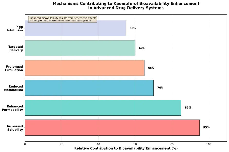
Figure 1 Mechanisms Contributing to Kaempferol Bioavailability Enhancement in Advanced Drug Delivery Systems.
Figure 2 presents comparative plasma concentration-time profiles demonstrating the impact of different delivery systems on kaempferol pharmacokinetics.
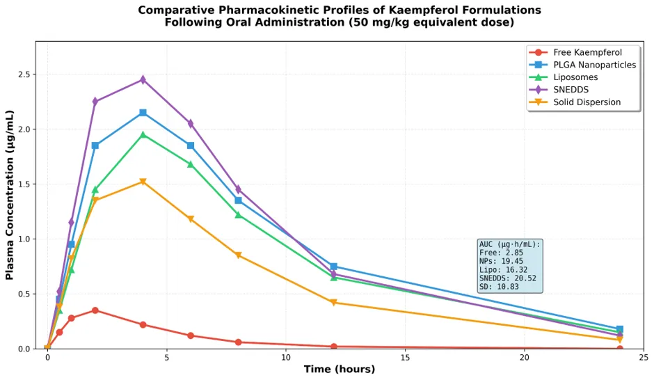
Figure 2 Comparative Pharmacokinetic Profiles of Kaempferol Formulations Following Oral Administration.
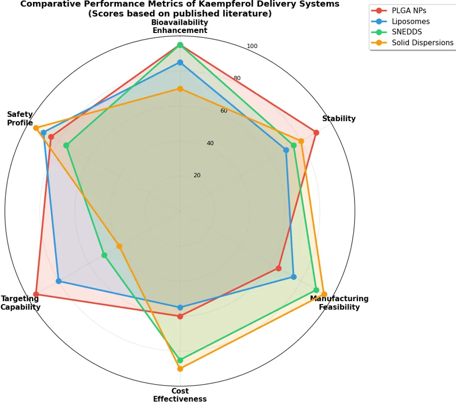
Figure 3 Comparative Performance Metrics of Kaempferol Delivery Systems.
Figure 4 illustrates molecular interactions between kaempferol and various delivery system components.
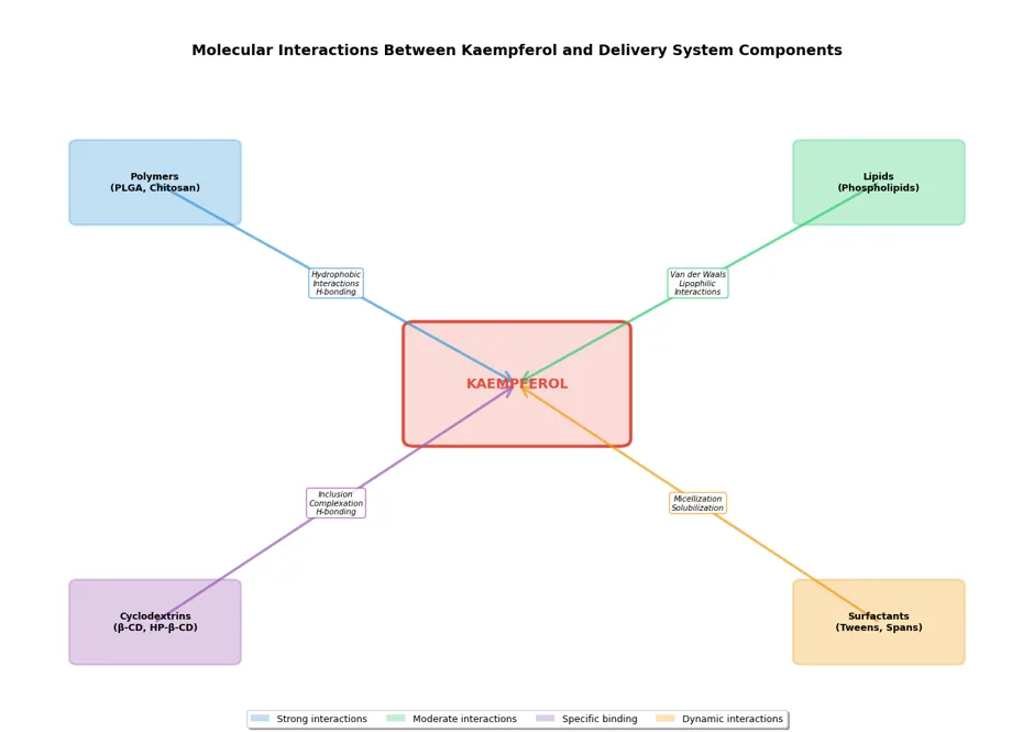
Figure 4 Molecular Interactions Between Kaempferol and Delivery System Components
CONCLUSION
Kaempferol's impressive pharmacological profile, encompassing antioxidant, anti-inflammatory, anticancer, neuroprotective, and cardioprotective properties, positions it as a promising therapeutic agent for multiple chronic diseases. However, fundamental biopharmaceutical limitations—poor aqueous solubility (~0.0175 mg/mL), low oral bioavailability (2-5%), extensive first-pass metabolism, P-gp-mediated efflux, and chemical instability—have historically constrained clinical translation.
This comprehensive review demonstrates that advanced drug delivery systems effectively address these challenges through multiple synergistic mechanisms. Nanotechnology-based approaches, particularly SNEDDS (7.2-fold bioavailability enhancement), PLGA nanoparticles (6.8-fold), and PEGylated liposomes (6.1-fold), offer the most substantial pharmacokinetic improvements. Lipid-based carriers provide additional advantages of GRAS status, scalability, and lymphatic transport. Simpler approaches including solid dispersions (3.8-fold enhancement) and cyclodextrin complexes (3.2-fold) present economically viable alternatives for nutraceutical and immediate-release pharmaceutical applications.
Critical evaluation reveals that optimal delivery system selection requires balancing multiple considerations:
Emerging technologies including stimuli-responsive systems, biomimetic carriers (cell membrane-coated nanoparticles, exosomes), theragnostic platforms, 3D printing for personalized medicine, and artificial intelligence-guided optimization promise to further advance kaempferol delivery systems. Integration of pharmacogenomic profiling may enable personalized formulation design based on individual metabolizer phenotypes and disease characteristics.
Despite extensive preclinical research demonstrating proof-of-concept, clinical translation remains limited. Moving forward, priorities include:
The convergence of pharmaceutical technology, materials science, nanotechnology, and systems biology provides unprecedented opportunities for optimizing kaempferol's therapeutic potential. Success requires systematic, rational approaches grounded in thorough understanding of physicochemical properties, biopharmaceutical barriers, formulation science principles, and clinical requirements. With continued research and development efforts, kaempferol formulations hold significant promise for successful translation from laboratory to clinical practice, ultimately improving patient outcomes across diverse therapeutic areas.
AUTHORS CONTRIBUTION
Aakash tripathi; Data analysis and wrote the Main manuscript; Vandana Sahani Ma’am; conceived of the presented idea and Prepare figures and supervision. All authors reviewed the manuscript.
FUNDING
This work was not funded
CONFLICT OF INTEREST
The authors have no relevant affiliations or financial involvement with any organization or entity with a financial interest in or financial conflict with the subject matter or materials discussed in the manuscript.
ETHICAL APPROVAL STATEMENT
This study did not require ethical board approval as it did not contain human or animal trail.
REFERENCES


Aakash Tripathi, Vandana Sahni, Shivanand Patil, Kaempferol Formulations and Drug Delivery Systems: Strategies to Improve Solubility and Bioavailability, Int. J. of Pharm. Sci., 2026, Vol 4, Issue 9, 763-789. https://doi.org/10.5281/zenodo.22336020
 10.5281/zenodo.22336020