View Article

  • SGLT2 Inhibitors as Anti fibrotic Agents : Molecular Mechanisms and Therapeutic Potential in Cardiac and Renal Fibrosis

  • College of Pharmaceutical Sciences, Government Medical College, Thiruvanandhapuram, Kerala, India-695011.

Abstract

Excessive extracellular matrix accumulation and increasing tissue dysfunction are the key features of fibrosis, a significant disease load in several organ systems. Even though it contributes to death worldwide, there are still few and poorly accepted treatment alternatives. Sodium-glucose cotransporter-2 (SGLT2) inhibitors which were initially designed as antidiabetic agents, have emerged as intriguing antifibrotic prospects. With a focus on TGF-?/Smad signalling, PI3K/AKT/mTOR activation, and oxidative stress, this review investigates the molecular mechanisms underlying fibrosis and how SGLT2 inhibitors modify these pathways to slow the progression of fibrosis in cardiac and renal tissues. Based on the evidence that is now available, SGLT2 inhibitors are a family of antifibrotic medicines that are therapeutically accessible, mechanistically versatile, and deserving of more research

Keywords

SGLT2 inhibitors; Fibrosis; TGF-?/Smad signaling; PI3K/AKT/mTOR pathway; Oxidative stress; Myofibroblast; Renal fibrosis; Myocardial fibrosis; Extracellular matrix; Antifibrotic therapy

Introduction

× Popup Image

Extracellular matrix (ECM) components, particularly collagen, build up excessively in fibrosis, a pathological process that causes organ failure and tissue scarring. It typically arises as a result of oxidative stress, prolonged damage, chronic inflammation, or abnormal wound healing. After an injury, tissue repair typically returns the structure to normal. However, persistent fibroblast and myofibroblast activation in fibrosis leads to excessive deposition of collagen and other extracellular matrix proteins, resulting in stiffness and disruption of normal tissue architecture.The heart, liver, kidneys, lungs, and peritoneum are among the many organs that are impacted by this process. Fibrogenesis is often caused by common mechanisms such as autoimmune reactions, oxidative stress, cellular death, and chronic infections, although the pathogenic triggers vary by organ. Fibrosis has a dramatic effect on clinical outcomes, increasing mortality and causing organ dysfunction, fibrotic diseases are responsible for roughly 45% of disease-related deaths. Due to ageing populations, environmental contaminants, and widespread epidemics, this frequency is probably even higher in poorer nations, making fibrosis a more prevalent problem. Only a few medications are available that only slightly slow the course of fibrosis, despite breakthroughs in our understanding of fibrosis processes and treatments. Patient compliance is frequently hampered by the side effects (such as nausea and gastrointestinal pain) and high cost of current medications, highlighting the critical need for innovative therapeutic approaches that target novel fibrosis mechanisms and pathways. In the field of anti-fibrotic research, sodium-glucose cotransporter 2 inhibitors (SGLT2i) have demonstrated to be effective and widespread use in the treatment of hyperglycemia. An increasing amount of evidence supports their potential as antifibrotic agents, making them a promising class for future fibrosis therapies 1.

Extracellular matrix (ECM) homeostasis and tissue structure are preserved by fibroblasts, which are diverse mesenchymal cells. Following tissue injury, they become activated into myofibroblasts, characterized by increased ECM production, stress fiber formation, and contractile activity. Persistent myofibroblast activation causes pathological fibrosis by excessive collagen deposition and tissue stiffening, whereas transitory stimulation promotes normal wound healing and tissue repair. Fibroblast–myofibroblast transition and fibrotic development are primarily regulated by mechanical variables like ECM stiffness and signalling pathways like transforming growth factor-β (TGF-β) 2.

By controlling fibroblast activation, ECM production, EMT, and several interrelated signalling pathways, TGF-β plays a critical role in the development and progression of fibrosis. Organ fibrosis and excessive tissue remodelling are caused by persistent stimulation of TGF-β signalling. The PI3K/AKT/mTOR pathway is a crucial molecular regulator of fibrosis and contributes to fibroblast activation, ECM accumulation, inflammation, and aberrant tissue remodelling. Therefore, targeting TGF-β-mediated pathways, particularly the Smad3/Smad7 balance, represents a promising therapeutic strategy for the management of fibrotic diseases3.

Fibrosis in the liver, kidney, lung and heart is linked to dysregulation of this signalling cascade. Thus, one intriguing therapeutic approach for preventing or treating fibrotic disorders is to target PI3K/AKT/mTOR signaling4.

One important pathogenic component in the growth and fibrosis of uterine fibroids is oxidative stress. Oxidative stress increases smooth muscle proliferation, inflammation, collagen production, and extracellular matrix buildup through ROS-mediated activation of PI3K/AKT/mTOR and TGF-β signalling pathways. Thus, focusing on oxidative stress and associated signalling pathways may offer promising treatment approaches to stop fibroid growth and lessen fibrosis5

A class of oral antidiabetic medications known as sodium-glucose cotransporter-2 (SGLT2) inhibitors was first created to treat type 2 diabetes mellitus by preventing renal glucose reabsorption and increasing urine glucose excretion. Empagliflozin, Dapagliflozin, and Canagliflozin are common agents. Because of its pleiotropic protective effects that go beyond glycaemic management, SGLT2 inhibitors have become a novel therapeutic approach for fibrosis in recent years. Their positive function in decreasing fibrosis in several organs, including as the heart, kidneys, liver, and lungs, has been shown in both experimental and clinical investigations. Both glucose-dependent and glucose-independent mechanisms, such as suppression of inflammatory cytokines like TNF-α and IL-6, inhibition of NLRP3 inflammasome activation, reduction of oxidative stress through activation of antioxidant pathways like Nrf2, and attenuation of profibrotic TGF-β/Smad signalling, are responsible for the antifibrotic effects of SGLT2 inhibitors. These substances also enhance mitochondrial function, lower tissue hypoxia, control autophagy and cellular metabolism, and prevent the epithelial-mesenchymal transition (EMT), which limits collagen formation, fibroblast activation, and extracellular matrix deposition. When taken as a whole, these processes reduce the development of organ fibrosis and demonstrate the potential of SGLT2 inhibitors as antifibrotic drugs 6.

2. Cellular and Molecular Mechanisms of Fibrosis

2.1 Fibroblast Activation and the Myofibroblast Transition

Fibroblasts are diverse mesenchymal cells found in almost every connective tissue, where they carry out vital homeostatic functions like ECM component creation, remodelling, and destruction. Fibroblasts exhibit a non-activated state in the undamaged condition, preserving structural support without producing an excessive amount of matrix. However, fibroblasts experience a crucial phenotypic change into myofibroblasts after tissue damage, which is fuelled by a mix of soluble mediators, mechanical signals, and cell-cell interactions.Myofibroblasts differ from their precursor fibroblasts in a number of ways: they produce significantly more extracellular matrix (ECM) proteins, especially type I and type III collagen; they form prominent cytoplasmic stress fibres made of alpha-smooth muscle actin (α-SMA); and they acquire contractile activity that allows for physical remodelling of the surrounding matrix. These characteristics are ideal for the requirements of acute wound healing, where temporary myofibroblast activation promotes wound closure and temporary matrix deposition before the cells eventually suffer apoptosis as the healing process comes to an end.When activation turns from transient to permanent, the pathogenic aspect of myofibroblast biology becomes apparent.

Myofibroblasts in fibrotic illness remain in a chronically active state that sustains excessive collagen deposition, progressive matrix stiffness, and disruption of normal tissue architecture instead of going through planned cell death after the lesion has healed. The ECM's mechanical characteristics take part in a self-reinforcing cycle: fibroblasts pathways intensify profibrotic signalling as matrix stiffness rises, further promoting myofibroblast activation and collagen synthesis. A key aspect of fibrotic growth is the reciprocal interaction between matrix mechanics and cellular behaviour, which makes it a crucial target for therapeutic intervention 2.

2.2 TGF-β Signaling in Fibrogenesis

In almost every organ system, transforming growth factor-beta (TGF-β) plays a crucial role in the development and maintenance of fibrosis. It is the most powerful and well-studied profibrotic cytokine discovered to date. It can coordinate fibroblast activation, extracellular matrix (ECM) synthesis, epithelial-to-mesenchymal transition (EMT), and the inhibition of ECM-degrading enzymes, all of which simultaneously promote matrix production and prevent its clearance. Receptor-regulated Smad proteins, primarily Smad2 and Smad3, undergo nuclear translocation and phosphorylation as part of the classical signalling cascade that is started by TGF-β. The type I receptor is phosphorylated upon ligand attachment to the TGF-β type II receptor, which in turn phosphorylates Smad2 and Smad3. Together with the common mediator Smad4, these activated Smads form heteromeric complexes that translocate to the nucleus and control the transcription of profibrotic target genes, such as those that encode collagen, fibronectin, and tissue inhibitor of metalloproteinases (TIMPs).The balance between Smad3, which drives profibrotic transcription, and Smad7, an inhibitory Smad that acts as a negative feedback regulator by targeting the TGF-β receptor complex for degradation, is a critical determinant of fibrotic outcome. Persistent TGF-β signaling is associated with suppression of Smad7 expression, shifting the Smad3/Smad7 balance in favour of sustained profibrotic gene activation.In addition to the canonical Smad pathway, TGF-β activates a number of non-Smad signalling cascades that promote fibrogenesis, such as the PI3K/AKT/mTOR axis, Rho-GTPase signalling and mitogen-activated protein kinase (MAPK) pathways. Beyond the direct transcriptional effects on ECM genes, these non-canonical pathways also amplify and diversify the profibrotic response, with TGF-β also impacting cell survival, proliferation, cytoskeletal architecture and metabolic reprogramming. The large number of downstream effectors of TGF-β explains the central role of this cytokine in fibrosis and the difficulty of targeting it therapeutically, since a broad inhibition of TGF-β activity risks compromising its essential functions in immune regulation and tissue homeostasis.Apart from the canonical Smad pathway, TGF-β also activates several non-Smad signalling cascades promoting fibrogenesis, such as the PI3K/AKT/mTOR axis, Rho-GTPase signalling and mitogen-activated protein kinase (MAPK) pathways. Besides direct transcriptional effects on ECM genes, TGF-β can also regulate cell survival, proliferation, cytoskeletal architecture and metabolic reprogramming. These non-canonical pathways contribute to, and diversify, the profibrotic response. TGF-β is important in fibrosis and hard to target therapeutically because of the many downstream effectors. This is due to the possible disruption of the essential role of TGF-β in immune modulation and tissue homeostasis via general TGF-β activity inhibition 3.
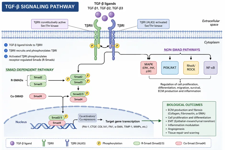


Figure 1 : Schematic Representation of the TGF-β Signalling Pathway and Its Role in Fibrosis


2.3 PI3K/AKT/mTOR Pathway Dysregulation

The phosphoinositide 3-kinase (PI3K)/protein kinase B (AKT)/mechanistic target of rapamycin (mTOR) signalling pathway is another important molecular axis in the pathophysiology of fibrosis. This pathway combines diverse upstream signals such as growth factors, cytokines and mechanical cues to modulate fundamental cellular processes including survival, proliferation, protein synthesis and metabolism. Dysregulation of PI3K/AKT/mTOR signalling has been shown to be involved in fibroblast activation, ECM accumulation, inflammatory cytokine production and abnormal tissue remodelling in fibrotic disease of the liver, kidneys, lungs and heart.

Activated PI3K phosphorylates phosphatidylinositol-4,5-bisphosphate (PIP2) into phosphatidylinositol-3,4,5-trisphosphate (PIP3), which attracts AKT to the plasma membrane for phosphorylation and activation. Activated AKT phosphorylates and activates mTOR complex, which regulates protein synthesis via its downstream effectors S6 kinase 1 (S6K1) and eukaryotic initiation factor 4E-binding protein 1 (4EBP1). In the context of fibrosis, mTOR C1 activation promotes the translational machinery required for high-level collagen and extracellular matrix protein synthesis, and AKT activation promotes fibroblast survival and resistance to apoptosis, thus maintaining the myofibroblast population.

The PI3K/AKT/mTOR pathway and TGF-β signalling are not parallel but closely related. TGF-β can activate PI3K/AKT signalling through both Smad-dependent and Smad-independent pathways, and AKT can reciprocally modulate Smad activity, leading to complex cross-regulatory networks that enhance profibrotic outcomes. mTOR signalling also participates in the regulation of autophagy, a cellular degradation pathway that exerts a double, context-dependent function in fibrosis: autophagy may support myofibroblast differentiation under some conditions, whereas dysregulation of autophagy results in the accumulation of damaged organelles and the maintenance of cellular stress responses that sustain fibrogenesis4.

2.4 Oxidative Stress and Reactive Oxygen Species in Fibrosis

A common aspect of fibrotic disease in all organ systems is oxidative stress, which is defined as an imbalance between the generation of reactive oxygen species (ROS) and the ability of cellular antioxidant defence systems. NADPH oxidases, mitochondrial electron transport chain malfunction, and inflammatory cell activation are the main sources of ROS, which accumulate and cause fibrogenesis by a variety of convergent pathways.

At the molecular level, ROS serve as second messengers that intensify profibrotic cascades started by upstream stimuli by activating the TGF-β/Smad and PI3K/AKT/mTOR signalling pathways.

ROS simultaneously increase latent TGF-β activation and TGF-β expression, creating a positive feedback loop where profibrotic signalling and oxidative stress support each other. In addition to these pathway-level effects, ROS directly harm lipids, proteins, and DNA in cells, resulting in inflammatory reactions and cellular death that further promote matrix deposition and fibroblast activation. Because ROS-mediated activation of PI3K/AKT/mTOR and TGF-β signalling enhances smooth muscle cell proliferation, inflammatory mediator production, collagen synthesis, and ECM buildup, the involvement of oxidative stress in uterine fibroids has been particularly extensively characterised. Antioxidant pathways, especially the nuclear factor erythroid 2-related factor 2 (Nrf2) antioxidant response element system, are crucial targets for antifibrotic intervention because the underlying role of oxidative stress in fibrogenesis is conserved throughout organ systems. Heme oxygenase-1 (HO-1), superoxide dismutase, and glutathione peroxidase are just a few of the cytoprotective and antioxidant enzymes that Nrf2, a transcription factor, activates to fight ROS buildup and the fibrogenic signals it produces5.

3. SGLT2 inhibitors: Pharmacology and Mechanisms of Action

3.1 Primary Pharmacological Mechanism

During the past few years there has been a lot of scientific and medical interest in a class of oral drugs called SGLT2 inhibitors (SGLT2i). Their origins lie in the research of the 1930's when it was found that phlorizin isolated from the bark of the roots of the apple tree had non-selective SGLT inhibitory activity. Later advances in medicinal chemistry allowed the modification of phlorizin, leading to development of several newer compounds collectively called SGLT2 inhibitors6.

Sodium-glucose cotransporter 1 (SGLT-1) and sodium-glucose cotransporter 2 (SGLT-2) are expressed in the epithelial cells of the renal tubules and are mainly responsible for glucose reabsorption in the kidneys. These transporters mediate the reabsorption of the filtered glucose by the glomerulus, and SGLT-2 is mainly involved in the S1 and S2 segments of the proximal tubules and is responsible for approximately 90% of this reabsorption. On the other hand, SGLT-1 is mostly found in the kidneys and gastrointestinal tract and contributes a relatively small amount 7. The SGLT2 inhibitors competitively interact with the glucose-binding sites on the SGLT-2 proteins in the renal tubules, resulting in decreased reabsorption of glucose and increased excretion of glucose, salt and water in the urine. These results help to lower blood sugar levels and decrease fluid overload 8. Sotagliflozin inhibits both SGLT-1 and SGLT-2 transporters. Four highly selective SGLT-2 inhibitors have received FDA approval namely empagliflozin, canagliflozin, dapagliflozin and ertugliflozin9.

3.2 Pleiotropic Effects Beyond Glycemic Control

The identification of benefits of SGLT2 inhibitors beyond blood glucose lowering has changed their clinical profile and has led to a great deal of research into their mechanisms of action in non-diabetic settings. Investigation into the broader biological effects of these agents was prompted by large cardiovascular outcome trials, such as EMPA-REG OUTCOME, CANVAS, and DAPA-HF, which showed reductions in cardiovascular mortality, heart failure hospitalisation, and renal disease progression that could not be explained solely by improved glycaemic control.Pleiotropic effects of SGLT2 inhibitors on fibrosis include a variety of inter-related pathways. They inhibit activation of the NLRP3 inflammasome, a multi-protein complex that activates and releases the potent inflammatory mediators IL-1β and IL-18, and inhibit the production of pro-inflammatory cytokines like tumour necrosis factor-alpha (TNF-α) and interleukin-6 (IL-6) thus decreasing systemic and tissue inflammation. They achieve this by activating the Nrf2 antioxidant pathway, which reduces ROS production and enhances cellular antioxidant capacity, thus reducing oxidative stress. They change mitochondrial function via increasing energy efficiency and decreasing mitochondrial ROS generation that contributes to oxidative stress and inflammatory signalling.They regulate autophagy and cellular metabolism in ways that may influence fibroblast survival and activation. In addition to this they reduce the transcriptional drive toward ECM synthesis and myofibroblast persistence by directly attenuating the TGF-β/Smad signalling pathway. Furthermore, SGLT2 inhibitors prevent epithelial cells from undergoing epithelial-mesenchymal transition (EMT), which can result in activated fibroblasts in fibrotic tissue 10-19.

4. Antifibrotic Effects of SGLT2 inhibitors Across Organ Systems

4.1 Renal fibrosis

Most chronic kidney diseases (CKDs) lead to renal fibrosis, which is characterised by an increase in interstitial space due to myofibroblast proliferation and excessive production of extracellular matrix (ECM). Renal function, specifically glomerular filtration rate (GFR) is negatively correlated with the degree of interstitial fibrosis20.

Adult polycystic kidney disease is one of the many hereditary disorders that cause renal fibrosis, but mutations in podocyte- or glomerular-associated genes like APOL1, WT1, NPHS1 and COL4A3 also cause it. Additionally, genetic association studies have revealed polymorphisms in genes regulating the renin-angiotensin-aldosterone system, DNA repair, TGFβ and reactive oxygen species (ROS) signalling, endocytosis, autophagy, apoptosis, and WNT signalling pathways 21-23.

Tubular epithelial injury is a common mechanism among various kidney disorders, highlighting the significance of epithelial damage in the onset of fibrosis. Proteinuria can be caused by glomerular filtration barrier dysfunction, direct medication nephrotoxicity, hypoxia, diabetes, or genetic disorders. When damaged tubular epithelial cells dedifferentiate, adopt a flattened morphology, and express injury markers such as mesenchymal markers such as vimentin, a phenomenon known as partial epithelial–mesenchymal transition (EMT) occurs. Experimental and clinical studies have demonstrated that injured proximal tubular epithelial cells play a central role in promoting kidney fibrosis. These cells activate immune cells that further enhance fibrotic responses and stimulate fibroblasts and pericytes through epithelial–mesenchymal transition24.

In mouse models local fibroblasts and pericytes are the main source of renal myofibroblasts and endothelial cells and epithelial cells and immune cells contribute little to the myofibroblast population 25. The interaction of mesenchymal cells, immunological cells such as macrophages, and damaged proximal tubular epithelial cells are the primary cause of renal fibrosis 26. All the various experimental models of kidney fibrosis developed to explore possible treatment strategies are unable to mimic the complexity of renal fibrotic disease in humans.

4.1.1 Effects of SGLT-2 Inhibitors on Renal Fibrosis

In chronic kidney disease (CKD), SGLT-2 inhibitors (SGLT-2i) have shown renoprotective effects in hyperglycaemic states that result in renal fibrosis by elevating matrix protein synthesis, pro-fibrotic signalling and impairing extracellular matrix turnover 27,28. SGLT-2i decreases blood glucose and slows renal fibrosis progression29,30. In addition to their glucose-lowering properties, there is a growing body of pre-clinical evidence suggesting antifibrotic effects of SGLT-2i through different mechanisms including reduction of hypoxia and oxidative stress, inhibition of inflammatory responses, stimulation of autophagy, correction of metabolic derangements and maintenance of capillary integrity .

Renal fibrosis is thought to be mostly caused by hypoxia31. Research by Professor Judit Hodrea and associates shown that dapagliflozin (DAPA) can reduce tubulointerstitial fibrosis by changing the hypoxic response of renal tubular cells 32. Empagliflozin (EMPA) has been shown to inhibit renal fibrosis in human proximal tubular HK-2 cells by downregulating the expression of hypoxia-inducible factor-1α (HIF-1α)33. Oxidative stress also plays a crucial role in fibrotic kidney injury, and several investigations have demonstrated that SGLT-2i reduce renal fibrosis by reducing oxidative stress levels 34-36.

Besides to this SGLT-2i exert anti-inflammatory effects that play a part in their protective role within the kidneys. For example, dapagliflozin inhibits the activation of the NOD-, LRR- and pyrin domain containing protein 3 (NLRP3) inflammasome in ischemia-reperfusion injury (IRI) models and thereby limits the development of fibrosis 37. In addition, SGLT-2i reduces the infiltration of inflammatory cells into renal tissue, particularly macrophages 38–40. Regulation of autophagy is another critical mechanism for their antifibrotic effects. Canagliflozin (CANA) has been shown to control autophagy through the signal transducer and activator of transcription 6 (STAT6) pathway41, while EMPA enhances renal function by modulating mitochondrial autophagy42.

Renal fibrosis is closely related to lipid toxicity and metabolic imbalance. Professor Zhang and colleagues discovered that SGLT-2i decreases the accumulation of lipids in renal tubular cells, which in turn slows the progression of fibrosis. These agents also prevent loss of the renal capillary network via a glucose transporter 2 (GLUT2)-dependent mechanism that inhibits endothelial cell loss and subsequent fibrosis following kidney injury. These results together highlight the different antifibrotic potential of SGLT-2 inhibitors in renal disease.

Transforming growth factor-β1 (TGF-β1) is a key cytokine in the pathogenesis of fibrosis. In a clinical study from Professor Tian and colleagues, SGLT2 inhibitors control renal TGF-β1 production and delay the progression of renal fibrosis. There are currently no clinical data, but several pre-clinical studies have shown the potential antifibrotic effects of SGLT2 inhibitors in kidney disease. A major drawback is the challenge of directly assessing renal fibrosis, because accurate measurement typically requires invasive procedures such as kidney biopsy.


Table 1 summarises the antifibrotic effects of SGLT2 inhibitors in models of renal fibrosis, emphasising the study types, experimental models, and key molecular pathways that contribute to the renoprotective effects of SGLT2 inhibitors.

Experimental models

Study type

Key findings

Reference

T1DM was induced by streptozotocin in adult male Wistar rats

In vitro

Diminished high glucose-induced protein O-GlcNAcylation and moderated the tubular response to hypoxia through the hypoxia-inducible factor pathway


32

Dall salt-sensitive rats with hypertensive kidney damage caused by a high salt diet.

Both in vitro and vivo

Acts as a renoprotective agent by suppressing EMT in the pathology of renal fibrosis via interaction with the SIRT3-FOXO3a pathway

34

The I/R fibrosis mice model

Both in vitro and vivo

Prevented activation of NLRP3 inflammasome and protected the kidney against fibrosis development

36

Ang II-induced renal fibrosis in rats

In vitro

Caused by reduced inflammatory infiltration and unrelated to the regulation of elevated blood pressure

37

5/6 nephrectomy-induced CKD in rats

Both in vitro and vivo

This outcome is attributable to the targeted modulation of the mTOR and mitophagy pathways, leading to the inhibition of CD206CD68 M2 macrophage polarization and the attenuation of inflammatory signals from CD8 effector T cells.

38


4.2 Myocardial fibrosis

Myocardial fibrosis, defined as excessive accumulation of ECM proteins in the cardiac interstitium, is often associated with cardiac diseases. In the case of MI, fibrosis is reparative, replacing dead cardiomyocytes with collagen-rich scar tissue, to maintain structural integrity and prevent sequelae such as heart rupture. On the other hand, hypertension, ageing, obesity and diabetes are the main causes of interstitial fibrosis, increased myocardial stiffness, decreased ventricular compliance, diastolic dysfunction and heart failure with intact ejection fraction.

4.2.1 Mechanism of myocardial fibrosis

Diffuse myocardial fibrosis is characterised by the activation of cardiac fibroblasts which release collagen-processing enzymes, growth factors, cytokines and extracellular matrix proteins leading to over deposition of collagen and myocardial stiffening 47,48. Resident cardiac fibroblasts, key regulators of this process 49, have been hard to identify due to the absence of very specific markers. Lineage tracing and single cell transcriptomic studies have shown that fibroblasts are a heterogenous population with distinct embryological origins, anatomical locations and functional properties50-53. Differences in proliferation capacity and profibrotic signalling have been reported between atrial and ventricular fibroblasts and between left and right ventricular fibroblast populations 54,55.

Single-cell studies identified multiple fibroblast and myofibroblast subpopulations with differential expression of classical markers such as Col1a1, indicating functional diversity during cardiac injury and repair56-58. Activated fibroblasts proliferate, express more periostin, expand the endoplasmic reticulum and differentiate to myofibroblasts that express α-smooth muscle actin 59. The stimulated ECM-producing fibroblasts can contribute to fibrosis without full myofibroblast transition 59,60. Other sources of myofibroblasts have been suggested, including fibrocytes and endothelium or epicardial-derived mesenchymal cells; experimental data suggest that local fibroblasts are the predominant donors after pressure overload or myocardial infarction61.

The type of heart damage and the surrounding microenvironment determine the fibroblasts phenotypic and function62. While ischaemic injury causes dynamic inflammatory and reparative responses, pressure overload primarily generates an ECM-synthesizing program63. Fibroblasts identify DAMPs and release cytokines, chemokines, and MMPs during the inflammatory phase following myocardial infarction. Fibroblasts grow and actively produce extracellular matrix (ECM) during the proliferative phase, and they express collagen-crosslinking enzymes such LOXs during scar maturation. Following damage, both profibrotic and antifibrotic fibroblast subgroups were found by single-cell transcriptome analyses64,65. Fibroblasts may have protective functions in addition to their profibrotic activity by preventing cardiomyocyte damage, maintaining ECM structure, and reducing inflammation via the TGFβ–SMAD3 pathway66.

4.2.2 Role other cardiac cells

By producing cytokines, growth factors, and profibrotic mediators that activate cardiac fibroblasts and control ECM turnover, other cardiac cell types, such as cardiomyocytes, immune cells, endothelial cells, and lymphatic cells, contribute to widespread myocardial fibrosis67,68. A significant part is played by inflammation, with T cells and macrophages encouraging collagen deposition and fibroblast activation via mediators such TGFβ, IFNγ.Following damage, cardiac fibroblasts also react to DAMPs and inflammasome activation, especially NLRP3, which increases the production of IL-1β 69. By secreting TGFβ, LOXs, galectin 3-related factors, and MMP-containing exosomes70-82, stressed cardiomyocytes further promote fibrosis. By secreting TGFβ and endothelin 1, encouraging leukocyte recruitment via ICAM1, and causing hypoxia by capillary rarefaction83-89, endothelial cells make an indirect contribution.Lymphatic failure exacerbates diffuse myocardial fibrosis by causing oedema, mechanical stress, and chronic inflammation.

Inflammatory cytokines, growth factors, neurohumoral mediators, and mechanical stress all activate cardiac fibroblasts via pathways including integrins, MAPK90-95. Aldosterone and angiotensin II stimulate collagen synthesis, fibroblast proliferation, and myofibroblast differentiation96-101. TGFβ1 is a key profibrotic mediator that increases the expression of fibrogenic genes by activating conventional SMAD-dependent and non-canonical signalling pathways, including as MAPK, TAK1, and RHOA102-109. Fibroblast activation and ECM synthesis are further increased by non-coding RNAs, metabolic reprogramming, mitochondrial malfunction, and oxidative stress110-126. Age linked to diabetes and ageing increase profibrotic signalling and collagen crosslinking via RAGE127.

Procollagens, LOXs, MMPs, TIMPs, and other ECM-regulating substances that regulate collagen synthesis, maturation, and degradation are secreted by activated fibroblasts128-130. Myocardial stiffness and chronic fibrosis are caused by excess collagen deposition and crosslinking, which are mediated by elevated PCPs, PCPE1, LOXs. Collagen turnover and ventricular remodelling are similarly impacted by altered MMP–TIMP balance; in HFrEF, increased MMP activity is associated with myocardial dilatation and systolic dysfunction.

4.2.3 Effects of SGLT-2 Inhibitors on Cardiac Fibrosis

SGLT-2 inhibitors (SGLT2i) have shown significant therapeutic effects in lowering cardiovascular mortality and managing heart failure131,132. A growing body of research indicates that these substances have cardioprotective benefits by reducing heart fibrosis via a variety of mechanisms. Excessive cytokine production, cardiac fibroblast activation and proliferation, increased extracellular matrix deposition, and the buildup of fibrotic material in the myocardial interstitium and perivascular areas are all facilitated by persistent hyperglycemia. Heart function is hampered by these pathological alterations, which decrease myocardial flexibility. SGLT2i helps reduce cardiac fibrosis caused by hyperglycemia by enhancing glycaemic regulation 132,133.

Apart from their glucose-lowering activity, SGLT2i also reduce myocardial fibrosis through several glucose-independent mechanisms, including anti-inflammatory effects, attenuation of oxidative stress, modulation of autophagy, and metabolic regulation. These agents suppress myocardial inflammation and fibrosis through regulation of signaling pathways involving Hypoxia-inducible factor-2α (HIF-2α)134, Signal transducer and activator of transcription 3 (STAT3)135, the NLRP3/Apoptosis-associated speck-like protein (ASC) inflammasome 136, Serum and glucocorticoid-regulated kinase 1 (SGK1) signaling 137, and NLRP3/Myeloid differentiation  response related pathways138. In addition, SGLT2i alleviate oxidative stress and fibrotic remodeling through pathways such as nuclear factor erythroid 2-related factor 2 (Nrf2)/Antioxidant response element (ARE) signaling139, Janus kinase/Signal transducer and activator of transcription (JAK/STAT) signaling140, and the Phosphoinositide 3-kinase (PI3K)/Protein kinase B (AKT)/Nrf2 pathway 141.

By controlling fibroblast activation and autophagic processes through mechanisms linked to TGF-β/Smad signalling and sodium-hydrogen exchanger (NHE) pathways, SGLT2i further reduces myocardial fibrosis. Additionally, a 2022 study showed that SGLT2i decreased ventricular fibrosis in experimental animals via modifying the mTOR signalling system, indicating a new treatment approach to myocardial fibrosis142-148.

The role of SGLT-2 inhibitors in reducing myocardial fibrosis has also been confirmed by clinical research. When evaluating cardiac fibrosis, extracellular volume fraction (ECV) is considered a crucial clinical indicator. Empagliflozin's effects on cardiac ECV were assessed in patients with type 2 diabetes mellitus (T2DM) and coronary artery disease (CAD) in a recent study by Professor Mason and colleagues. After six months of treatment, the empagliflozin group showed a decrease in ECV compared to the placebo group, indicating improvement in cardiac fibrosis and ventricular remodelling 149.

A prevalent pathological outcome in many cardiovascular conditions, such as heart failure, myocardial infarction, and cardiomyopathy, is myocardial fibrosis. Reduction of cardiac fibrosis is a major factor in the therapeutic success of SGLT2i in these circumstances, as numerous clinical investigations have consistently demonstrated. However, because it requires sophisticated methods, including histological investigation, imaging techniques, and biomarker analysis, accurate diagnosis of cardiac fibrosis remains difficult. The development of relevant clinical research is nevertheless hampered by these methods complexity and expensive expense150-161.


Table 2: Experimental Models and Molecular Mechanisms of Emerging Therapeutic Targets in Myocardial Fibrosis.

Experimental models

Study type

Key findings

Reference

Coronary artery ligation-induced myocardial infarction in mice

Both in vitro and in vivo

Decreased cardiac fibrosis by modulating macrophage polarization through STAT3 signaling

149

Alloxan-induced diabetic myocardial fibrosis in rabbits

Both in vitro and in vivo

Improved left ventricular diastolic dysfunction and reduced fibrosis through regulation of SGK1 signaling

151

Doxorubicin-induced myocardial fibrosis in mice

Both in vitro and in vivo

Suppressed ferroptosis, fibrosis, apoptosis, and inflammation through NLRP3 and MyD88-related pathways, resulting in improved cardiac function

152

KK-Ay mice (genetic type 2 diabetes model)

In vitro

Reduced oxidative stress and fibrosis by inhibiting the TGF-β/Smad pathway and activating Nrf2/ARE signaling

153

Chronic myocardial ischemia induced by left circumflex artery constriction in Yorkshire swine

Both in vitro and in vivo

Attenuated fibrosis through suppression of Jak/STAT signaling, activation of AMPK, and enhancement of antioxidant signaling

154

Doxorubicin-induced cardiac fibrosis in rats

Both in vitro and in vivo

Reduced oxidative stress, mitochondrial dysfunction, fibrosis, hypertrophy, and inflammation through PI3K/AKT/Nrf2 signaling

155

Aortic constriction-induced congestive heart failure in rabbits

In vitro

Improved myocardial fibrosis by suppressing the TGF-β1/Smad signaling pathway

156

High-fat diet-induced myocardial hypertrophy and fibrosis in mice

Both in vitro and in vivo

Lowered TGF-β2 expression in cardiomyocytes through inhibition of NHE-1 activity

157

Human atrial fibroblasts

Both in vitro and in vivo

Inhibition of NHE reduced phosphorylated PLC expression and IP3 production, thereby decreasing ER Ca²⁺ release, extracellular Ca²⁺ influx, and profibrotic fibroblast activity

158

Coronary artery ligation-induced myocardial infarction in rats

Both in vitro and in vivo

Regulated excessive autophagy through inhibition of NHE1 activity in myocardial cells

159

Aortic constriction-induced cardiac hypertrophy in rats

In vivo

Reduced mTOR pathway activation, alleviated ER stress and unfolded protein response (UPR), and subsequently decreased left ventricular fibrosis and adverse cardiac remodelling

161


REFERENCES

  1. Chen X, Zhang J, Guo L, Wu C, Zhou J, Xu M, et al. Decoding organ fibrosis: mechanistic insights and emerging therapeutic strategies. Signal transduction and targeted therapy [Internet]. 2026 June;11(1):82. Available from: https://pubmed.ncbi.nlm.nih.gov/41786685/
  2. Fereshteh Sadat Younesi, Miller AE, Barker TH, Fabio, Hinz B. Fibroblast and myofibroblast activation in normal tissue repair and fibrosis. Nature reviews Molecular cell biology. 2024 Apr 8;.
  3. Meng X, Nikolic-Paterson DJ, Lan HY. TGF-β: the master regulator of fibrosis. Nature Reviews Nephrology [Internet]. 2016 Apr 25;12(6):325–38. Available from: https://pubmed.ncbi.nlm.nih.gov/27108839/
  4. Emad Shamsan, Maged Almezgagi, Gamah M, Khan N, Qasem A, Liu Chuanchuan, et al. The role of PI3k/AKT signaling pathway in attenuating liver fibrosis: a comprehensive review. Frontiers in Medicine. 2024 Mar 25;11.
  5. Llorens-Cebrià C, den M Van, Vergara A, Conxita Jacobs-Cachá, Soler MJ. Antioxidant Roles of SGLT2 Inhibitors in the Kidney. Biomolecules [Internet]. 2022 Jan 16 [cited 2024 Nov 30];12(1):143–3. Available from: https://www.mdpi.com/2218-273X/12/1/143
  6. Vrhovac I, Balen Eror D, Klessen D, Burger C, Breljak D, Kraus O, et al. Localizations of Na+-d-glucose cotransporters SGLT1 and SGLT2 in human kidney and of SGLT1 in human small intestine, liver, lung, and heart. Pflügers Archiv - European Journal of Physiology. 2014 Oct 11;467(9):1881–98.
  7. Maccari R, Ottanà R. Sodium-Glucose Cotransporter Inhibitors as Antidiabetic Drugs: Current Development and Future Perspectives. Journal of Medicinal Chemistry. 2022 Aug 4;65(16):10848–81.
  8. Zelniker TA, Braunwald E. Mechanisms of Cardiorenal Effects of Sodium-Glucose Cotransporter 2 Inhibitors. Journal of the American College of Cardiology. 2020 Feb;75(4):422–34.
  9. Perry RJ, Shulman GI. Sodium-glucose cotransporter-2 inhibitors: Understanding the mechanisms for therapeutic promise and persisting risks. The Journal of Biological Chemistry [Internet]. 2020 Oct 16;295(42):14379–90. Available from: https://www.ncbi.nlm.nih.gov/pmc/articles/PMC7573269/
  10. Salvatore T, Galiero R, Caturano A, Rinaldi L, Di Martino A, Albanese G, et al. An Overview of the Cardiorenal Protective Mechanisms of SGLT2 Inhibitors. International Journal of Molecular Sciences. 2022 Mar 26;23(7):3651.
  11. Li S, Levy WC. Impact of SGLT2 Inhibitors on Serum Sodium in Heart Failure With Reduced Ejection Fraction. JACC: Heart Failure. 2022 Apr;.
  12. Salvatore T, Caturano A, Galiero R, Di Martino A, Albanese G, Vetrano E, et al. Cardiovascular Benefits from Gliflozins: Effects on Endothelial Function. Biomedicines. 2021 Sept 29;9(10):1356.
  13. Vallon V, Gerasimova M, Rose MA, Masuda T, Satriano J, Mayoux E, et al. SGLT2 inhibitor empagliflozin reduces renal growth and albuminuria in proportion to hyperglycemia and prevents glomerular hyperfiltration in diabetic Akita mice. American Journal of Physiology-Renal Physiology. 2014 Jan 15;306(2):F194–204.
  14. Yaribeygi H, Atkin SL, Butler AE, Sahebkar A. Sodium-glucose cotransporter inhibitors and oxidative stress: An update. Journal of Cellular Physiology. 2018 Nov 15;234(4):3231–7.
  15. Yaribeygi H, Butler AE, Barreto GE, Sahebkar A. Antioxidative potential of antidiabetic agents: A possible protective mechanism against vascular complications in diabetic patients. Journal of Cellular Physiology. 2018 Sept 7;234(3):2436–46.
  16. Rosalba La Grotta, Paola de Candia, Olivieri F, Giulia Matacchione, Giuliani A, Maria Rita Rippo, et al. Anti-inflammatory effect of SGLT-2 inhibitors via uric acid and insulin. Cellular and molecular life sciences. 2022 May 1;79(5).
  17. La Grotta R, Frigé C, Matacchione G, Olivieri F, de Candia P, Ceriello A, et al. Repurposing SGLT-2 Inhibitors to Target Aging: Available Evidence and Molecular Mechanisms. International Journal of Molecular Sciences [Internet]. 2022 Jan 1;23(20):12325. Available from: https://www.mdpi.com/1422-0067/23/20/12325
  18. Yaribeygi H, Maleki M, Nasimi F, Butler AE, Jamialahmadi T, Sahebkar A. Sodium‐glucose co‐transporter 2 inhibitors and hematopoiesis. Journal of Cellular Physiology. 2022 Aug 11;237(10):3778–87.
  19. Androutsakos T, Nasiri-Ansari N, Bakasis AD, Kyrou I, Efstathopoulos E, Randeva HS, et al. SGLT-2 Inhibitors in NAFLD: Expanding Their Role beyond Diabetes and Cardioprotection. International Journal of Molecular Sciences. 2022 Mar 13;23(6):3107.
  20. Li P, Gupta S, Mothi SS, Rennke HG, Leaf DE, Waikar SS, et al. Histopathologic Correlates of Kidney Function: Insights From Nephrectomy Specimens. American Journal of Kidney Diseases [Internet]. 2020 Oct 21;77(3):336–45. Available from: https://www.sciencedirect.com/science/article/pii/S0272638620310477
  21. Tziastoudi M, Theoharides TC, Nikolaou E, Efthymiadi M, Theodoros Eleftheriadis, Stefanidis I. Key Genetic Components of Fibrosis in Diabetic Nephropathy: An Updated Systematic Review and Meta-Analysis. International Journal of Molecular Sciences. 2022 Dec 5;23(23):15331–1.
  22. Devuyst O. Genetic Variants and Risk of Chronic Kidney Disease. Peritoneal Dialysis International. 2014 Mar 1;34(2):150–0.
  23. Anastasios Fountoglou, Constantinos Deltas, Ekaterini Siomou, Evangelia Dounousi. Genome-wide association studies reconstructing chronic kidney disease. Nephrology Dialysis Transplantation. 2023 Dec 20;.
  24. Kuppe C, Ibrahim MM, Kranz J, Zhang X, Ziegler S, Perales-Patón J, et al. Decoding myofibroblast origins in human kidney fibrosis. Nature. 2020 Nov 11;589(7841):281–6.
  25. Kuppe C, Ramirez Flores RO, Li Z, Hayat S, Levinson RT, Liao X, et al. Spatial multi-omic map of human myocardial infarction. Nature. 2022 Aug 10;608(7924):766–77.
  26. Schreibing F, Anslinger TM, Kramann R. Fibrosis in Pathology of Heart and Kidney: From Deep RNA-Sequencing to Novel Molecular Targets. Circulation Research. 2023 Apr 14;132(8):1013–33.
  27. Ravindran S, Munusamy S. Renoprotective mechanisms of sodium‐glucose co‐transporter 2 (SGLT2) inhibitors against the progression of diabetic kidney disease. Journal of Cellular Physiology. 2021 Oct 29;.
  28. Humphreys BD. Mechanisms of Renal Fibrosis. Annual Review of Physiology. 2018 Feb 10;80(1):309–26.
  29. Aroor AR, Das NA, Carpenter AJ, Habibi J, Jia G, Ramirez-Perez FI, et al. Glycemic control by the SGLT2 inhibitor empagliflozin decreases aortic stiffness, renal resistivity index and kidney injury. Cardiovascular Diabetology. 2018 July 30;17(1).
  30. Li J, Liu H, Takagi S, Nitta K, Kitada M, Srivastava SP, et al. Renal protective effects of empagliflozin via inhibition of EMT and aberrant glycolysis in proximal tubules. JCI Insight. 2020 Mar 26;5(6).
  31. Tanaka T, Nangaku M. The role of hypoxia, increased oxygen consumption, and hypoxia-inducible factor-1 alpha in progression of chronic kidney disease. Current Opinion in Nephrology and Hypertension. 2010 Jan;19(1):43–50.
  32. Judit Hodrea, Balogh D, Hosszu A, Lionetti L, Balázs Besztercei, Sandor Koszegi, et al. Reduced O-GlcNAcylation and tubular hypoxia contribute to the antifibrotic effect of SGLT2 inhibitor dapagliflozin in the diabetic kidney. 4. 2020 Apr 1;318(4):F1017–29.
  33. Ndibalema A, Kabuye D, Wen S, Li L, Li X, Fan Q. <p>Empagliflozin Protects Against Proximal Renal Tubular Cell Injury Induced by High Glucose via Regulation of Hypoxia-Inducible Factor 1-Alpha</p>. Diabetes, Metabolic Syndrome and Obesity: Targets and Therapy. 2020 June;Volume 13(13):1953–67.
  34. Wang Z, Zhai J, Zhang T, He L, Ma S, Zuo Q, et al. Canagliflozin ameliorates epithelial-mesenchymal transition in high-salt diet-induced hypertensive renal injury through restoration of sirtuin 3 expression and the reduction of oxidative stress. Biochemical and Biophysical Research Communications. 2023 Apr;653(653):53–61.
  35. Zeng S, Delic D, Chu C, Xiong Y, Luo T, Chen X, et al. Antifibrotic effects of low dose SGLT2 Inhibition with empagliflozin in comparison to Ang II receptor blockade with telmisartan in 5/6 nephrectomised rats on high salt diet. Biomedicine & Pharmacotherapy = Biomedecine & Pharmacotherapie [Internet]. 2022 Feb 1;146(146):112606. Available from: https://pubmed.ncbi.nlm.nih.gov/34968924/
  36. Ke Q, Shi C, Yunhui Lv, Wang L, Luo J, Jiang L, et al. SGLT2 inhibitor counteracts NLRP3 inflammasome via tubular metabolite itaconate in fibrosis kidney. ˜The œFASEB journal. 2021 Dec 17;36(1).
  37. Castoldi G, Carletti R, Ippolito S, Colzani M, Barzaghi F, Stella A, et al. Renal Anti-Fibrotic Effect of Sodium Glucose Cotransporter 2 Inhibition in Angiotensin II-Dependent Hypertension. American Journal of Nephrology [Internet]. 2020 Jan 7;51(2):119–29. Available from: https://karger.com/ajn/article-abstract/51/2/119/33008/Renal-Anti-Fibrotic-Effect-of-Sodium-Glucose?redirectedFrom=fulltext
  38. Wu H, Hocher B. Empagliflozin Reduces Kidney Fibrosis and Improves Kidney Function by Alternative Macrophage Activation in Rats With 5/6-Nephrectomy. Journal of the American Society of Nephrology. 2022 Nov;33(11S):588–8.
  39. Castoldi G, Carletti R, Ippolito S, Massimiliano Colzani, Barzaghi F, Stella A, et al. Sodium-glucose cotransporter 2 inhibition prevents renal fibrosis in cyclosporine nephropathy. Acta diabetologica. 2021 Mar 24;8(58).
  40. Yang Y, Li Q, Ling Y, Leng L, Ma Y, Xue L, et al. m6A eraser FTO modulates autophagy by targeting SQSTM1/P62 in the prevention of canagliflozin against renal fibrosis. Frontiers in Immunology. 2023 Jan 4;13(13):1094556–6.
  41. Zhang Z, Ni L, Zhang L, Zha D, Hu C, Zhang L, et al. Empagliflozin Regulates the AdipoR1/p-AMPK/p-ACC Pathway to Alleviate Lipid Deposition in Diabetic Nephropathy. Diabetes, Metabolic Syndrome and Obesity: Targets and Therapy. 2021 Jan;Volume 14(14):227–40.
  42. Zhang Y, Nakano D, Guan Y, Hitomi H, Uemura A, Masaki T, et al. A sodium-glucose cotransporter 2 inhibitor attenuates renal capillary injury and fibrosis by a vascular endothelial growth factor–dependent pathway after renal injury in mice. Kidney International. 2018 Sept;94(3):524–35.
  43. Tian Y, Chen X, Liang X, Wu X, Yao C. SGLT2 inhibitors attenuate nephrin loss and enhance TGF-β1 secretion in type 2 diabetes patients with albuminuria: a randomized clinical trial. Scientific Reports [Internet]. 2022 Sept 20;12(1). Available from: https://www.ncbi.nlm.nih.gov/pmc/articles/PMC9489863/
  44. Frangogiannis NG. Cardiac fibrosis: Cell biological mechanisms, molecular pathways and therapeutic opportunities. Molecular Aspects of Medicine. 2019 Feb;65:70–99.
  45. Berk BC, Fujiwara K, Lehoux S. ECM remodeling in hypertensive heart disease. Journal of Clinical Investigation. 2007 Mar 1;117(3):568–75.
  46. Gao X, White D, Dart AM, Du XJ. Post-infarct cardiac rupture: Recent insights on pathogenesis and therapeutic interventions. 134. 2012 May 1;134(2):156–79.
  47. Travers JG, Kamal FA, Robbins J, Yutzey KE, Blaxall BC. Cardiac Fibrosis. Circulation Research. 2016 Mar 18;118(6):1021–40.
  48. Humeres C, Frangogiannis NG. Fibroblasts in the Infarcted, Remodeling, and Failing Heart. JACC: Basic to Translational Science. 2019 June;4(3):449–67.
  49. Kong P, Christia P, Frangogiannis NG. The pathogenesis of cardiac fibrosis. Cellular and Molecular Life Sciences. 2013 May 7;71(4):549–74.
  50. Tallquist MD, Molkentin JD. Redefining the identity of cardiac fibroblasts. Nature Reviews Cardiology [Internet]. 2017 Aug 1 [cited 2022 Mar 17];14(8):484–91. Available from: https://www.nature.com/articles/nrcardio.2017.57
  51. Pinto AR, Ilinykh A, Ivey MJ, Kuwabara JT, D’Antoni ML, Debuque R, et al. Revisiting Cardiac Cellular Composition. Circulation Research. 2016 Feb 5;118(3):400–9.
  52. Tallquist MD. Cardiac Fibroblast Diversity. Annual Review of Physiology. 2020 Feb 10;82(1):63–78.
  53. Burstein B, Libby E, Calderone A, Nattel S. Differential Behaviors of Atrial Versus Ventricular Fibroblasts. Circulation. 2008 Apr;117(13):1630–41.
  54. Powers JC, Sabri A, Al-Bataineh D, Chotalia D, Guo X, Tsipenyuk F, et al. Differential microRNA-21 and microRNA-221 Upregulation in the Biventricular Failing Heart Reveals Distinct Stress Responses of Right Versus Left Ventricular Fibroblasts. Circulation: Heart Failure. 2020 Jan;13(1).
  55. Farbehi N, Patrick R, Aude Dorison, Munira Xaymardan, Vaibhao Janbandhu, Wystub-Lis K, et al. Single-cell expression profiling reveals dynamic flux of cardiac stromal, vascular and immune cells in health and injury. eLife. 2019 Mar 26;8(30).
  56. Gladka MM, Molenaar B, de Ruiter H, van der Elst S, Tsui H, Versteeg D, et al. Single-Cell Sequencing of the Healthy and Diseased Heart Reveals Cytoskeleton-Associated Protein 4 as a New Modulator of Fibroblasts Activation. Circulation [Internet]. 2018 July 10 [cited 2021 Apr 28];138(2):166–80. Available from: https://pubmed.ncbi.nlm.nih.gov/29386203/
  57. Ivey MJ, Tallquist MD. Defining the Cardiac Fibroblast. Circulation Journal. 2016;80(11):2269–76.
  58. Alex L, Russo I, Holoborodko V, Frangogiannis NG. Characterization of a mouse model of obesity-related fibrotic cardiomyopathy that recapitulates features of human heart failure with preserved ejection fraction. American Journal of Physiology-Heart and Circulatory Physiology. 2018 Oct 1;315(4):H934–49.
  59. Furtado MB, Costa MW, Rosenthal NA. The cardiac fibroblast: Origin, identity and role in homeostasis and disease. Differentiation. 2016 Sept;92(3):93–101.
  60. Moore-Morris T, Guimarães-Camboa N, Banerjee I, Zambon AC, Kisseleva T, Velayoudon A, et al. Resident fibroblast lineages mediate pressure overload–induced cardiac fibrosis. Journal of Clinical Investigation. 2014 June 17;124(7):2921–34.
  61. Kanisicak O, Khalil H, Ivey MJ, Karch J, Maliken BD, Correll RN, et al. Genetic lineage tracing defines myofibroblast origin and function in the injured heart. Nature Communications [Internet]. 2016 July 22 [cited 2022 Aug 5];7(1):12260. Available from: https://www.nature.com/articles/ncomms12260
  62. Hartupee J, Mann DL. Neurohormonal activation in heart failure with reduced ejection fraction. Nature Reviews Cardiology [Internet]. 2018 Oct 6;14(1):30–8. Available from: https://www.ncbi.nlm.nih.gov/pmc/articles/PMC5286912/
  63. Paulus WJ, Tschöpe C. A Novel Paradigm for Heart Failure With Preserved Ejection Fraction. Journal of the American College of Cardiology. 2013 July;62(4):263–71.
  64. Frangogiannis NG. The Extracellular Matrix in Ischemic and Nonischemic Heart Failure. Circulation Research. 2019 June 21;125(1):117–46.
  65. van Nieuwenhoven FA, Turner NA. The role of cardiac fibroblasts in the transition from inflammation to fibrosis following myocardial infarction. Vascular Pharmacology. 2013 Mar;58(3):182–8.
  66. Tellone E, Galtieri A, Russo A, Ficarra S. Protective Effects of the Caffeine Against Neurodegenerative Diseases. Current Medicinal Chemistry. 2019 Oct 23;26(27):5137–51.
  67. Swirski FK, Nahrendorf M. Cardioimmunology: the immune system in cardiac homeostasis and disease. Nature Reviews Immunology [Internet]. 2018 Dec 1 [cited 2021 Feb 12];18(12):733–44. Available from: https://www.nature.com/articles/s41577-018-0065-8?WT.feed_name=subjects_cardiovascular-diseases#:~:text=After%20myocardial%20infarction%20or%20in
  68. Westermann D, Lindner D, Kasner M, Zietsch C, Savvatis K, Escher F, et al. Cardiac Inflammation Contributes to Changes in the Extracellular Matrix in Patients With Heart Failure and Normal Ejection Fraction. Circulation: Heart Failure. 2011 Jan;4(1):44–52.
  69. Prabhu SD, Frangogiannis NG. The Biological Basis for Cardiac Repair After Myocardial Infarction. Circulation Research. 2016 June 24;119(1):91–112.
  70. Shen D, Chen J. Cardiac Macrophages Promote Polarization of Macrophages toward M2 Phenotype to Improve Myocardial Remodeling via NGAL after Myocardial Infarction. Cell Biochemistry and Biophysics. 2025 Mar 26;.
  71. Bansal SS, Ismahil MA, Goel M, Patel B, Hamid T, Rokosh G, et al. Activated T Lymphocytes are Essential Drivers of Pathological Remodeling in Ischemic Heart Failure. Circulation: Heart Failure. 2017 Mar;10(3).
  72. Laroumanie F, Douin-Echinard V, Pozzo J, Lairez O, Tortosa F, Vinel C, et al. CD4+ T cells promote the transition from hypertrophy to heart failure during chronic pressure overload. Circulation [Internet]. 2014 May 27 [cited 2020 Feb 20];129(21):2111–24. Available from: https://www.ncbi.nlm.nih.gov/pubmed/24657994
  73. Nevers T, Salvador AM, Velázquez F, Njabulo Ngwenyama, Carrillo‐Salinas F, Aronovitz M, et al. Th1 effector T cells selectively orchestrate cardiac fibrosis in nonischemic heart failure. Journal of Experimental Medicine. 2017 Oct 2;214(11):3311–29.
  74. Geral Baldeviano, Barin J, Talor M, Srinivasan S, Chen P, Djahida Bedja, et al. Interleukin-17A is dispensable for myocarditis but essential for the progression to dilated cardiomyopathy (135.25). The Journal of Immunology. 2010 Apr 1;184(1_Supplement):135.25–135.25.
  75. Anzai A, Choi JN, He S, Fenn AM, Manfred Nairz, Rattik S, et al. The infarcted myocardium solicits GM-CSF for the detrimental oversupply of inflammatory leukocytes. jexpmed. 2017 Nov 6;214(11):3293–310.
  76. Kruglov EA, Nathanson RA, Nguyen T, Dranoff JA. Secretion of MCP-1/CCL2 by bile duct epithelia induces myofibroblastic transdifferentiation of portal fibroblasts. American Journal of Physiology-Gastrointestinal and Liver Physiology. 2006 Apr;290(4):G765–71.
  77. Turner NA. Inflammatory and fibrotic responses of cardiac fibroblasts to myocardial damage associated molecular patterns (DAMPs). Journal of Molecular and Cellular Cardiology. 2016 May;94:189–200.
  78. Kawaguchi M, Takahashi M, Hata T, Kashima Y, Usui F, Morimoto H, et al. Inflammasome activation of cardiac fibroblasts is essential for myocardial ischemia/reperfusion injury. Circulation [Internet]. 2011 Feb 15 [cited 2023 Feb 2];123(6):594–604. Available from: https://pubmed.ncbi.nlm.nih.gov/21282498/
  79. Arne Yndestad, Øystein Sandanger, Jong WMC, Pål Aukrust, Zuurbier CJ. Response to letter from Toldo et al. on “NLRP3 inflammasome activation during myocardial ischemia reperfusion is cardioprotective”. Biochemical and Biophysical Research Communications. 2016 Apr 29;474(2):328–9.
  80. Chen B, Frangogiannis NG. Chemokines in Myocardial Infarction. Journal of Cardiovascular Translational Research. 2020 May 15;14(1):35–52.
  81. Kuhn TC, Knobel J, Burkert-Rettenmaier S, Li X, Meyer IS, Jungmann A, et al. Secretome Analysis of Cardiomyocytes Identifies PCSK6 (Proprotein Convertase Subtilisin/Kexin Type 6) as a Novel Player in Cardiac Remodeling After Myocardial Infarction. Circulation. 2020 May 19;141(20):1628–44.
  82. Bienvenu LA, Reichelt ME, Morgan J, Fletcher EK, Bell JR, Rickard AJ, et al. Cardiomyocyte Mineralocorticoid Receptor Activation Impairs Acute Cardiac Functional Recovery After Ischemic Insult. Hypertension. 2015 Nov;66(5):970–7.
  83. Sheng Z, Pennica D, Wood WI, Chien KR. Cardiotrophin-1 displays early expression in the murine heart tube and promotes cardiac myocyte survival. 1996 Feb 1;122(2):419–28.
  84. Martínez‐Martínez E, Brugnolaro C, Ibarrola J, Ravassa S, Mathieu Buonafine, Begoña López, et al. CT-1 (Cardiotrophin-1)-Gal-3 (Galectin-3) Axis in Cardiac Fibrosis and Inflammation. Hypertension. 2019 Mar 1;73(3):602–11.
  85. Ribeiro-Rodrigues TM, Laundos TL, Pereira-Carvalho R, Batista-Almeida D, Pereira R, Coelho-Santos V, et al. Exosomes secreted by cardiomyocytes subjected to ischaemia promote cardiac angiogenesis. Cardiovascular Research. 2017 June 19;113(11):1338–50.
  86. Li Y, Lui KO, Zhou B. Reassessing endothelial-to-mesenchymal transition in cardiovascular diseases. Nature Reviews Cardiology [Internet]. 2018 Aug 1 [cited 2022 June 21];15(8):445–56. Available from: https://www.nature.com/articles/s41569-018-0023-y
  87. Sun X, Nkennor B, Mastikhina O, Soon K, Nunes SS. Endothelium-mediated contributions to fibrosis. Seminars in Cell & Developmental Biology [Internet]. 2019 Nov 29;101:78–86. Available from: https://www.sciencedirect.com/science/article/pii/S1084952119300394
  88. Grady SR, McGuire EJ. Intercellular adhesive selectivity. III. Species selectivity of embryonic liver intercellular adhesion. Journal of Cell Biology. 1976 Oct 1;71(1):96–106.
  89. Palazzo AJ, Jones SP, Girod WG, Anderson DC, Granger DN, Lefer DJ. Myocardial ischemia-reperfusion injury in CD18- and ICAM-1-deficient mice. American Journal of Physiology-Heart and Circulatory Physiology. 1998 Dec 1;275(6):H2300–7.
  90. Chua CC, Diglio CA, Siu BB, Chua BHL. Angiotensin II induces TGF-β1 production in rat heart endothelial cells. Biochimica et Biophysica Acta (BBA) - Molecular Cell Research. 1994 Aug;1223(1):141–7.
  91. Adiarto S, Heiden S, Vignon-Zellweger N, Nakayama K, Yagi K, Yanagisawa M, et al. ET-1 from endothelial cells is required for complete angiotensin II-induced cardiac fibrosis and hypertrophy. Life Sciences. 2012 Oct;91(13–14):651–7.
  92. Mohammed SF, Redfield MM. Response to Letters Regarding Article, “Coronary Microvascular Rarefaction and Myocardial Fibrosis in Heart Failure With Preserved Ejection Fraction”. Circulation. 2015 Oct 20;132(16).
  93. Brakenhielm E, González A, Díez J. Role of Cardiac Lymphatics in Myocardial Edema and Fibrosis. Journal of the American College of Cardiology. 2020 Aug;76(6):735–44.
  94. Kong D, Kong X, Wang L. Effect of cardiac lymph flow obstruction on cardiac collagen synthesis and interstitial fibrosis. Physiological Research. 2006;253–8.
  95. Henri O, Pouehe C, Houssari M, Galas L, Nicol L, Edwards-Lévy F, et al. Selective Stimulation of Cardiac Lymphangiogenesis Reduces Myocardial Edema and Fibrosis Leading to Improved Cardiac Function Following Myocardial Infarction. Circulation. 2016 Apr 12;133(15):1484–97.
  96. Tomasek JJ, Gabbiani G, Hinz B, Chaponnier C, Brown RA. Myofibroblasts and mechano-regulation of Connective Tissue Remodelling. Nature Reviews Molecular Cell Biology. 2002 May;3(5):349–63.
  97. Brakenhielm E, Alitalo K. Cardiac lymphatics in health and disease. Nature Reviews Cardiology. 2018 Oct 17;16(1):56–68.
  98. MacKenna DA, Dolfi F, Vuori K, Ruoslahti E. Extracellular signal-regulated kinase and c-Jun NH2-terminal kinase activation by mechanical stretch is integrin-dependent and matrix-specific in rat cardiac fibroblasts. Journal of Clinical Investigation. 1998 Jan 15;101(2):301–10.
  99. Rahaman SO, Grove LM, Paruchuri S, Southern BD, Abraham S, Niese KA, et al. TRPV4 mediates myofibroblast differentiation and pulmonary fibrosis in mice. The Journal of Clinical Investigation [Internet]. 2014 Dec 1 [cited 2022 Mar 17];124(12):5225–38. Available from: https://pubmed.ncbi.nlm.nih.gov/25365224/
  100. Herum KM, Lunde IG, Skrbic B, Florholmen G, Behmen D, Sjaastad I, et al. Syndecan-4 signaling via NFAT regulates extracellular matrix production and cardiac myofibroblast differentiation in response to mechanical stress. Journal of molecular and cellular cardiology [Internet]. 2013 [cited 2019 Oct 31];54:73–81. Available from: https://www.ncbi.nlm.nih.gov/pubmed/23178899
  101. Leask A. Integrinβ1: A Mechanosignaling Sensor Essential for Connective Tissue Deposition by Fibroblasts. Advances in Wound Care. 2013 May;2(4):160–6.
  102. Shimizu T, Liao JK. Rho Kinases and Cardiac Remodeling. Circulation Journal [Internet]. 2016 [cited 2020 Jan 13];80(7):1491–8. Available from: https://www.ncbi.nlm.nih.gov/pmc/articles/PMC5563468/
  103. Wang J, Chen H, Seth A, McCulloch CA. Mechanical force regulation of myofibroblast differentiation in cardiac fibroblasts. American Journal of Physiology Heart and Circulatory Physiology. 2003 Nov 1;285(5):H1871–81.
  104. Crabos M, Roth M, Hahn AW, Erne P. Characterization of angiotensin II receptors in cultured adult rat cardiac fibroblasts. Coupling to signaling systems and gene expression. Journal of Clinical Investigation. 1994 June 1;93(6):2372–8.
  105. W Schorb, Booz GW, Dostal DE, Conrad K, Chang KH, Baker KM. Angiotensin II is mitogenic in neonatal rat cardiac fibroblasts. Circulation Research. 1993 June 1;72(6):1245–54.
  106. Lijnen P, Petrov V. Induction of cardiac fibrosis by aldosterone. Journal of Molecular and Cellular Cardiology [Internet]. 2000 June 1 [cited 2022 Aug 21];32(6):865–79. Available from: https://pubmed.ncbi.nlm.nih.gov/10888242/
  107. Rickard AJ, Morgan J, Tesch G, Funder JW, Fuller PJ, Young MJ. Deletion of Mineralocorticoid Receptors From Macrophages Protects Against Deoxycorticosterone/Salt-Induced Cardiac Fibrosis and Increased Blood Pressure. Hypertension. 2009 Sept;54(3):537–43.
  108. Neumann S, Huse K, Semrau R, Anno Diegeler, Gebhardt R, Gayane Hrachia Buniatian, et al. Aldosterone and <scp>d</scp> -Glucose Stimulate the Proliferation of Human Cardiac Myofibroblasts In Vitro. Hypertension. 2002 Mar 1;39(3):756–60.
  109. Brilla CG, Zhou G, Matsubara L, Weber KT. Collagen Metabolism in Cultured Adult Rat Cardiac Fibroblasts: Response to Angiotensin II and Aldosterone. Journal of Molecular and Cellular Cardiology. 1994 July;26(7):809–20.
  110. Edgley AJ, Krum H, Kelly DJ. Targeting Fibrosis for the Treatment of Heart Failure: A Role for Transforming Growth Factor-β. Cardiovascular Therapeutics [Internet]. 2010 Sept 23 [cited 2019 Sept 27];30(1):e30–40. Available from: https://onlinelibrary.wiley.com/doi/pdf/10.1111/j.1755-5922.2010.00228.x
  111. Hanna A, Frangogiannis NG. The Role of the TGF-β Superfamily in Myocardial Infarction. Frontiers in Cardiovascular Medicine [Internet]. 2019 Sept 18 [cited 2020 Aug 10];6. Available from: https://www.ncbi.nlm.nih.gov/pmc/articles/PMC6760019/
  112. Inkinen K, Wolff H, Lindroos P, Ahonen J. Connective Tissue Growth Factor and Its Correlation to Other Growth Factors in Experimental Granulation Tissue. Connective Tissue Research. 2003 Jan;44(1):19–29.
  113. Nagaraju CK, Robinson EL, Mouna Abdesselem, Sander Trenson, Dries E, Gilbert G, et al. Myofibroblast Phenotype and Reversibility of Fibrosis in Patients With End-Stage Heart Failure. Journal of the American College of Cardiology. 2019 May 1;73(18):2267–82.
  114. Liu C, Lim ST, Teo MHY, Tan MSY, Kulkarni MD, Qiu B, et al. Collaborative Regulation of LRG1 by TGF-β1 and PPAR-β/δ Modulates Chronic Pressure Overload–Induced Cardiac Fibrosis. Circulation: Heart Failure. 2019 Dec;12(12).
  115. Chothani S, Schäfer S, Adami E, Viswanathan S, Widjaja AA, Langley SR, et al. Widespread Translational Control of Fibrosis in the Human Heart by RNA-Binding Proteins. Circulation. 2019 Sept 10;140(11):937–51.
  116. Yan H, Wang Y, Li J, Qu X, Huang Y, Hale J, et al. Distinct Roles of TET Proteins in the Regulation of Normal and Disordered Human Erythropoiesis. Blood. 2015 Dec 3;126(23):159–9.
  117. Piccoli MT, Bär C, Thum T. Non-coding RNAs as modulators of the cardiac fibroblast phenotype. Journal of Molecular and Cellular Cardiology. 2016 Mar 1;92:75–81.
  118. Zhang Y, Huang XR, Wei LH, Chung AC, Yu CM, Lan HY. miR-29b as a Therapeutic Agent for Angiotensin II-induced Cardiac Fibrosis by Targeting TGF-β/Smad3 signaling. Molecular Therapy. 2014 May;22(5):974–85.
  119. Zhao X, Wang K, Liao Y, Zeng Q, Li Y, Hu F, et al. MicroRNA-101a Inhibits Cardiac Fibrosis Induced by Hypoxia via Targeting TGFβRI on Cardiac Fibroblasts. Cellular Physiology and Biochemistry. 2015;35(1):213–26.
  120. Thum T, Gross C, Fiedler J, Fischer T, Kissler S, Bussen M, et al. MicroRNA-21 contributes to myocardial disease by stimulating MAP kinase signalling in fibroblasts. Nature. 2008 Nov 30;456(7224):980–4.
  121. Liang H, Zhang C, Ban T, Liu Y, Mei L, Piao X, et al. A novel reciprocal loop between microRNA-21 and TGFβRIII is involved in cardiac fibrosis. The International Journal of Biochemistry & Cell Biology. 2012 Dec;44(12):2152–60.
  122. Tao H, Cao W, Yang JJ, Shi KH, Zhou X, Liu LP, et al. Long noncoding RNA H19 controls DUSP5/ERK1/2 axis in cardiac fibroblast proliferation and fibrosis. Cardiovascular Pathology. 2016 Sept;25(5):381–9.
  123. Zhao X, Kwan JYY, Yip K, Liu PP, Liu FF. Targeting metabolic dysregulation for fibrosis therapy. Nature Reviews Drug Discovery. 2019 Sept 23;19(1):57–75.
  124. Gibb AA, Lazaropoulos MP, Elrod JW. Myofibroblasts and Fibrosis. Circulation Research. 2020 July 17;127(3):427–47.
  125. Xie N, Tan Z, Banerjee S, Cui H, Ge J, Liu RM, et al. Glycolytic Reprogramming in Myofibroblast Differentiation and Lung Fibrosis. American Journal of Respiratory and Critical Care Medicine. 2015 Dec 15;192(12):1462–74.
  126. Kottmann RM, Kulkarni AA, Smolnycki KA, Lyda E, Dahanayake T, Salibi R, et al. Lactic Acid Is Elevated in Idiopathic Pulmonary Fibrosis and Induces Myofibroblast Differentiation via pH-Dependent Activation of Transforming Growth Factor-β. American Journal of Respiratory and Critical Care Medicine. 2012 Oct 15;186(8):740–51.
  127. Ha H, Yu MR, Lee HB. High glucose–induced PKC activation mediates TGF-β1 and fibronectin synthesis by peritoneal mesothelial cells. Kidney International. 2001 Feb;59(2):463–70.
  128. Jiang L, Xiao L, Sugiura H, Huang X, Ali A, Kuro-o M, et al. Metabolic reprogramming during TGFβ1-induced epithelial-to-mesenchymal transition. Oncogene. 2014 Oct 6;34(30):3908–16.
  129. de Paz-Lugo P, Lupiáñez JA, Meléndez-Hevia E. High glycine concentration increases collagen synthesis by articular chondrocytes in vitro: acute glycine deficiency could be an important cause of osteoarthritis. Amino Acids [Internet]. 2018 [cited 2020 May 6];50(10):1357–65. Available from: https://www.ncbi.nlm.nih.gov/pmc/articles/PMC6153947/
  130. Comstock JP, Udenfriend S. Effect of Lactate on Collagen Proline Hydroxylase Activity in Cultured L-929 Fibroblasts. Proceedings of the National Academy of Sciences. 1970 June;66(2):552–7.
  131. Russo I, Frangogiannis NG. Diabetes-associated cardiac fibrosis: Cellular effectors, molecular mechanisms and therapeutic opportunities. Journal of Molecular and Cellular Cardiology. 2016 Jan;90:84–93.
  132. Weber KT, Sun Y, Bhattacharya SK, Ahokas RA, Gerling IC. Myofibroblast-mediated mechanisms of pathological remodelling of the heart. Nature Reviews Cardiology. 2012 Dec 4;10(1):15–26.
  133. Goldsmith EC, Bradshaw A D, Zile MR, Spinale FG. Myocardial fibroblast–matrix interactions and potential therapeutic targets. Journal of Molecular and Cellular Cardiology. 2014 May 1;70:92–9.
  134. L??pez B, Gonz??lez A, D??ez J. Role of matrix metalloproteinases in hypertension-associated cardiac fibrosis. Current Opinion in Nephrology and Hypertension. 2004 Mar;13(2):197–204.
  135. Katwa LC, Campbell SE, Tyagi SC, Soon Jin Lee, Cicila GT, Weber KT. Cultured Myofibroblasts Generate Angiotensin Peptidesde Novo. Journal of Molecular and Cellular Cardiology. 1997 May 1;29(5):1375–86.
  136. López B, González A, Lindner D, Westermann D, Ravassa S, Beaumont J, et al. Osteopontin-mediated myocardial fibrosis in heart failure: a role for lysyl oxidase? Cardiovascular Research. 2013 Apr 25;99(1):111–20.
  137. Alandi IV, Nieman M, Molkentin JD, Blaxall BC. Inhibiting Fibronectin Improves Cardiac Function in a Mouse Model of Heart Failure. The FASEB Journal. 2016 Apr;30(S1).
  138. Begoña López, Arantxa González, Beaumont J, Ramón Querejeta, Larman M, Díez J. Identification of a Potential Cardiac Antifibrotic Mechanism of Torasemide in Patients With Chronic Heart Failure. Journal of the American College of Cardiology. 2007 Aug 1;50(9):859–67.
  139. Yang J, Konstantinos Savvatis, Jong Soon Kang, Fan P, Zhong H, Schwartz K, et al. Targeting LOXL2 for cardiac interstitial fibrosis and heart failure treatment. nature commun. 2016 Dec 1;7(1).
  140. Shizu R, Shindo S, Yoshida T, Numazawa S. MicroRNA-122 Down-Regulation Is Involved in Phenobarbital-Mediated Activation of the Constitutive Androstane Receptor. Rishi A, editor. PLoS ONE. 2012 July 18;7(7):e41291.
  141. Heymans S, Schroen B, Vermeersch P, Milting H, Gao F, Kassner A, et al. Increased Cardiac Expression of Tissue Inhibitor of Metalloproteinase-1 and Tissue Inhibitor of Metalloproteinase-2 Is Related to Cardiac Fibrosis and Dysfunction in the Chronic Pressure-Overloaded Human Heart. Circulation. 2005 Aug 23;112(8):1136–44.
  142. Herum K, Lunde I, McCulloch A, Christensen G. The Soft- and Hard-Heartedness of Cardiac Fibroblasts: Mechanotransduction Signaling Pathways in Fibrosis of the Heart. Journal of Clinical Medicine [Internet]. 2017 May 19 [cited 2019 Oct 31];6(5):53. Available from: https://www.mdpi.com/2077-0383/6/5/53/htm
  143. Brown E, Heerspink HJL, Cuthbertson DJ, Wilding JPH. SGLT2 inhibitors and GLP-1 receptor agonists: established and emerging indications. The Lancet. 2021 July;398(10296):262–76.
  144. Long Q, Li L, Yang H, Lu Y, Yang H, Zhu Y, et al. SGLT2 inhibitor, canagliflozin, ameliorates cardiac inflammation in experimental autoimmune myocarditis. International Immunopharmacology. 2022 Sept;110:109024.
  145. Tian J, Zhang M, Suo M, Liu D, Wang X, Liu M, et al. Dapagliflozin alleviates cardiac fibrosis through suppressing EndMT and fibroblast activation via AMPKα/TGF‐β/Smad signalling in type 2 diabetic rats. Journal of Cellular and Molecular Medicine. 2021 June 25;25(16):7642–59.
  146. Zelniker TA, Braunwald E. Clinical Benefit of Cardiorenal Effects of Sodium-Glucose Cotransporter 2 Inhibitors. Journal of the American College of Cardiology. 2020 Feb;75(4):435–47.
  147. Yang Z, Li T, Xian J, Chen J, Huang Y, Zhang Q, et al. <scp>SGLT2</scp> inhibitor dapagliflozin attenuates cardiac fibrosis and inflammation by reverting the <scp>HIF</scp> ‐2α signaling pathway in arrhythmogenic cardiomyopathy. The FASEB Journal. 2022 June 17;36(7).
  148. Lee TM, Chang NC, Lin SZ. Dapagliflozin, a selective SGLT2 Inhibitor, attenuated cardiac fibrosis by regulating the macrophage polarization via STAT3 signaling in infarcted rat hearts. Free Radical Biology & Medicine [Internet]. 2017 Mar 1;104:298–310. Available from: https://pubmed.ncbi.nlm.nih.gov/28132924/
  149. Ye Y, Bajaj M, Yang HC, Perez-Polo JR, Birnbaum Y. SGLT-2 Inhibition with Dapagliflozin Reduces the Activation of the Nlrp3/ASC Inflammasome and Attenuates the Development of Diabetic Cardiomyopathy in Mice with Type 2 Diabetes. Further Augmentation of the Effects with Saxagliptin, a DPP4 Inhibitor. Cardiovascular Drugs and Therapy. 2017 Apr;31(2):119–32.
  150. Lee SG, Kim D, Lee JJ, Lee HJ, Moon R, Lee YJ, et al. Dapagliflozin attenuates diabetes-induced diastolic dysfunction and cardiac fibrosis by regulating SGK1 signaling. BMC Medicine. 2022 Sept 7;20(1).
  151. Quagliariello V, Michelino De Laurentiis, Rea D, Barbieri A, Maria Gaia Monti, Carbone A, et al. The SGLT-2 inhibitor empagliflozin improves myocardial strain, reduces cardiac fibrosis and pro-inflammatory cytokines in non-diabetic mice treated with doxorubicin. Cardiovascular Diabetology. 2021 July 23;20(1).
  152. Li C, Zhang J, Xue M, Li X, Han F, Liu X, et al. SGLT2 inhibition with empagliflozin attenuates myocardial oxidative stress and fibrosis in diabetic mice heart. Cardiovascular Diabetology. 2019 Feb 2;18(1).
  153. Sabe SA, Xu CM, Sabra M, Harris DD, Malhotra A, Aboulgheit A, et al. Canagliflozin Improves Myocardial Perfusion, Fibrosis, and Function in a Swine Model of Chronic Myocardial Ischemia. Journal of the American Heart Association. 2023 Jan 3;12(1).
  154. Hsieh PL, Chu PM, Cheng HC, Huang YT, Chou WC, Tsai KL, et al. Dapagliflozin Mitigates Doxorubicin-Caused Myocardium Damage by Regulating AKT-Mediated Oxidative Stress, Cardiac Remodeling, and Inflammation. International journal of molecular sciences. 2022 Sept 4;23(17):10146–6.
  155. Chen X, Yang Q, Bai W, Yao W, Liu L, Xing Y, et al. Dapagliflozin Attenuates Myocardial Fibrosis by Inhibiting the TGF-β1/Smad Signaling Pathway in a Normoglycemic Rabbit Model of Chronic Heart Failure. Frontiers in pharmacology [Internet]. 2022 May 13 [cited 2024 Apr 25];13. Available from: https://www.ncbi.nlm.nih.gov/pmc/articles/PMC9136228/
  156. Osaka N, Mori Y, Michishige Terasaki, Munenori Hiromura, Saito T, Hironori Yashima, et al. Luseogliflozin inhibits high glucose-induced TGF-β2 expression in mouse cardiomyocytes by suppressing NHE-1 activity. Journal of International Medical Research. 2022 May 1;50(5).
  157. Chung CC, Lin YK, Chen YC, Kao YH, Yeh YH, Nguyen Ngoc Trang, et al. Empagliflozin suppressed cardiac fibrogenesis through sodium-hydrogen exchanger inhibition and modulation of the calcium homeostasis. Cardiovascular diabetology. 2023 Feb 6;22(1).
  158. Jiang K, Xu Y, Wang D, Chen F, Tu Z, Qian J, et al. Cardioprotective mechanism of SGLT2 inhibitor against myocardial infarction is through reduction of autosis. Protein & Cell. 2021 Jan 8;5(5).
  159. Kang S, Verma S, Teng G, Belke D, Svystonyuk D, Guzzardi D, et al. DIRECT EFFECTS OF EMPAGLIFLOZIN ON EXTRACELLULAR MATRIX REMODELING IN HUMAN CARDIAC FIBROBLASTS: NOVEL TRANSLATIONAL CLUES TO EMPA-REG OUTCOME. Canadian Journal of Cardiology. 2017 Oct;33(10):S169.
  160. Pang B, Zhang LL, Li B, Sun FX, Wang Z Da. The sodium glucose co-transporter 2 inhibitor ertugliflozin for Alzheimer’s disease: Inhibition of brain insulin signaling disruption-induced tau hyperphosphorylation. Physiology & Behavior. 2023 May;263(11):114134.
  161. Plata Mosquera CA. Empagliflozin reduces extracellular volume and myocardial sodium content in patients with heart failure and preserved ejection fraction and correlates with reverse cardiac remodeling (EMPAG CMR-HF). European Heart Journal. 2025 Nov;46(Supplement_1).

Reference

  1. Chen X, Zhang J, Guo L, Wu C, Zhou J, Xu M, et al. Decoding organ fibrosis: mechanistic insights and emerging therapeutic strategies. Signal transduction and targeted therapy [Internet]. 2026 June;11(1):82. Available from: https://pubmed.ncbi.nlm.nih.gov/41786685/
  2. Fereshteh Sadat Younesi, Miller AE, Barker TH, Fabio, Hinz B. Fibroblast and myofibroblast activation in normal tissue repair and fibrosis. Nature reviews Molecular cell biology. 2024 Apr 8;.
  3. Meng X, Nikolic-Paterson DJ, Lan HY. TGF-β: the master regulator of fibrosis. Nature Reviews Nephrology [Internet]. 2016 Apr 25;12(6):325–38. Available from: https://pubmed.ncbi.nlm.nih.gov/27108839/
  4. Emad Shamsan, Maged Almezgagi, Gamah M, Khan N, Qasem A, Liu Chuanchuan, et al. The role of PI3k/AKT signaling pathway in attenuating liver fibrosis: a comprehensive review. Frontiers in Medicine. 2024 Mar 25;11.
  5. Llorens-Cebrià C, den M Van, Vergara A, Conxita Jacobs-Cachá, Soler MJ. Antioxidant Roles of SGLT2 Inhibitors in the Kidney. Biomolecules [Internet]. 2022 Jan 16 [cited 2024 Nov 30];12(1):143–3. Available from: https://www.mdpi.com/2218-273X/12/1/143
  6. Vrhovac I, Balen Eror D, Klessen D, Burger C, Breljak D, Kraus O, et al. Localizations of Na+-d-glucose cotransporters SGLT1 and SGLT2 in human kidney and of SGLT1 in human small intestine, liver, lung, and heart. Pflügers Archiv - European Journal of Physiology. 2014 Oct 11;467(9):1881–98.
  7. Maccari R, Ottanà R. Sodium-Glucose Cotransporter Inhibitors as Antidiabetic Drugs: Current Development and Future Perspectives. Journal of Medicinal Chemistry. 2022 Aug 4;65(16):10848–81.
  8. Zelniker TA, Braunwald E. Mechanisms of Cardiorenal Effects of Sodium-Glucose Cotransporter 2 Inhibitors. Journal of the American College of Cardiology. 2020 Feb;75(4):422–34.
  9. Perry RJ, Shulman GI. Sodium-glucose cotransporter-2 inhibitors: Understanding the mechanisms for therapeutic promise and persisting risks. The Journal of Biological Chemistry [Internet]. 2020 Oct 16;295(42):14379–90. Available from: https://www.ncbi.nlm.nih.gov/pmc/articles/PMC7573269/
  10. Salvatore T, Galiero R, Caturano A, Rinaldi L, Di Martino A, Albanese G, et al. An Overview of the Cardiorenal Protective Mechanisms of SGLT2 Inhibitors. International Journal of Molecular Sciences. 2022 Mar 26;23(7):3651.
  11. Li S, Levy WC. Impact of SGLT2 Inhibitors on Serum Sodium in Heart Failure With Reduced Ejection Fraction. JACC: Heart Failure. 2022 Apr;.
  12. Salvatore T, Caturano A, Galiero R, Di Martino A, Albanese G, Vetrano E, et al. Cardiovascular Benefits from Gliflozins: Effects on Endothelial Function. Biomedicines. 2021 Sept 29;9(10):1356.
  13. Vallon V, Gerasimova M, Rose MA, Masuda T, Satriano J, Mayoux E, et al. SGLT2 inhibitor empagliflozin reduces renal growth and albuminuria in proportion to hyperglycemia and prevents glomerular hyperfiltration in diabetic Akita mice. American Journal of Physiology-Renal Physiology. 2014 Jan 15;306(2):F194–204.
  14. Yaribeygi H, Atkin SL, Butler AE, Sahebkar A. Sodium-glucose cotransporter inhibitors and oxidative stress: An update. Journal of Cellular Physiology. 2018 Nov 15;234(4):3231–7.
  15. Yaribeygi H, Butler AE, Barreto GE, Sahebkar A. Antioxidative potential of antidiabetic agents: A possible protective mechanism against vascular complications in diabetic patients. Journal of Cellular Physiology. 2018 Sept 7;234(3):2436–46.
  16. Rosalba La Grotta, Paola de Candia, Olivieri F, Giulia Matacchione, Giuliani A, Maria Rita Rippo, et al. Anti-inflammatory effect of SGLT-2 inhibitors via uric acid and insulin. Cellular and molecular life sciences. 2022 May 1;79(5).
  17. La Grotta R, Frigé C, Matacchione G, Olivieri F, de Candia P, Ceriello A, et al. Repurposing SGLT-2 Inhibitors to Target Aging: Available Evidence and Molecular Mechanisms. International Journal of Molecular Sciences [Internet]. 2022 Jan 1;23(20):12325. Available from: https://www.mdpi.com/1422-0067/23/20/12325
  18. Yaribeygi H, Maleki M, Nasimi F, Butler AE, Jamialahmadi T, Sahebkar A. Sodium?glucose co?transporter 2 inhibitors and hematopoiesis. Journal of Cellular Physiology. 2022 Aug 11;237(10):3778–87.
  19. Androutsakos T, Nasiri-Ansari N, Bakasis AD, Kyrou I, Efstathopoulos E, Randeva HS, et al. SGLT-2 Inhibitors in NAFLD: Expanding Their Role beyond Diabetes and Cardioprotection. International Journal of Molecular Sciences. 2022 Mar 13;23(6):3107.
  20. Li P, Gupta S, Mothi SS, Rennke HG, Leaf DE, Waikar SS, et al. Histopathologic Correlates of Kidney Function: Insights From Nephrectomy Specimens. American Journal of Kidney Diseases [Internet]. 2020 Oct 21;77(3):336–45. Available from: https://www.sciencedirect.com/science/article/pii/S0272638620310477
  21. Tziastoudi M, Theoharides TC, Nikolaou E, Efthymiadi M, Theodoros Eleftheriadis, Stefanidis I. Key Genetic Components of Fibrosis in Diabetic Nephropathy: An Updated Systematic Review and Meta-Analysis. International Journal of Molecular Sciences. 2022 Dec 5;23(23):15331–1.
  22. Devuyst O. Genetic Variants and Risk of Chronic Kidney Disease. Peritoneal Dialysis International. 2014 Mar 1;34(2):150–0.
  23. Anastasios Fountoglou, Constantinos Deltas, Ekaterini Siomou, Evangelia Dounousi. Genome-wide association studies reconstructing chronic kidney disease. Nephrology Dialysis Transplantation. 2023 Dec 20;.
  24. Kuppe C, Ibrahim MM, Kranz J, Zhang X, Ziegler S, Perales-Patón J, et al. Decoding myofibroblast origins in human kidney fibrosis. Nature. 2020 Nov 11;589(7841):281–6.
  25. Kuppe C, Ramirez Flores RO, Li Z, Hayat S, Levinson RT, Liao X, et al. Spatial multi-omic map of human myocardial infarction. Nature. 2022 Aug 10;608(7924):766–77.
  26. Schreibing F, Anslinger TM, Kramann R. Fibrosis in Pathology of Heart and Kidney: From Deep RNA-Sequencing to Novel Molecular Targets. Circulation Research. 2023 Apr 14;132(8):1013–33.
  27. Ravindran S, Munusamy S. Renoprotective mechanisms of sodium?glucose co?transporter 2 (SGLT2) inhibitors against the progression of diabetic kidney disease. Journal of Cellular Physiology. 2021 Oct 29;.
  28. Humphreys BD. Mechanisms of Renal Fibrosis. Annual Review of Physiology. 2018 Feb 10;80(1):309–26.
  29. Aroor AR, Das NA, Carpenter AJ, Habibi J, Jia G, Ramirez-Perez FI, et al. Glycemic control by the SGLT2 inhibitor empagliflozin decreases aortic stiffness, renal resistivity index and kidney injury. Cardiovascular Diabetology. 2018 July 30;17(1).
  30. Li J, Liu H, Takagi S, Nitta K, Kitada M, Srivastava SP, et al. Renal protective effects of empagliflozin via inhibition of EMT and aberrant glycolysis in proximal tubules. JCI Insight. 2020 Mar 26;5(6).
  31. Tanaka T, Nangaku M. The role of hypoxia, increased oxygen consumption, and hypoxia-inducible factor-1 alpha in progression of chronic kidney disease. Current Opinion in Nephrology and Hypertension. 2010 Jan;19(1):43–50.
  32. Judit Hodrea, Balogh D, Hosszu A, Lionetti L, Balázs Besztercei, Sandor Koszegi, et al. Reduced O-GlcNAcylation and tubular hypoxia contribute to the antifibrotic effect of SGLT2 inhibitor dapagliflozin in the diabetic kidney. 4. 2020 Apr 1;318(4):F1017–29.
  33. Ndibalema A, Kabuye D, Wen S, Li L, Li X, Fan Q. <p>Empagliflozin Protects Against Proximal Renal Tubular Cell Injury Induced by High Glucose via Regulation of Hypoxia-Inducible Factor 1-Alpha</p>. Diabetes, Metabolic Syndrome and Obesity: Targets and Therapy. 2020 June;Volume 13(13):1953–67.
  34. Wang Z, Zhai J, Zhang T, He L, Ma S, Zuo Q, et al. Canagliflozin ameliorates epithelial-mesenchymal transition in high-salt diet-induced hypertensive renal injury through restoration of sirtuin 3 expression and the reduction of oxidative stress. Biochemical and Biophysical Research Communications. 2023 Apr;653(653):53–61.
  35. Zeng S, Delic D, Chu C, Xiong Y, Luo T, Chen X, et al. Antifibrotic effects of low dose SGLT2 Inhibition with empagliflozin in comparison to Ang II receptor blockade with telmisartan in 5/6 nephrectomised rats on high salt diet. Biomedicine & Pharmacotherapy = Biomedecine & Pharmacotherapie [Internet]. 2022 Feb 1;146(146):112606. Available from: https://pubmed.ncbi.nlm.nih.gov/34968924/
  36. Ke Q, Shi C, Yunhui Lv, Wang L, Luo J, Jiang L, et al. SGLT2 inhibitor counteracts NLRP3 inflammasome via tubular metabolite itaconate in fibrosis kidney. ?The ?FASEB journal. 2021 Dec 17;36(1).
  37. Castoldi G, Carletti R, Ippolito S, Colzani M, Barzaghi F, Stella A, et al. Renal Anti-Fibrotic Effect of Sodium Glucose Cotransporter 2 Inhibition in Angiotensin II-Dependent Hypertension. American Journal of Nephrology [Internet]. 2020 Jan 7;51(2):119–29. Available from: https://karger.com/ajn/article-abstract/51/2/119/33008/Renal-Anti-Fibrotic-Effect-of-Sodium-Glucose?redirectedFrom=fulltext
  38. Wu H, Hocher B. Empagliflozin Reduces Kidney Fibrosis and Improves Kidney Function by Alternative Macrophage Activation in Rats With 5/6-Nephrectomy. Journal of the American Society of Nephrology. 2022 Nov;33(11S):588–8.
  39. Castoldi G, Carletti R, Ippolito S, Massimiliano Colzani, Barzaghi F, Stella A, et al. Sodium-glucose cotransporter 2 inhibition prevents renal fibrosis in cyclosporine nephropathy. Acta diabetologica. 2021 Mar 24;8(58).
  40. Yang Y, Li Q, Ling Y, Leng L, Ma Y, Xue L, et al. m6A eraser FTO modulates autophagy by targeting SQSTM1/P62 in the prevention of canagliflozin against renal fibrosis. Frontiers in Immunology. 2023 Jan 4;13(13):1094556–6.
  41. Zhang Z, Ni L, Zhang L, Zha D, Hu C, Zhang L, et al. Empagliflozin Regulates the AdipoR1/p-AMPK/p-ACC Pathway to Alleviate Lipid Deposition in Diabetic Nephropathy. Diabetes, Metabolic Syndrome and Obesity: Targets and Therapy. 2021 Jan;Volume 14(14):227–40.
  42. Zhang Y, Nakano D, Guan Y, Hitomi H, Uemura A, Masaki T, et al. A sodium-glucose cotransporter 2 inhibitor attenuates renal capillary injury and fibrosis by a vascular endothelial growth factor–dependent pathway after renal injury in mice. Kidney International. 2018 Sept;94(3):524–35.
  43. Tian Y, Chen X, Liang X, Wu X, Yao C. SGLT2 inhibitors attenuate nephrin loss and enhance TGF-β1 secretion in type 2 diabetes patients with albuminuria: a randomized clinical trial. Scientific Reports [Internet]. 2022 Sept 20;12(1). Available from: https://www.ncbi.nlm.nih.gov/pmc/articles/PMC9489863/
  44. Frangogiannis NG. Cardiac fibrosis: Cell biological mechanisms, molecular pathways and therapeutic opportunities. Molecular Aspects of Medicine. 2019 Feb;65:70–99.
  45. Berk BC, Fujiwara K, Lehoux S. ECM remodeling in hypertensive heart disease. Journal of Clinical Investigation. 2007 Mar 1;117(3):568–75.
  46. Gao X, White D, Dart AM, Du XJ. Post-infarct cardiac rupture: Recent insights on pathogenesis and therapeutic interventions. 134. 2012 May 1;134(2):156–79.
  47. Travers JG, Kamal FA, Robbins J, Yutzey KE, Blaxall BC. Cardiac Fibrosis. Circulation Research. 2016 Mar 18;118(6):1021–40.
  48. Humeres C, Frangogiannis NG. Fibroblasts in the Infarcted, Remodeling, and Failing Heart. JACC: Basic to Translational Science. 2019 June;4(3):449–67.
  49. Kong P, Christia P, Frangogiannis NG. The pathogenesis of cardiac fibrosis. Cellular and Molecular Life Sciences. 2013 May 7;71(4):549–74.
  50. Tallquist MD, Molkentin JD. Redefining the identity of cardiac fibroblasts. Nature Reviews Cardiology [Internet]. 2017 Aug 1 [cited 2022 Mar 17];14(8):484–91. Available from: https://www.nature.com/articles/nrcardio.2017.57
  51. Pinto AR, Ilinykh A, Ivey MJ, Kuwabara JT, D’Antoni ML, Debuque R, et al. Revisiting Cardiac Cellular Composition. Circulation Research. 2016 Feb 5;118(3):400–9.
  52. Tallquist MD. Cardiac Fibroblast Diversity. Annual Review of Physiology. 2020 Feb 10;82(1):63–78.
  53. Burstein B, Libby E, Calderone A, Nattel S. Differential Behaviors of Atrial Versus Ventricular Fibroblasts. Circulation. 2008 Apr;117(13):1630–41.
  54. Powers JC, Sabri A, Al-Bataineh D, Chotalia D, Guo X, Tsipenyuk F, et al. Differential microRNA-21 and microRNA-221 Upregulation in the Biventricular Failing Heart Reveals Distinct Stress Responses of Right Versus Left Ventricular Fibroblasts. Circulation: Heart Failure. 2020 Jan;13(1).
  55. Farbehi N, Patrick R, Aude Dorison, Munira Xaymardan, Vaibhao Janbandhu, Wystub-Lis K, et al. Single-cell expression profiling reveals dynamic flux of cardiac stromal, vascular and immune cells in health and injury. eLife. 2019 Mar 26;8(30).
  56. Gladka MM, Molenaar B, de Ruiter H, van der Elst S, Tsui H, Versteeg D, et al. Single-Cell Sequencing of the Healthy and Diseased Heart Reveals Cytoskeleton-Associated Protein 4 as a New Modulator of Fibroblasts Activation. Circulation [Internet]. 2018 July 10 [cited 2021 Apr 28];138(2):166–80. Available from: https://pubmed.ncbi.nlm.nih.gov/29386203/
  57. Ivey MJ, Tallquist MD. Defining the Cardiac Fibroblast. Circulation Journal. 2016;80(11):2269–76.
  58. Alex L, Russo I, Holoborodko V, Frangogiannis NG. Characterization of a mouse model of obesity-related fibrotic cardiomyopathy that recapitulates features of human heart failure with preserved ejection fraction. American Journal of Physiology-Heart and Circulatory Physiology. 2018 Oct 1;315(4):H934–49.
  59. Furtado MB, Costa MW, Rosenthal NA. The cardiac fibroblast: Origin, identity and role in homeostasis and disease. Differentiation. 2016 Sept;92(3):93–101.
  60. Moore-Morris T, Guimarães-Camboa N, Banerjee I, Zambon AC, Kisseleva T, Velayoudon A, et al. Resident fibroblast lineages mediate pressure overload–induced cardiac fibrosis. Journal of Clinical Investigation. 2014 June 17;124(7):2921–34.
  61. Kanisicak O, Khalil H, Ivey MJ, Karch J, Maliken BD, Correll RN, et al. Genetic lineage tracing defines myofibroblast origin and function in the injured heart. Nature Communications [Internet]. 2016 July 22 [cited 2022 Aug 5];7(1):12260. Available from: https://www.nature.com/articles/ncomms12260
  62. Hartupee J, Mann DL. Neurohormonal activation in heart failure with reduced ejection fraction. Nature Reviews Cardiology [Internet]. 2018 Oct 6;14(1):30–8. Available from: https://www.ncbi.nlm.nih.gov/pmc/articles/PMC5286912/
  63. Paulus WJ, Tschöpe C. A Novel Paradigm for Heart Failure With Preserved Ejection Fraction. Journal of the American College of Cardiology. 2013 July;62(4):263–71.
  64. Frangogiannis NG. The Extracellular Matrix in Ischemic and Nonischemic Heart Failure. Circulation Research. 2019 June 21;125(1):117–46.
  65. van Nieuwenhoven FA, Turner NA. The role of cardiac fibroblasts in the transition from inflammation to fibrosis following myocardial infarction. Vascular Pharmacology. 2013 Mar;58(3):182–8.
  66. Tellone E, Galtieri A, Russo A, Ficarra S. Protective Effects of the Caffeine Against Neurodegenerative Diseases. Current Medicinal Chemistry. 2019 Oct 23;26(27):5137–51.
  67. Swirski FK, Nahrendorf M. Cardioimmunology: the immune system in cardiac homeostasis and disease. Nature Reviews Immunology [Internet]. 2018 Dec 1 [cited 2021 Feb 12];18(12):733–44. Available from: https://www.nature.com/articles/s41577-018-0065-8?WT.feed_name=subjects_cardiovascular-diseases#:~:text=After%20myocardial%20infarction%20or%20in
  68. Westermann D, Lindner D, Kasner M, Zietsch C, Savvatis K, Escher F, et al. Cardiac Inflammation Contributes to Changes in the Extracellular Matrix in Patients With Heart Failure and Normal Ejection Fraction. Circulation: Heart Failure. 2011 Jan;4(1):44–52.
  69. Prabhu SD, Frangogiannis NG. The Biological Basis for Cardiac Repair After Myocardial Infarction. Circulation Research. 2016 June 24;119(1):91–112.
  70. Shen D, Chen J. Cardiac Macrophages Promote Polarization of Macrophages toward M2 Phenotype to Improve Myocardial Remodeling via NGAL after Myocardial Infarction. Cell Biochemistry and Biophysics. 2025 Mar 26;.
  71. Bansal SS, Ismahil MA, Goel M, Patel B, Hamid T, Rokosh G, et al. Activated T Lymphocytes are Essential Drivers of Pathological Remodeling in Ischemic Heart Failure. Circulation: Heart Failure. 2017 Mar;10(3).
  72. Laroumanie F, Douin-Echinard V, Pozzo J, Lairez O, Tortosa F, Vinel C, et al. CD4+ T cells promote the transition from hypertrophy to heart failure during chronic pressure overload. Circulation [Internet]. 2014 May 27 [cited 2020 Feb 20];129(21):2111–24. Available from: https://www.ncbi.nlm.nih.gov/pubmed/24657994
  73. Nevers T, Salvador AM, Velázquez F, Njabulo Ngwenyama, Carrillo?Salinas F, Aronovitz M, et al. Th1 effector T cells selectively orchestrate cardiac fibrosis in nonischemic heart failure. Journal of Experimental Medicine. 2017 Oct 2;214(11):3311–29.
  74. Geral Baldeviano, Barin J, Talor M, Srinivasan S, Chen P, Djahida Bedja, et al. Interleukin-17A is dispensable for myocarditis but essential for the progression to dilated cardiomyopathy (135.25). The Journal of Immunology. 2010 Apr 1;184(1_Supplement):135.25–135.25.
  75. Anzai A, Choi JN, He S, Fenn AM, Manfred Nairz, Rattik S, et al. The infarcted myocardium solicits GM-CSF for the detrimental oversupply of inflammatory leukocytes. jexpmed. 2017 Nov 6;214(11):3293–310.
  76. Kruglov EA, Nathanson RA, Nguyen T, Dranoff JA. Secretion of MCP-1/CCL2 by bile duct epithelia induces myofibroblastic transdifferentiation of portal fibroblasts. American Journal of Physiology-Gastrointestinal and Liver Physiology. 2006 Apr;290(4):G765–71.
  77. Turner NA. Inflammatory and fibrotic responses of cardiac fibroblasts to myocardial damage associated molecular patterns (DAMPs). Journal of Molecular and Cellular Cardiology. 2016 May;94:189–200.
  78. Kawaguchi M, Takahashi M, Hata T, Kashima Y, Usui F, Morimoto H, et al. Inflammasome activation of cardiac fibroblasts is essential for myocardial ischemia/reperfusion injury. Circulation [Internet]. 2011 Feb 15 [cited 2023 Feb 2];123(6):594–604. Available from: https://pubmed.ncbi.nlm.nih.gov/21282498/
  79. Arne Yndestad, Øystein Sandanger, Jong WMC, Pål Aukrust, Zuurbier CJ. Response to letter from Toldo et al. on “NLRP3 inflammasome activation during myocardial ischemia reperfusion is cardioprotective”. Biochemical and Biophysical Research Communications. 2016 Apr 29;474(2):328–9.
  80. Chen B, Frangogiannis NG. Chemokines in Myocardial Infarction. Journal of Cardiovascular Translational Research. 2020 May 15;14(1):35–52.
  81. Kuhn TC, Knobel J, Burkert-Rettenmaier S, Li X, Meyer IS, Jungmann A, et al. Secretome Analysis of Cardiomyocytes Identifies PCSK6 (Proprotein Convertase Subtilisin/Kexin Type 6) as a Novel Player in Cardiac Remodeling After Myocardial Infarction. Circulation. 2020 May 19;141(20):1628–44.
  82. Bienvenu LA, Reichelt ME, Morgan J, Fletcher EK, Bell JR, Rickard AJ, et al. Cardiomyocyte Mineralocorticoid Receptor Activation Impairs Acute Cardiac Functional Recovery After Ischemic Insult. Hypertension. 2015 Nov;66(5):970–7.
  83. Sheng Z, Pennica D, Wood WI, Chien KR. Cardiotrophin-1 displays early expression in the murine heart tube and promotes cardiac myocyte survival. 1996 Feb 1;122(2):419–28.
  84. Martínez?Martínez E, Brugnolaro C, Ibarrola J, Ravassa S, Mathieu Buonafine, Begoña López, et al. CT-1 (Cardiotrophin-1)-Gal-3 (Galectin-3) Axis in Cardiac Fibrosis and Inflammation. Hypertension. 2019 Mar 1;73(3):602–11.
  85. Ribeiro-Rodrigues TM, Laundos TL, Pereira-Carvalho R, Batista-Almeida D, Pereira R, Coelho-Santos V, et al. Exosomes secreted by cardiomyocytes subjected to ischaemia promote cardiac angiogenesis. Cardiovascular Research. 2017 June 19;113(11):1338–50.
  86. Li Y, Lui KO, Zhou B. Reassessing endothelial-to-mesenchymal transition in cardiovascular diseases. Nature Reviews Cardiology [Internet]. 2018 Aug 1 [cited 2022 June 21];15(8):445–56. Available from: https://www.nature.com/articles/s41569-018-0023-y
  87. Sun X, Nkennor B, Mastikhina O, Soon K, Nunes SS. Endothelium-mediated contributions to fibrosis. Seminars in Cell & Developmental Biology [Internet]. 2019 Nov 29;101:78–86. Available from: https://www.sciencedirect.com/science/article/pii/S1084952119300394
  88. Grady SR, McGuire EJ. Intercellular adhesive selectivity. III. Species selectivity of embryonic liver intercellular adhesion. Journal of Cell Biology. 1976 Oct 1;71(1):96–106.
  89. Palazzo AJ, Jones SP, Girod WG, Anderson DC, Granger DN, Lefer DJ. Myocardial ischemia-reperfusion injury in CD18- and ICAM-1-deficient mice. American Journal of Physiology-Heart and Circulatory Physiology. 1998 Dec 1;275(6):H2300–7.
  90. Chua CC, Diglio CA, Siu BB, Chua BHL. Angiotensin II induces TGF-β1 production in rat heart endothelial cells. Biochimica et Biophysica Acta (BBA) - Molecular Cell Research. 1994 Aug;1223(1):141–7.
  91. Adiarto S, Heiden S, Vignon-Zellweger N, Nakayama K, Yagi K, Yanagisawa M, et al. ET-1 from endothelial cells is required for complete angiotensin II-induced cardiac fibrosis and hypertrophy. Life Sciences. 2012 Oct;91(13–14):651–7.
  92. Mohammed SF, Redfield MM. Response to Letters Regarding Article, “Coronary Microvascular Rarefaction and Myocardial Fibrosis in Heart Failure With Preserved Ejection Fraction”. Circulation. 2015 Oct 20;132(16).
  93. Brakenhielm E, González A, Díez J. Role of Cardiac Lymphatics in Myocardial Edema and Fibrosis. Journal of the American College of Cardiology. 2020 Aug;76(6):735–44.
  94. Kong D, Kong X, Wang L. Effect of cardiac lymph flow obstruction on cardiac collagen synthesis and interstitial fibrosis. Physiological Research. 2006;253–8.
  95. Henri O, Pouehe C, Houssari M, Galas L, Nicol L, Edwards-Lévy F, et al. Selective Stimulation of Cardiac Lymphangiogenesis Reduces Myocardial Edema and Fibrosis Leading to Improved Cardiac Function Following Myocardial Infarction. Circulation. 2016 Apr 12;133(15):1484–97.
  96. Tomasek JJ, Gabbiani G, Hinz B, Chaponnier C, Brown RA. Myofibroblasts and mechano-regulation of Connective Tissue Remodelling. Nature Reviews Molecular Cell Biology. 2002 May;3(5):349–63.
  97. Brakenhielm E, Alitalo K. Cardiac lymphatics in health and disease. Nature Reviews Cardiology. 2018 Oct 17;16(1):56–68.
  98. MacKenna DA, Dolfi F, Vuori K, Ruoslahti E. Extracellular signal-regulated kinase and c-Jun NH2-terminal kinase activation by mechanical stretch is integrin-dependent and matrix-specific in rat cardiac fibroblasts. Journal of Clinical Investigation. 1998 Jan 15;101(2):301–10.
  99. Rahaman SO, Grove LM, Paruchuri S, Southern BD, Abraham S, Niese KA, et al. TRPV4 mediates myofibroblast differentiation and pulmonary fibrosis in mice. The Journal of Clinical Investigation [Internet]. 2014 Dec 1 [cited 2022 Mar 17];124(12):5225–38. Available from: https://pubmed.ncbi.nlm.nih.gov/25365224/
  100. Herum KM, Lunde IG, Skrbic B, Florholmen G, Behmen D, Sjaastad I, et al. Syndecan-4 signaling via NFAT regulates extracellular matrix production and cardiac myofibroblast differentiation in response to mechanical stress. Journal of molecular and cellular cardiology [Internet]. 2013 [cited 2019 Oct 31];54:73–81. Available from: https://www.ncbi.nlm.nih.gov/pubmed/23178899
  101. Leask A. Integrinβ1: A Mechanosignaling Sensor Essential for Connective Tissue Deposition by Fibroblasts. Advances in Wound Care. 2013 May;2(4):160–6.
  102. Shimizu T, Liao JK. Rho Kinases and Cardiac Remodeling. Circulation Journal [Internet]. 2016 [cited 2020 Jan 13];80(7):1491–8. Available from: https://www.ncbi.nlm.nih.gov/pmc/articles/PMC5563468/
  103. Wang J, Chen H, Seth A, McCulloch CA. Mechanical force regulation of myofibroblast differentiation in cardiac fibroblasts. American Journal of Physiology Heart and Circulatory Physiology. 2003 Nov 1;285(5):H1871–81.
  104. Crabos M, Roth M, Hahn AW, Erne P. Characterization of angiotensin II receptors in cultured adult rat cardiac fibroblasts. Coupling to signaling systems and gene expression. Journal of Clinical Investigation. 1994 June 1;93(6):2372–8.
  105. W Schorb, Booz GW, Dostal DE, Conrad K, Chang KH, Baker KM. Angiotensin II is mitogenic in neonatal rat cardiac fibroblasts. Circulation Research. 1993 June 1;72(6):1245–54.
  106. Lijnen P, Petrov V. Induction of cardiac fibrosis by aldosterone. Journal of Molecular and Cellular Cardiology [Internet]. 2000 June 1 [cited 2022 Aug 21];32(6):865–79. Available from: https://pubmed.ncbi.nlm.nih.gov/10888242/
  107. Rickard AJ, Morgan J, Tesch G, Funder JW, Fuller PJ, Young MJ. Deletion of Mineralocorticoid Receptors From Macrophages Protects Against Deoxycorticosterone/Salt-Induced Cardiac Fibrosis and Increased Blood Pressure. Hypertension. 2009 Sept;54(3):537–43.
  108. Neumann S, Huse K, Semrau R, Anno Diegeler, Gebhardt R, Gayane Hrachia Buniatian, et al. Aldosterone and <scp>d</scp> -Glucose Stimulate the Proliferation of Human Cardiac Myofibroblasts In Vitro. Hypertension. 2002 Mar 1;39(3):756–60.
  109. Brilla CG, Zhou G, Matsubara L, Weber KT. Collagen Metabolism in Cultured Adult Rat Cardiac Fibroblasts: Response to Angiotensin II and Aldosterone. Journal of Molecular and Cellular Cardiology. 1994 July;26(7):809–20.
  110. Edgley AJ, Krum H, Kelly DJ. Targeting Fibrosis for the Treatment of Heart Failure: A Role for Transforming Growth Factor-β. Cardiovascular Therapeutics [Internet]. 2010 Sept 23 [cited 2019 Sept 27];30(1):e30–40. Available from: https://onlinelibrary.wiley.com/doi/pdf/10.1111/j.1755-5922.2010.00228.x
  111. Hanna A, Frangogiannis NG. The Role of the TGF-β Superfamily in Myocardial Infarction. Frontiers in Cardiovascular Medicine [Internet]. 2019 Sept 18 [cited 2020 Aug 10];6. Available from: https://www.ncbi.nlm.nih.gov/pmc/articles/PMC6760019/
  112. Inkinen K, Wolff H, Lindroos P, Ahonen J. Connective Tissue Growth Factor and Its Correlation to Other Growth Factors in Experimental Granulation Tissue. Connective Tissue Research. 2003 Jan;44(1):19–29.
  113. Nagaraju CK, Robinson EL, Mouna Abdesselem, Sander Trenson, Dries E, Gilbert G, et al. Myofibroblast Phenotype and Reversibility of Fibrosis in Patients With End-Stage Heart Failure. Journal of the American College of Cardiology. 2019 May 1;73(18):2267–82.
  114. Liu C, Lim ST, Teo MHY, Tan MSY, Kulkarni MD, Qiu B, et al. Collaborative Regulation of LRG1 by TGF-β1 and PPAR-β/δ Modulates Chronic Pressure Overload–Induced Cardiac Fibrosis. Circulation: Heart Failure. 2019 Dec;12(12).
  115. Chothani S, Schäfer S, Adami E, Viswanathan S, Widjaja AA, Langley SR, et al. Widespread Translational Control of Fibrosis in the Human Heart by RNA-Binding Proteins. Circulation. 2019 Sept 10;140(11):937–51.
  116. Yan H, Wang Y, Li J, Qu X, Huang Y, Hale J, et al. Distinct Roles of TET Proteins in the Regulation of Normal and Disordered Human Erythropoiesis. Blood. 2015 Dec 3;126(23):159–9.
  117. Piccoli MT, Bär C, Thum T. Non-coding RNAs as modulators of the cardiac fibroblast phenotype. Journal of Molecular and Cellular Cardiology. 2016 Mar 1;92:75–81.
  118. Zhang Y, Huang XR, Wei LH, Chung AC, Yu CM, Lan HY. miR-29b as a Therapeutic Agent for Angiotensin II-induced Cardiac Fibrosis by Targeting TGF-β/Smad3 signaling. Molecular Therapy. 2014 May;22(5):974–85.
  119. Zhao X, Wang K, Liao Y, Zeng Q, Li Y, Hu F, et al. MicroRNA-101a Inhibits Cardiac Fibrosis Induced by Hypoxia via Targeting TGFβRI on Cardiac Fibroblasts. Cellular Physiology and Biochemistry. 2015;35(1):213–26.
  120. Thum T, Gross C, Fiedler J, Fischer T, Kissler S, Bussen M, et al. MicroRNA-21 contributes to myocardial disease by stimulating MAP kinase signalling in fibroblasts. Nature. 2008 Nov 30;456(7224):980–4.
  121. Liang H, Zhang C, Ban T, Liu Y, Mei L, Piao X, et al. A novel reciprocal loop between microRNA-21 and TGFβRIII is involved in cardiac fibrosis. The International Journal of Biochemistry & Cell Biology. 2012 Dec;44(12):2152–60.
  122. Tao H, Cao W, Yang JJ, Shi KH, Zhou X, Liu LP, et al. Long noncoding RNA H19 controls DUSP5/ERK1/2 axis in cardiac fibroblast proliferation and fibrosis. Cardiovascular Pathology. 2016 Sept;25(5):381–9.
  123. Zhao X, Kwan JYY, Yip K, Liu PP, Liu FF. Targeting metabolic dysregulation for fibrosis therapy. Nature Reviews Drug Discovery. 2019 Sept 23;19(1):57–75.
  124. Gibb AA, Lazaropoulos MP, Elrod JW. Myofibroblasts and Fibrosis. Circulation Research. 2020 July 17;127(3):427–47.
  125. Xie N, Tan Z, Banerjee S, Cui H, Ge J, Liu RM, et al. Glycolytic Reprogramming in Myofibroblast Differentiation and Lung Fibrosis. American Journal of Respiratory and Critical Care Medicine. 2015 Dec 15;192(12):1462–74.
  126. Kottmann RM, Kulkarni AA, Smolnycki KA, Lyda E, Dahanayake T, Salibi R, et al. Lactic Acid Is Elevated in Idiopathic Pulmonary Fibrosis and Induces Myofibroblast Differentiation via pH-Dependent Activation of Transforming Growth Factor-β. American Journal of Respiratory and Critical Care Medicine. 2012 Oct 15;186(8):740–51.
  127. Ha H, Yu MR, Lee HB. High glucose–induced PKC activation mediates TGF-β1 and fibronectin synthesis by peritoneal mesothelial cells. Kidney International. 2001 Feb;59(2):463–70.
  128. Jiang L, Xiao L, Sugiura H, Huang X, Ali A, Kuro-o M, et al. Metabolic reprogramming during TGFβ1-induced epithelial-to-mesenchymal transition. Oncogene. 2014 Oct 6;34(30):3908–16.
  129. de Paz-Lugo P, Lupiáñez JA, Meléndez-Hevia E. High glycine concentration increases collagen synthesis by articular chondrocytes in vitro: acute glycine deficiency could be an important cause of osteoarthritis. Amino Acids [Internet]. 2018 [cited 2020 May 6];50(10):1357–65. Available from: https://www.ncbi.nlm.nih.gov/pmc/articles/PMC6153947/
  130. Comstock JP, Udenfriend S. Effect of Lactate on Collagen Proline Hydroxylase Activity in Cultured L-929 Fibroblasts. Proceedings of the National Academy of Sciences. 1970 June;66(2):552–7.
  131. Russo I, Frangogiannis NG. Diabetes-associated cardiac fibrosis: Cellular effectors, molecular mechanisms and therapeutic opportunities. Journal of Molecular and Cellular Cardiology. 2016 Jan;90:84–93.
  132. Weber KT, Sun Y, Bhattacharya SK, Ahokas RA, Gerling IC. Myofibroblast-mediated mechanisms of pathological remodelling of the heart. Nature Reviews Cardiology. 2012 Dec 4;10(1):15–26.
  133. Goldsmith EC, Bradshaw A D, Zile MR, Spinale FG. Myocardial fibroblast–matrix interactions and potential therapeutic targets. Journal of Molecular and Cellular Cardiology. 2014 May 1;70:92–9.
  134. L??pez B, Gonz??lez A, D??ez J. Role of matrix metalloproteinases in hypertension-associated cardiac fibrosis. Current Opinion in Nephrology and Hypertension. 2004 Mar;13(2):197–204.
  135. Katwa LC, Campbell SE, Tyagi SC, Soon Jin Lee, Cicila GT, Weber KT. Cultured Myofibroblasts Generate Angiotensin Peptidesde Novo. Journal of Molecular and Cellular Cardiology. 1997 May 1;29(5):1375–86.
  136. López B, González A, Lindner D, Westermann D, Ravassa S, Beaumont J, et al. Osteopontin-mediated myocardial fibrosis in heart failure: a role for lysyl oxidase? Cardiovascular Research. 2013 Apr 25;99(1):111–20.
  137. Alandi IV, Nieman M, Molkentin JD, Blaxall BC. Inhibiting Fibronectin Improves Cardiac Function in a Mouse Model of Heart Failure. The FASEB Journal. 2016 Apr;30(S1).
  138. Begoña López, Arantxa González, Beaumont J, Ramón Querejeta, Larman M, Díez J. Identification of a Potential Cardiac Antifibrotic Mechanism of Torasemide in Patients With Chronic Heart Failure. Journal of the American College of Cardiology. 2007 Aug 1;50(9):859–67.
  139. Yang J, Konstantinos Savvatis, Jong Soon Kang, Fan P, Zhong H, Schwartz K, et al. Targeting LOXL2 for cardiac interstitial fibrosis and heart failure treatment. nature commun. 2016 Dec 1;7(1).
  140. Shizu R, Shindo S, Yoshida T, Numazawa S. MicroRNA-122 Down-Regulation Is Involved in Phenobarbital-Mediated Activation of the Constitutive Androstane Receptor. Rishi A, editor. PLoS ONE. 2012 July 18;7(7):e41291.
  141. Heymans S, Schroen B, Vermeersch P, Milting H, Gao F, Kassner A, et al. Increased Cardiac Expression of Tissue Inhibitor of Metalloproteinase-1 and Tissue Inhibitor of Metalloproteinase-2 Is Related to Cardiac Fibrosis and Dysfunction in the Chronic Pressure-Overloaded Human Heart. Circulation. 2005 Aug 23;112(8):1136–44.
  142. Herum K, Lunde I, McCulloch A, Christensen G. The Soft- and Hard-Heartedness of Cardiac Fibroblasts: Mechanotransduction Signaling Pathways in Fibrosis of the Heart. Journal of Clinical Medicine [Internet]. 2017 May 19 [cited 2019 Oct 31];6(5):53. Available from: https://www.mdpi.com/2077-0383/6/5/53/htm
  143. Brown E, Heerspink HJL, Cuthbertson DJ, Wilding JPH. SGLT2 inhibitors and GLP-1 receptor agonists: established and emerging indications. The Lancet. 2021 July;398(10296):262–76.
  144. Long Q, Li L, Yang H, Lu Y, Yang H, Zhu Y, et al. SGLT2 inhibitor, canagliflozin, ameliorates cardiac inflammation in experimental autoimmune myocarditis. International Immunopharmacology. 2022 Sept;110:109024.
  145. Tian J, Zhang M, Suo M, Liu D, Wang X, Liu M, et al. Dapagliflozin alleviates cardiac fibrosis through suppressing EndMT and fibroblast activation via AMPKα/TGF?β/Smad signalling in type 2 diabetic rats. Journal of Cellular and Molecular Medicine. 2021 June 25;25(16):7642–59.
  146. Zelniker TA, Braunwald E. Clinical Benefit of Cardiorenal Effects of Sodium-Glucose Cotransporter 2 Inhibitors. Journal of the American College of Cardiology. 2020 Feb;75(4):435–47.
  147. Yang Z, Li T, Xian J, Chen J, Huang Y, Zhang Q, et al. <scp>SGLT2</scp> inhibitor dapagliflozin attenuates cardiac fibrosis and inflammation by reverting the <scp>HIF</scp> ?2α signaling pathway in arrhythmogenic cardiomyopathy. The FASEB Journal. 2022 June 17;36(7).
  148. Lee TM, Chang NC, Lin SZ. Dapagliflozin, a selective SGLT2 Inhibitor, attenuated cardiac fibrosis by regulating the macrophage polarization via STAT3 signaling in infarcted rat hearts. Free Radical Biology & Medicine [Internet]. 2017 Mar 1;104:298–310. Available from: https://pubmed.ncbi.nlm.nih.gov/28132924/
  149. Ye Y, Bajaj M, Yang HC, Perez-Polo JR, Birnbaum Y. SGLT-2 Inhibition with Dapagliflozin Reduces the Activation of the Nlrp3/ASC Inflammasome and Attenuates the Development of Diabetic Cardiomyopathy in Mice with Type 2 Diabetes. Further Augmentation of the Effects with Saxagliptin, a DPP4 Inhibitor. Cardiovascular Drugs and Therapy. 2017 Apr;31(2):119–32.
  150. Lee SG, Kim D, Lee JJ, Lee HJ, Moon R, Lee YJ, et al. Dapagliflozin attenuates diabetes-induced diastolic dysfunction and cardiac fibrosis by regulating SGK1 signaling. BMC Medicine. 2022 Sept 7;20(1).
  151. Quagliariello V, Michelino De Laurentiis, Rea D, Barbieri A, Maria Gaia Monti, Carbone A, et al. The SGLT-2 inhibitor empagliflozin improves myocardial strain, reduces cardiac fibrosis and pro-inflammatory cytokines in non-diabetic mice treated with doxorubicin. Cardiovascular Diabetology. 2021 July 23;20(1).
  152. Li C, Zhang J, Xue M, Li X, Han F, Liu X, et al. SGLT2 inhibition with empagliflozin attenuates myocardial oxidative stress and fibrosis in diabetic mice heart. Cardiovascular Diabetology. 2019 Feb 2;18(1).
  153. Sabe SA, Xu CM, Sabra M, Harris DD, Malhotra A, Aboulgheit A, et al. Canagliflozin Improves Myocardial Perfusion, Fibrosis, and Function in a Swine Model of Chronic Myocardial Ischemia. Journal of the American Heart Association. 2023 Jan 3;12(1).
  154. Hsieh PL, Chu PM, Cheng HC, Huang YT, Chou WC, Tsai KL, et al. Dapagliflozin Mitigates Doxorubicin-Caused Myocardium Damage by Regulating AKT-Mediated Oxidative Stress, Cardiac Remodeling, and Inflammation. International journal of molecular sciences. 2022 Sept 4;23(17):10146–6.
  155. Chen X, Yang Q, Bai W, Yao W, Liu L, Xing Y, et al. Dapagliflozin Attenuates Myocardial Fibrosis by Inhibiting the TGF-β1/Smad Signaling Pathway in a Normoglycemic Rabbit Model of Chronic Heart Failure. Frontiers in pharmacology [Internet]. 2022 May 13 [cited 2024 Apr 25];13. Available from: https://www.ncbi.nlm.nih.gov/pmc/articles/PMC9136228/
  156. Osaka N, Mori Y, Michishige Terasaki, Munenori Hiromura, Saito T, Hironori Yashima, et al. Luseogliflozin inhibits high glucose-induced TGF-β2 expression in mouse cardiomyocytes by suppressing NHE-1 activity. Journal of International Medical Research. 2022 May 1;50(5).
  157. Chung CC, Lin YK, Chen YC, Kao YH, Yeh YH, Nguyen Ngoc Trang, et al. Empagliflozin suppressed cardiac fibrogenesis through sodium-hydrogen exchanger inhibition and modulation of the calcium homeostasis. Cardiovascular diabetology. 2023 Feb 6;22(1).
  158. Jiang K, Xu Y, Wang D, Chen F, Tu Z, Qian J, et al. Cardioprotective mechanism of SGLT2 inhibitor against myocardial infarction is through reduction of autosis. Protein & Cell. 2021 Jan 8;5(5).
  159. Kang S, Verma S, Teng G, Belke D, Svystonyuk D, Guzzardi D, et al. DIRECT EFFECTS OF EMPAGLIFLOZIN ON EXTRACELLULAR MATRIX REMODELING IN HUMAN CARDIAC FIBROBLASTS: NOVEL TRANSLATIONAL CLUES TO EMPA-REG OUTCOME. Canadian Journal of Cardiology. 2017 Oct;33(10):S169.
  160. Pang B, Zhang LL, Li B, Sun FX, Wang Z Da. The sodium glucose co-transporter 2 inhibitor ertugliflozin for Alzheimer’s disease: Inhibition of brain insulin signaling disruption-induced tau hyperphosphorylation. Physiology & Behavior. 2023 May;263(11):114134.
  161. Plata Mosquera CA. Empagliflozin reduces extracellular volume and myocardial sodium content in patients with heart failure and preserved ejection fraction and correlates with reverse cardiac remodeling (EMPAG CMR-HF). European Heart Journal. 2025 Nov;46(Supplement_1).

Photo
Shabna Rasheed
Corresponding author

College of Pharmaceutical Sciences, Government Medical College, Thiruvanandhapuram, Kerala, India-695011.

Photo
Dr. Rajalakshmi
Co-author

College of Pharmaceutical Sciences, Government Medical College, Thiruvanandhapuram, Kerala, India-695011.

Shabna Rasheed, Dr. Rajalakshmi, SGLT2 Inhibitors as Antifibrotic Agents: Molecular Mechanisms and Therapeutic Potential in Cardiac and Renal Fibrosis Review article, Int. J. of Pharm. Sci., 2026, Vol 4, Issue 9, 836-859, https://doi.org/10.5281/zenodo.22655398

More related articles
Computational Drug Discovery of Urs-12-ene Derivat...
M.Dhanalakshmi, R. Naveen, S. Narmatha, N.Nandhini, P. Muthulaksh...
CB-Dock2: An Emerging Platform for Protein–Ligan...
Dhinesh Kumar Sambasivam, Sowmian P., Soumian T. V, Naveen D., Aj...
More related articles
Computational Drug Discovery of Urs-12-ene Derivatives as Potential Antidiabetic...
M.Dhanalakshmi, R. Naveen, S. Narmatha, N.Nandhini, P. Muthulakshmi, M. Muthazhagi, S.Mohamed Halith...
CB-Dock2: An Emerging Platform for Protein–Ligand Blind Docking and Structure-...
Dhinesh Kumar Sambasivam, Sowmian P., Soumian T. V, Naveen D., Ajay Kumaran P., Dhayagesh R. , Sures...
Computational Drug Discovery of Urs-12-ene Derivatives as Potential Antidiabetic...
M.Dhanalakshmi, R. Naveen, S. Narmatha, N.Nandhini, P. Muthulakshmi, M. Muthazhagi, S.Mohamed Halith...
CB-Dock2: An Emerging Platform for Protein–Ligand Blind Docking and Structure-...
Dhinesh Kumar Sambasivam, Sowmian P., Soumian T. V, Naveen D., Ajay Kumaran P., Dhayagesh R. , Sures...