

We use cookies to ensure our website works properly and to personalise your experience. Cookies policy
Arunai College of Pharmacy.
Obesity is a major global health concern associated with several metabolic disorders, including type 2 diabetes mellitus, dyslipidemia, and cardiovascular diseases. Pancreatic lipase plays a key role in the digestion and absorption of dietary fats, making it an important target for anti-obesity therapy. The present study aimed to evaluate the in vitro pancreatic lipase inhibitory activity of Clitoria ternatea leaf extract and to characterize its functional groups using Fourier Transform Infrared (FT-IR) spectroscopy. The leaves were shade-dried, powdered, and extracted by the maceration method using 70% ethanol. Preliminary phytochemical screening was carried out to identify major phytoconstituents. Pancreatic lipase inhibitory activity was assessed by the titrimetric method using olive oil as the natural substrate, with orlistat as the positive control. The assay was performed in triplicate at concentrations of 50, 100, 150, 200 and 250 mcg/ml, and the percentage inhibition was calculated from the mean titration values. The extract exhibited concentration-dependent inhibition of pancreatic lipase, with percentage inhibition values of 19.6%, 24.2%, 30.3%, 39.3, and 45.4% respectively, while orlistat showed 80.3% inhibition. FT-IR analysis confirmed the presence of functional groups corresponding to bioactive phytochemicals that may contribute to the observed inhibitory activity. These findings indicate that Clitoria ternatea leaf extract possesses promising pancreatic lipase inhibitory potential and may serve as natural source for the development of anti-obesity agents. Furthur studies are required to isolate the active constituents and validate their efficacy through in vivo investigations.
Lipases are enzymes responsible for breaking down triglycerides into free fatty acids and glycerol. Different types of lipases are found in various tissues such as the liver, adipose tissue, blood vessels, and pancreas. Among them, pancreatic lipase is the major digestive enzyme involved in fat digestion inside the small intestine. It hydrolyzes dietary fats into absorbable forms like monoglycerides and fatty acids. Bile salts help this process by emulsifying fats and increasing the surface area for enzyme action. Lipase activity depends on factors such as pH, temperature, substrate concentration, and inhibitors.
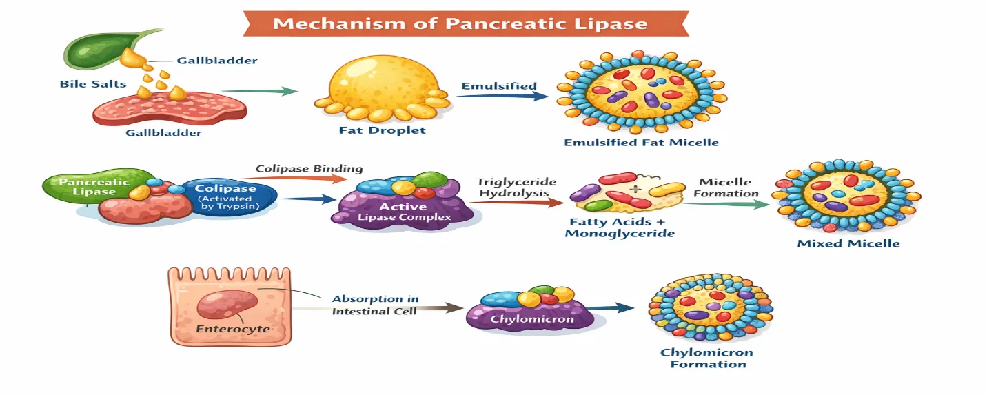
Pancreatic lipase plays an important physiological role in digestion and absorption of fats and fat-soluble vitamins. Increased lipase levels are usually associated with acute pancreatitis, gall bladder disease, pancreatic duct obstruction, kidney dysfunction, and certain medications. Decreased lipase activity occurs in conditions like chronic pancreatitis, cystic fibrosis, pancreatic insufficiency, severe malnutrition, and pancreatic surgery. These conditions impair fat digestion and lead to symptoms such as steatorrhea, bloating, abdominal pain, weight loss, and nutrient deficiencies. The mechanism of pancreatic lipase action begins with emulsification of dietary fats by bile salts. Colipase stabilizes the lipase enzyme on the lipid surface, enabling triglyceride hydrolysis at specific positions. This reaction produces free fatty acids and monoglycerides, which form micelles with bile salts and are later absorbed in the intestine. The absorbed products are reassembled into triglycerides and transported through chylomicrons.
The introduction also discusses diagnostic methods used to evaluate pancreatic disorders. Blood lipase tests measure serum lipase levels using enzymatic methods. Amylase tests are supportive investigations for pancreatic diseases. Stool tests, particularly fecal elastase tests, help detect pancreatic insufficiency. Imaging techniques such as ultrasound, CT scan, MRI, and MRCP assist in identifying pancreatic inflammation, necrosis, duct obstruction, and other abnormalities associated with altered lipase levels. Celiac disease testing is also important because intestinal damage can indirectly reduce pancreatic enzyme secretion. major focus of the introduction is the therapeutic importance of pancreatic lipase inhibition. Inhibition of pancreatic lipase reduces the digestion and absorption of dietary fats, thereby decreasing caloric intake. This forms the basis for anti-obesity therapy. Drugs such as Orlistat act through this mechanism. Lipase inhibition is useful in managing obesity, overweight conditions, type-2 diabetes mellitus, dyslipidemia, metabolic syndrome, non-alcoholic fatty liver disease (NAFLD), and polycystic ovary syndrome (PCOD). Weight reduction achieved through lipase inhibition improves insulin sensitivity, lipid profiles, and overall metabolic health.The project particularly focuses on Clitoria ternatea (Butterfly pea), a medicinal plant belonging to the family Fabaceae. The plant is rich in phytochemicals such as flavonoids, alkaloids, tannins, saponins, anthocyanins, and phenolic compounds. These bioactive constituents are known for several pharmacological activities including antioxidant, anti-inflammatory, antimicrobial, antidiabetic, hepatoprotective, Neuro protective, and anti lipase activities.
The introduction further highlights that natural products are gaining importance as safer alternatives to synthetic anti-obesity drugs because many synthetic drugs cause adverse effects. Previous studies reported that plant-derived polyphenols and flavonoids can inhibit pancreatic lipase activity effectively. Therefore, evaluating the lipase inhibitory activity of Clitoria ternatea extract may help identify potential natural anti-obesity agents.The study also includes FT-IR spectroscopy for identifying functional groups present in the plant extract. FT-IR works by measuring the absorption of infrared radiation by molecular bonds. Each functional group produces characteristic peaks, helping identify the phytochemical constituents responsible for biological activity. Antimicrobial, Antidiabetic, hepatoprotective, neuroprotective, and anti lipase activities.The introduction further highlights that natural products are gaining importance as safer alternatives to synthetic anti-obesity drugs because many synthetic drugs cause adverse effects. Previous studies reported that plant-derived polyphenols and flavonoids.
INHIBITION OF PANCREATIC LIPASE ENZYME:
Extract preparation:
The Clitoria Ternetea leaves was collected, shade dried and grinded to get powdered plant material. The powdered plant material is soaked 70% ethanol for 24- 72 hrs at room temperature. The solution is filtered, evaporated and dried at vacuum dryer to get crude extract.
Reagent preparation:
Disodium hydrogen phosphate: 1.42gm Potassium dihydrogen phosphate: 0.27gm
Dissolve both salts in approx. 40 ml distilled water. Check and adjust the pH to 7.7 using 0.1 M Hcl or0.1 M NaOH if necessary. Make up the volume to 50 ml with distilled water
Dissolve 20mg pancreatic lipase in 10 ml of 200mM phosphate buffer (pH7.7). Keep the enzyme solution on ice until use.
Mix 190ml ethanol in 10 ml of distilled water.
1gm NaOH pellet is dissolved in 400 ml of distilled water, heat it if it is not dissolved. Make upto 500 ml using distilled water.
Sample preparation:
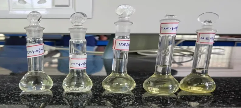
Add 100 mg of sample in 100 ml of 95% ethanol to prepare 1000 mcg/ml stock solution. From this prepare following dilution.
|
S.NO |
CONCENTRATION |
STOCK SOLUTION |
SOLVENT |
|
1. |
50mcg/ml |
0.5ml |
9.5ml |
|
2. |
100mcg/ml |
1ml |
9ml |
|
3. |
150mcg/ml |
1.5ml |
8.5ml |
|
4. |
200mcg/ml |
2ml |
8ml |
|
5. |
250mcg/ml |
2.5ml |
7.5ml |
PROCEDURE:
|
S. No |
Sample |
First titration value |
Second titration value |
Third titration value |
Mean value |
|
1. |
50 mcg/ml |
2.1 ml |
2.3 ml |
2.1 ml |
2.16 ml |
|
2. |
100 mcg/ml |
2.1 ml |
2.3 ml |
2 ml |
2.13 ml |
|
3. |
150 mcg/ml |
2 ml |
2.3 ml |
2 ml |
2.10 ml |
|
4. |
200 mcg/ml |
2 ml |
2.2 ml |
1.9 ml |
2.03 ml |
|
5. |
250 mcg/ml |
2 ml |
2.1 ml |
1.9 ml |
2.0 ml |
|
6. |
Standard |
1.8 ml |
1.9 ml |
1.6 ml |
1.76 ml |
|
7. |
Control |
2.2 ml |
2.4 ml |
2.3 ml |
2.30 ml |
|
8. |
Blank |
1.7 ml |
1.7 ml |
1.5 ml |
1.63 ml |
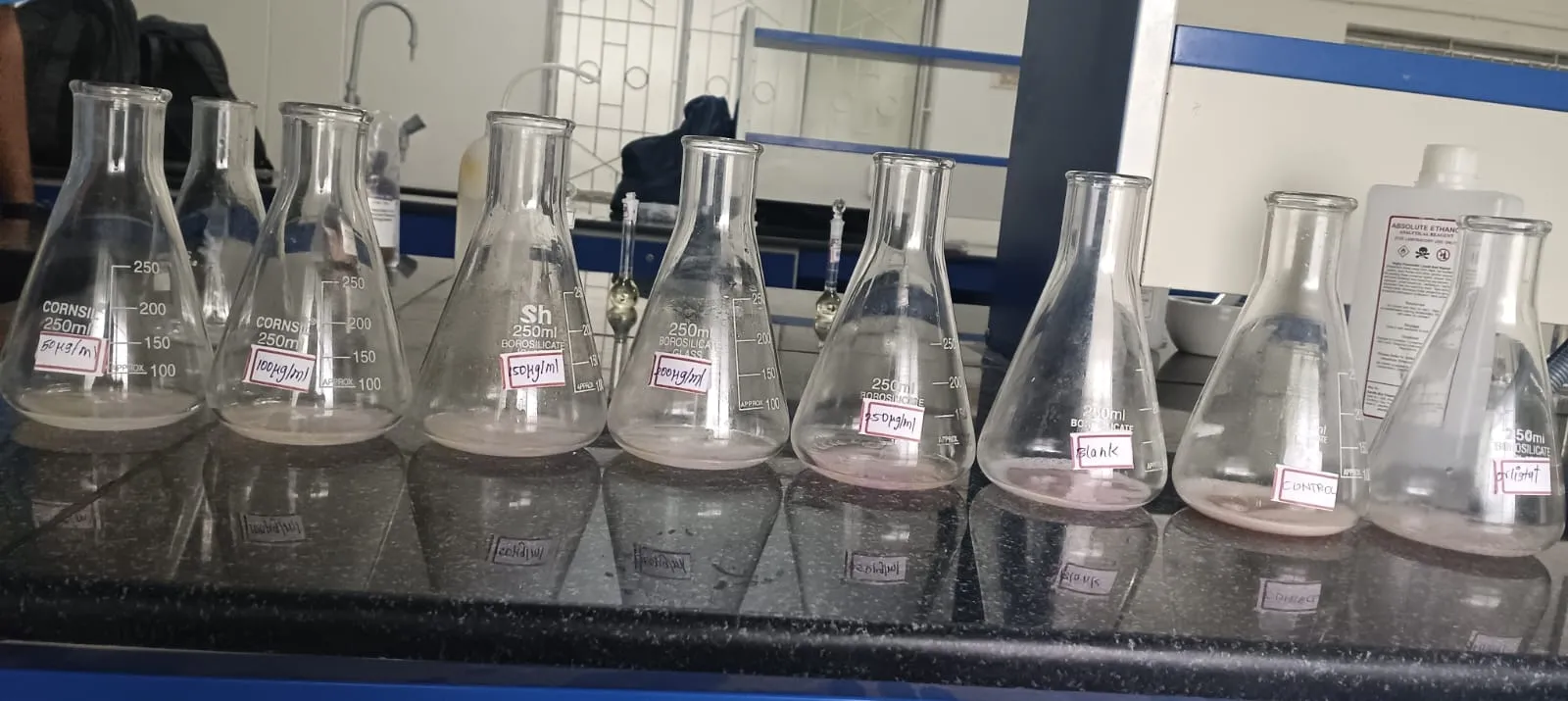
The blank titration value was subtracted from all the other titration values to eliminate the contribution of background acidity arising from the reagents, buffers, olive oil.
|
S. No |
Sample |
First titration corrected value |
Second titration corrected value |
Third titration corrected value |
Corrected titration value |
|
1. |
50 mcg/ml |
0.4 ml |
0.6 ml |
0.6 ml |
0.53 ml |
|
2. |
100 mcg/ml |
0.4 ml |
0.6 ml |
0.5 ml |
0.50 ml |
|
|
150 mcg/ml |
0.3 ml |
0.6 ml |
0.5 ml |
0.46 ml |
|
4. |
200 mcg/ml |
0.3 ml |
0.5 ml |
0.4 ml |
0.40 ml |
|
5. |
250 mcg/ml |
0.3 ml |
0.4 ml |
0.4 ml |
0.36 ml |
|
6. |
Standard |
0.1 ml |
0.2 ml |
0.1 ml |
0.13 ml |
|
7. |
Control |
0.5 ml |
0.7 ml |
0.8 ml |
0.66 ml |
Calculation of Percentage Inhibition:
Inhibition (%) = [EA without inhibitor – EA with inhibitor / EA without inhibitor] × 100
Where:
EA without inhibitor = Enzyme activity of control (no inhibitor)
EA with inhibitor = Enzyme activity in presence of test sample [44].
First titration:
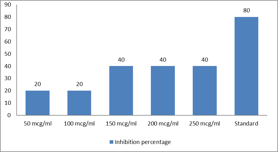
Inhibition % = [0.5 - 0.4 / 0.5] × 100 = 0.1 / 0.5 × 100 = 0.2 × 100 = 20
Inhibition% = [0.5 - 0.4 / 0.5] × 100 = 0.1 / 0.5× 100 = 0.2 × 100 = 20
Inhibition% = [0.5 – 0.3 / 0.5] × 100 = 0.2 / 0.5× 100 = 0.4 × 100 = 40
Inhibition% = [0.5 - 0.3 / 0.5] × 100 = 0.2 / 0.5 × 100 = 0.4 × 100 = 40
Inhibition% = [0.5 - 0.3 / 0.5] × 100 = 0.2 / 0.5× 100 = 0.4 × 100 = 40
Inhibition% = [0.5 - 0.1 / 0.5] × 100 = 0.4 / 0.5 × 100 = 0.8 × 100 = 80
Second titration:
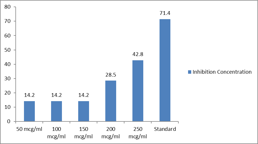
Inhibition % = [0.7 - 0.6 / 0.7] × 100 = 0.1 / 0.7 × 100 = 0.142 × 100 = 14.2
Inhibition% = [0.7 - 0.6 / 0.7] × 100 = 0.1 / 0.7 × 100 = 0.142 × 100 = 14.2
Inhibition% = [0.7 - 0.6 / 0.7] × 100 = 0.1 / 0.7 × 100 = 0.142 × 100 = 14.2
Inhibition% = [0.7 - 0.5 / 0.7] × 100 = 0.2 / 0.7 × 100 = 0.285 × 100 = 28.5
Inhibition% = [0.7 - 0.4 / 0.7] × 100 = 0.3 / 0.7 × 100 = 0.428 × 100 = 42.8
Inhibition% = [0.7 - 0.2 / 0.7] × 100 = 0.5 / 0.7 × 100 = 0.714 × 100 = 71.4
Third titration:
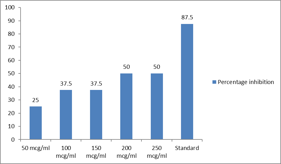
Inhibition % = [0.8 – 0.6 / 0.8] × 100 = 0.2 / 0.8 × 100 = 0.250 × 100 = 25
Inhibition% = [0.8 - 0.5 / 0.8] × 100 = 0.3 / 0.8 × 100 = 0.375 × 100 = 37.5
Inhibition% = [0.8 – 0.5 / 0.8] × 100 = 0.3 / 0.8 × 100 = 0.375 × 100 = 37.5
Inhibition% = [0.8 – 0.4 / 0.8] × 100 = 0.4 / 0.8 × 100 = 0.500 × 100 = 50
Inhibition% = [0.8 - 0.4 / 0.8] × 100 = 0.4 / 0.8 × 100 = 0.500 × 100 = 50
Inhibition% = [0.8 - 0.1 / 0.8] × 100 = 0.7 / 0.8 × 100 = 0.875 × 100 = 87.
Mean titration:
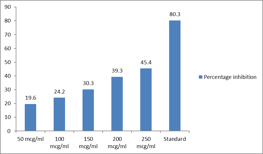
Inhibition % = [0.66 - 0.53 / 0.66] × 100 = 0.13 / 0.66 × 100 = 0.196 × 100 = 19.6
Inhibition% = [0.66 - 0.50 / 0.66] × 100 = 0.16 / 0.66 × 100 = 0.242 × 100 = 24.2
Inhibition% = [0.66 – 0.46 / 0.66] × 100 = 0.20 /0.66 × 100 = 0.303 × 100 = 30.3
Inhibition% = [0.66 - 0.40 / 0.66] × 100 = 0.26 / 0.66 × 100 = 0.393 × 100 = 39.3
Inhibition% = [0.66 - 0.36 / 0.66] × 100 = 0.30 / 0.66 × 100 = 0.454 × 100 = 45.4
Inhibition% = [0.66 – 0.13 / 0.66] × 100 = 0.53 / 0.66 × 100 = 0.803 × 100 = 80.3
First titration inhibition:
Second titration inhibition:
Third titration inhibition:
Mean titration inhibition:
RESULT
The pancreatic lipase inhibitory activity of Clitoria ternatea leaf extract was evaluated by the titrimetric assay method using olive oil as natural substrate. The assay was performed in triplicate, and the percentage inhibition was calculated using mean values of three independent experiments. The percentage inhibition of sample at concentrations of 50 mcg/ml, 100 mcg/ml, 150 mcg/ml, 200 mcg/ml and 250 mcg/ml are 19.6%, 24.2%, 30.3%, 39.3% and 45.4% respectively. The percentage inhibition of standard (orlistat) is 80.3%. The results showed that the Clitoria ternatea leaf extract exhibited concentration-dependent inhibition of pancreatic lipase activity. As the concentration of the extract increased, the percentage inhibition also increased.
REFERENCES


Dr. V. Suresh, Dr. S. K. Senthilkumar R. Asharaf Ali, C. Asma, A. Aziza, P. Chandrika, E. Deivamani, Assessment of the Anti-Lipase Activity of Clitoria Ternatea Linn. Extract Using an In Vitro Titrimetric Assay Method, Int. J. of Pharm. Sci., 2026, Vol 4, Issue 7, 6068-6075, https://doi.org/10.5281/zenodo.21721568
 10.5281/zenodo.21721568