

We use cookies to ensure our website works properly and to personalise your experience. Cookies policy
1,2,6 Department of Chemical and Biological Engineering, Brigham Young University, Provo, UT, U.S.A.
3 Department of Chemistry and Biochemistry, Brigham Young University, Provo, UT, U.S.A.
4,5 Department of Physics and Astronomy, Brigham Young University, Provo, UT, U.S.A.
This study describes a carrier having submicron, uniform and non-aggregated poly lactic acid (PLA) spheres loaded with the anticancer drug 5-fluorouracil (5FU) and with 9 nm superparamagnetic iron oxide nanoparticles (SPIONs) for magnetically guided drug delivery and local controlled release. Using a water/organic/water (w/o/w) double-emulsion process, we produced uniformly spherical microparticles smaller than 2 µm in diameter with well-dispersed SPIONs that retained superparamagnetic behavior after encapsulation. 5FU loading efficiency was determined to be 94%. Biological activity and chemical integrity was confirmed for the 5FU released from the product. Drug release kinetics showed faster release within the first day followed by sustained, slower release over 63 days with a cumulative release reaching 70% of loaded drug. Drug release was faster at 37°C compared to 21°C. PBS at pH 7.4 and 5.4 promoted faster release than did distilled water at pH 7.0. Release was prolonged from these PLA systems compared to other systems employing PLGA. This research introduces a rigorously optimized microcarrier system distinguished by sub-2-µm superparamagnetic PLA or PLGA microspheres of uniform morphology containing phase-dispersed SPIONs and exhibiting long-term controlled release, offering a transformative framework for magnetically directed drug delivery using high-gradient systems such as Halbach arrays.
1.1 Background
Primary symptoms of head and neck tumors, commonly called head and neck squamous cell carcinomas (HNSCCs), often share symptoms with benign illnesses such as pain in the throat and swelling [1, 2]. These misleading symptoms, along with inefficient standard screening methods, often delay accurate diagnosis [3]. Once appropriately diagnosed, therapy consists of surgical resection (if found in an early stage) followed by both chemotherapy and radiotherapy. Surgical removal is often difficult and sometimes impractical because of the high density of nerves, blood vessels, and other anatomical structures essential to speaking, eating, and breathing. Post-surgical treatments impose intense patient discomfort due to local radiation treatment or to whole-body side effects of chemotherapy and sometimes remain ineffective due to resistance of tumors to treatment [4, 5].
The American Cancer Society estimates that a total of over 2 million new cancer cases are expected to occur in the United States in 2025 [6]. With such a high rate of cancer occurrence, innovation in cancer therapy is highly desirable. One effective and widely used drug in cancer therapy is 5-fluorouracil (5FU) [7, 8, 9], which has broad activity against eukaryotic and prokaryotic cells [10]. However, the general toxicity of 5FU to the human body restricts its dosage and its modes of application [11, 12]. Biocompatible carriers are used to greatly slow the release of toxic drugs like 5FU as the carrier circulates, thus sparing general non-targeted tissues from cytotoxic exposure [13, 14, 15]. Moreover, accumulation of the carriers at the target tissue site and increase in release rate can produce a local concentration above cytotoxic level. The composition of the drug carrier determines the temporal release characteristics of the delivery system. By varying key parameters of the carriers’ size and composition, one may increase or decrease the drug release rate from the system to best fit a particular application. Additionally, drug sequestration and controlled release are vital features of anti-cancer drug delivery systems. Many drug delivery systems employ polymeric carriers to simultaneously control the rate of degradation and to protect the drug from rapid clearance and metabolic degradation before reaching the target area [16]. For anti-cancer drug delivery, these polymeric structures are often composed of biodegradable polyesters such as polylactic acid (PLA), polyglycolic acid (PGA), or poly(lactic-co-glycolic) acid (PLGA). The PLA used in our study prolongs the release window compared to systems employing PLGA [17].
1.2 Polymeric Double Emulsion Particles for Controlled Drug Release
While release may be controlled through shape (dimensions), composition, and molecular weight of polymer, the loading and retention rely on a more complex system of meta-stable phase separation in multiple emulsions, and on the stability of those phases during drying [18, 19, 20]. Small polymeric non-aggregated spherical drug carriers can be made via a double emulsion process, which combines a single emulsion within an additional second phase to entrap a large number of very small aqueous droplets of the first emulsion within larger individual droplets of organic phase of the second emulsion, as illustrated in Figure 1 [18, 21, 22]. An advantage to this entrapment method is that each phase of the emulsion may contain a different key chemical or drug, also depicted in Figure 1. This advantage may ensure more effective protection and sequestration of a water-soluble anti-cancer drug like 5-fluorouracil (5FU) during its transport through the body to a target location, while simultaneously achieving greater control over the degradation mechanism via the shape and size of the resulting polymer particle [20, 23]. When poly(vinyl alcohol) (PVA) is used as a surfactant to stabilize the emulsions, residual PVA at the resulting particle surface also reduces opsonization in the circulator system and thus promotes a reduced clearance rate [24].

Figure 1. Illustration of a double emulsion. This production begins with a water phase (white) containing dissolved drug 5FU (black) which is then dispersed in a degradable polymer dissolved in a hydrophobic solvent (organic phase, gray) which also contains dispersed SPIONs to form a single w/o emulsion. This single emulsion is then dispersed in a final phase of water (white) to form the double emulsion, following which the solvent and water are slowly removed by careful evaporation from the phases to form individual spherical particles.
Carriers composed of polyesters like PLA and PLGA degrade in the human body environment by erosion, releasing their drug payload through polymer chain cleavage via hydrolysis reactions with water [25]. These polyesters are commonly used for applications in the human body due to their biodegradability and biocompatibility of the final products [26, 27, 28]. PLA and PLGA are sufficiently polar that water diffuses into these particles; thus, hydrolytic polymer degradation occurs in the interior and on the surface of PLA or PLGA particles. However, PLA, being less polar than PLGA, degrades at a slower rate and releases drug more slowly than similar particles composed of PLGA.
In vivo, the drug is protected by the encapsulating polymer from any inactivation by metabolic enzymes or from rapid renal clearance until the polymeric carrier begins degradation, resulting in slow drug release over time as degradation progresses. Additionally, PLA and PLGA particles are deemed to be stable and protective during storage in dry conditions [29, 30, 31].
PLGA has been studied to determine its potential as a carrier for cancer therapy using 5FU. Prior experiments evaluating such PLGA nanoparticles in squamous cell carcinoma therapy found them to be effective in sustaining a prolonged release of the encapsulated drug [32]. These experiments also mentioned that 5FU-based release from PLGA nanoparticles promoted a more prolonged release than similar release of curcumin, another anti-cancer drug. Additional studies have been performed to optimize the production of uniformly sized PLGA nanoparticles for 5FU release [33].
The use of hybrid polymer systems which include PLGA for colorectal cancer therapy has also been found to be highly impactful in minimizing systemic toxicity and improving tumor cell death or growth inhibition [34, 35, 36, 37, 38]. One of these studies involved the use of poly(3-hydroxybutane-co-3-hydroxyvalerate acid)/poly(lactic-co-glycolic acid) nanoparticles for release of 5FU and oxaliplatin, which demonstrated the potential for polymeric delivery systems while minimizing acidic byproducts. This prior study proposed a trade-off of polymeric cancer drug delivery where one may reduce acidic byproducts at the cost of reduced entrapment efficiency [38].
A similar study found that PLGA particles may be synthesized at a minimum size of 125 nm in diameter using a double emulsion water/organic/water (w/o/w) method with PVA as a surfactant [37]. Hybrid polymeric nanoparticles consisting of PLGA and Eudragit FS have also been studied to incorporate a pH-responsive release into the system [36]. The particles were produced by a solid/oil/water double emulsion with a final average diameter of 150 µm and were found to suppress 5FU release in the acidic environment of the stomach and to induce sustained release in the alkaline environment of the colon. This finding suggested minimal systemic absorption of 5FU during transit through the stomach, which could have otherwise resulted in severe side effects.
Another study showed growth inhibition of cancerous tumors via the delivery of epidermal growth factor (EGF) through functionalized particles composed of PLGA. The authors found simultaneous EGF receptor-targeted therapy and 5FU chemotherapy to be effective and superior to monotherapeutic methods [35]. A similar study evaluating EGF receptor-targeted therapy using PLGA/Poly(ethylene glycol) (PEG) found similar results, emphasizing the crucial sustained release characteristics of PLGA controlled by polymer degradation [34]. None of these prior studies compared release from PLGA to that from PLA particles, which latter particles would be expected to slow the release rate and extend the release window.
1.3 Superparamagnetic Iron Oxide Nanoparticles
There is current interest in using superparamagnetic nanoparticles for therapeutic intervention due to their facile manipulation by magnetic fields. Superparamagnetic nanoparticles are characterized by two requirements. First, the particles exhibit a strong magnetic moment while in the presence of an external magnetic field, so they can be spatially manipulated. Second, their magnetization curve shows no hysteresis, meaning that when removed from an external magnetic field the sample exhibits zero magnetization. This characteristic prevents superparamagnetic materials from magnetically agglomerating in the absence of an external magnetic field. Superparamagnetic properties have been observed in non-toxic superparamagnetic iron oxide (Fe3O4) nanoparticles (SPIONs), prompting their broad development for many biomedical applications including magnetically guided therapies [39, 40, 41]. Studies of the toxicity of SPIONs indicate low or no cytotoxicity of SPIONs for both in vitro animal cells and human stem cells; furthermore, they reveal both a short-term and long-term biocompatibility with no apparent acute or chronic toxicity in mice in vivo [42, 43, 44].
By pairing an anti-cancer drug with SPIONs, the location of drug release may be spatially manipulated by an external magnetic field. When injected into the vasculature, drug-loaded superparamagnetic microcarriers may be magnetically directed with high-gradient electromagnets or permanent magnets to a target volume, such as to produce accumulation in a tumor [45, 46]. The head and neck regions are ideal for magnetically guided drug delivery because of facile placement of multiple external devices in Halbach arrays to create orthogonal magnetic fields for 3D focusing. A Halbach array contains a particular orientation of magnetic fields in 2D and 3D orientations to apply forces directed to volumes other than sole attraction to the surface of the magnet. This can be used to push magnetic nanoparticles to deeper tissue locations than only pull them toward the skin [47]. This design has a maximum working depth of about 10 cm (which is within working distance for head and neck tumors) and purports sufficient magnetic force to trap microcarriers containing SPIONs in the capillaries of a tumor, whereupon they can release their therapeutic payload.
Such forces may push and perhaps lodge particles to the internal lumen side of a capillary when tangential magnetic forces exceed the very small axial fluid drag force acting on sufficiently small particles. For example, Stokes law indicates that the axial drag force from blood plasma on a 1-µm (diameter) spherical particle in typical capillary flow (0.07 cm/s) is on the order of 10-11 N. Magnetic forces on SPIONs from Halbach arrays can exceed these fluid forces. Al-Jamal et al. studied the effect of SPION loading on blood circulation in mice and extrapolated his findings to humans. This study employed the finite element model of Nacev et al. using blood and blood vessel information from the Phase I clinical study of Lübbe et al. [48, 49, 50]. These studies concluded that external magnetic forces would be sufficient to achieve successful targeting in humans, further transitioning magnetic drug delivery research from its preclinical study stage to proposed clinical applications. Importantly, when the magnetic drug carriers are smaller than 2 µm in diameter, they do not block capillaries. These small particles could be magnetically pushed onto the walls of capillaries within a targeted tissue, such as a tumor, and then held in place by magnetic forces of a Halbach array.
The addition of superparamagnetic material such as magnetite (Fe3O4) nanoparticles to the composition of a drug carrier confers these very useful magnetic properties. However, superparamagnetism is rarely observed in particles > 25 nm in diameter [51]. Thus, numerous SPIONs of sufficiently small size (< 25 nm) must be loaded into the matrix of a larger particle which also carries the therapeutic.
Many studies have been conducted to synthesize PLA microspheres for sustained drug release by double emulsion technologies; but of those that contain SPIONs, there are no reports of PLA microspheres under 5 µm in diameter [20, 22, 52, 53, 54, 55, 56, 57, 58, 59, 60]. Previous research that combined iron oxide with PLA carriers typically used larger non-superparamagnetic iron oxide cores rather than dispersed SPIONs [55, 61, 62, 63], which may lead to magnetic self-aggregation in blood. The literature reports a few examples of PLA microspheres under 2 µm in diameter produced by double emulsion techniques containing anticancer drug and of microspheres composed of a SPION core and drug-loaded PLA shell with about a 10 µm diameter [59, 64]. Among these, Ayyanaar et al. [17] demonstrated that PLGA microspheres loaded with iron oxide nanoparticles could achieve pH-responsive release of 5FU, establishing an important conceptual basis for magnetically responsive polymeric carriers. However, no prior study has evaluated this microcarrier design within the context of magnetic targeting using Halbach arrays. Successful application of such arrays requires particles that are sub-2-µm, uniformly spherical, nonaggregated, and superparamagnetic at physiological temperature. These requirements guided the engineering and characterization objectives of the present study.
The methods presented herein systematically produce individual (non-aggregated PLA and PLGA particles of < 2 µm diameter incorporating monodisperse ~9 nm SPIONs, quantifies their superparamagnetic behavior, and evaluates extended 5FU release over 65 days. Through optimization of double emulsion parameters, we achieve distinct, nonaggregated microspheres with the potential for safe delivery and controlled release of highly toxic drugs to tumors.
This work builds upon prior concept studies while using PLA and introducing critical advances in particle engineering, magnetic characterization, and long-term drug release. Application of this work with a Halbach array provides the potential for both a protective and biocompatible barrier to general and non-specific release to the body while allowing superparamagnetically-directed accumulation of the carrier at the desired site of delivery and controlled drug release over time. Once at the focal volume of the Halbach array, they may contact each other while lodging on the capillary well.
2. MATERIALS AND METHODS
2.1 Materials
Polyvinyl alcohol (PVA) was purchased from Thermo Scientific with a molecular weight of 146,000-186,000 g/mol and hydrolysis of 99.3-100%. PLA was purchased from JAREES 3D Printer Filament and was measured by viscometry [65] to have a molecular weight of 127,000 g/mol. PLGA 50:50 was purchased from Sigma-Aldrich with a molecular weight of 45,000 g/mol. 5FU was purchased from Tokyo Chemical Industry. Methylene chloride (MeCl2) was purchased from Fisher Chemical.
2.2 SPION Synthesis
SPIONs were synthesized using the thermal decomposition method published by Sun et al. with a target size of around 9 nm in diameter [66]. Briefly, the particles were prepared by combining tris(acetylacetonato)iron(III) (Fe(acac)3), oleic acid, oleylamine, and 1,2-hexadecanediol with constant magnetic stirring and a flow of nitrogen. The mixture was heated to 200 °C for 2 hours and then heated to 300 °C and refluxed for 1 hour. The resulting particles were then rinsed 5 times with ethanol and placed in a vacuum desiccator to dry for 24 hours to form a powder.
2.3 Double Emulsion Synthesis
Our goal was to produce double-emulsion w/o/w non-aggregated microspheres with a target size of less than 2 µm in diameter when dried. Various experimental methods were examined and optimized to achieve a consistent and uniform particle size.
The standard process to form double emulsion microspheres proceeded as follows. The first (inner) water phase was formed by dissolving 5FU at saturation concentration (12 mg/mL) into a solution of PVA in distilled water. The organic phase was prepared by dissolving 3.0 g of PLA (or PLGA) into 17 mL of MeCl2. A suspension of SPIONs in MeCl2 was made by dispersing 10 mg of SPIONs (9 nm in diameter) into 20 mL of MeCl2 by sonication for 1 hour in a sonicating bath (~1 W/cm2, Sonicor, SC-100, Wallingford, CT, USA). Then, 1.0 g of the SPION dispersion was added to 4.0 g of the PLA/MeCl2, resulting in 0.088 mg of SPIONs per mg of organic phase and 0.094 mg of PLA per mg of organic phase. This mixture was then vortexed for one minute.
The w/o emulsion was formed by adding 0.125 g of the 5FU solution (first, or inner aqueous phase) to 4.875 g of the organic phase, and then emulsifying the phases for one minute to form submicron droplets by one of the following 3 techniques: a magnetic stir bar (800 rpm), sonicating probe (Sonics and Materials Inc., Model CV26, Newtown, CT, USA), or mechanical emulsifier (Ultra Turrax, T25 Basic with Model S25N-8G probe, IKA, Wilmington, NC, USA).
A second (outer) aqueous phase was prepared consisting of a surfactant dissolved in distilled water. Then, 4.0 g of the second (outer) aqueous solution was added to 1.0 g of the 5FU-polymer w/o microemulsion, followed by similar emulsification.
The resulting w/o/w emulsions were dried by various methods to determine the best method to produce small non-aggregated particles. These methods were freeze drying (FreeZone 2.5, F4413E-2, LABCONCO Kansas City, MO, USA), evaporative drying in a fume hood with a stir bar inside the vessel, and quiescent drying in a fume hood. In this last (and most favorable) method, the emulsion was left to dry with no stirring in a fume hood for 48 hours to slowly evaporate the MeCl2. The mass lost during this quiescent evaporation was recorded. The remaining content was washed three times by magnetic collection and decanting; then, distilled water was added back to replace the lost mass. The microspheres were then redispersed by the same method of sonication and again quiescently evaporated at room temperature in a fume hood for 48 hours. The effect of geometry of the drying container was also examined, including 20 mL scintillation vials, petri dishes, and watch glasses.
The mass ratios of the above standard formulation were 0.5:19.5:80.0 (inside-aqueous-phase: organic-phase: outside-aqueous-phase). The effects (upon particle size) of changing these phase ratios in the above procedure were examined by varying the mass ratio of the inner water phase to the organic phase in the w/o emulsion and by varying the mass ratio of the w/o emulsion to the outer water phase in the w/o/w emulsion.
The surfactant species in the water phases were varied to determine which produced optimal results. Candidate surfactants were PVA, sodium dodecyl sulfate (SDS), and Brij O20 in the inner and outer aqueous phases of the double emulsion. PVA was examined because it was used as a surfactant in the procedure published by Delie et al. which encapsulated material within PLA microparticles [22]. SDS was examined due to its hydrophilicity and long hydrocarbon chain which could inhibit aqueous droplets from merging with one another. Brij was examined because it is a polyethylene glycol-based surfactant similar to that used in the procedure published by Debotton et al. for encapsulating drug in double emulsions [20].
In some experiments, the concentrations of surfactant in the inner and outer water phases of the double emulsion were also varied, as was the concentration of 5FU in the inner water phase.
2.4 Particle Analysis
The SPIONs were characterized for size and shape by transmission electron microscopy (TEM, Tecnai, TF-20, FEI, Hillsboro, OR, USA). Magnetic properties were measured by vibrating sample magnetometry (VSM, Quantum Design, San Diego, CA, USA) using field cooling and zero field cooling (FC/ZFC) measurements. Finally, magnetization loops were collected and examined for possible magnetic hysteresis.
The resulting double emulsion particles were dried and examined by scanning electron microscopy using Apreo (SEM, Apreo, C Low-Vacuum, ThermoScientific, Waltham, MA, USA) and Verios (SEM, Verios G4 UC, Low-Vacuum, ThermoScientific, Waltham, MA, USA) microscopes. ImageJ (NIH, Bethesda, MD, USA) was used to determine the diameter size of the product double emulsion particles. From this data, the maximum and minimum diameters, average diameter, and the polydispersity index were calculated for the various samples. The polydispersity index was calculated as the standard deviation of the particle population diameter divided by the mean average diameter of the particle population.
2.5 Drug Loading Efficiency
The absorbance spectrum of 5FU was measured from 250 to 290 nm using a UV-VIS Spectrometer (Agilent Technologies, Cary 60 UV-VIS, West Chester, OH, USA), and the midpoint of a broad absorbance peak for 5FU was observed at 270 nm, consistent with literature values [13, 14, 67]. A calibration curve of 5FU absorbance at 270 nm was found to be linear over a range of 0 to 64 µg/mL (corresponding to 0 to 1.24 absorbance units).
The mass of encapsulated 5FU was calculated using the concentration of 5FU present in liquid double emulsion outer water phase measured using the UV-VIS spectrometer and subtracting that amount from the total mass of 5FU used in the loading procedure. This outer phase was easily accessible by magnetic separation: a strong magnet placed adjacent to the container following emulsification pulled the emulsified phase to the side of the container, leaving clear liquid that could be collected by pipet. Then, the drug loading efficiency was calculated using Equation 1:
Drug Loading Efficiency= Mass of Encapsulated 5FUMass of 5FU Used in Loading Procedure 1

2.6 Release of 5FU
The mass of released 5FU was calculated using the absorbance calibration at 270 nm. Specifically, dried microspheres (25 mg) were added to 5 mL of the release medium in a 6 mL scintillation vial. The contents were then sonicated for 1 minute using the sonicating probe at an amplitude of ~1.5 W/cm2 to fully disperse the microspheres in the medium. Vials containing the microsphere dispersion were secured in a rotating frame (45 RPM) to maintain continual slow convection within the vials and prevent the microspheres from settling. At each specified time point, a vial was removed from the rotating frame and placed on top of a strong magnet for 1 hour to magnetically separate the microspheres from the release medium. After separation, 3 mL of the release medium were transferred to an optical cuvette via pipette to measure absorbance at 270 nm, and the concentration was calculated. With few exceptions, the cuvette and its contents were discarded, and 3 mL of fresh release medium were added to the sample vial. The sample was then vortexed for 30 seconds and returned to the rotating frame until the next time point.
For the study reported herein, 4 formulations were prepared from particles produced by the ideal preparation protocol (see Results section). One formulation was made from PLA using 2.0% PVA in the outer water phase of the double emulsion. This is designated as the “base” formulation (named PLA@2.0), and the other 3 formulations are variations of this base formulation. Formulations of PLA with 0.5% and 1.0% are designated PLA@0.5 and PLA@1.0 respectively. When evaluating release from PLGA, these microparticles were made using 2.0% PVA in the outer water phase (PLGA@2.0).
Controlled drug release was evaluated by varying the environmental conditions of release medium, its pH, and the temperature of the release environment. These environmental variations were done only for the base formulation (PLA@2.0). Thus, there were four key parameters of the release experiment: 1) the concentration of PVA in the outer water phase of the double emulsion prepared during formulation, 2) the polymer used in the organic phase of the double emulsion during formulation, 3) the release medium and its pH, and 4) the temperature of the release environment. The standard release case to which others are compared is 5FU release from the base formulation (PLA@2.0) in PBS with a pH of 7.4 and a temperature of 37°C. This standard release was compared to drug release in which only a single parameter (formulation or environment) was varied. These experiments are listed in Table 1 which presents the microsphere name, formulation, environment of release, and temperature of release. The average diameter and polydispersity index (standard deviation of the diameter divided by the mean diameter) were determined from SEM images of the particles, processed with ImageJ software. These data are presented in the Results section and are listed in Table 1.
At least three release experiments were performed for each configuration in Table 1. Samples were taken at exact times so that a paired comparison analysis could be done, with 3 sets of paired data for each formulation or condition. Statistical analysis was done using conventional paired comparison and Student-t statistics to calculate one-sided T-values and p-values. Statistical significance was attributed to data when p < 0.05. For clarity in the plots presented hereafter, the data point signifies the mean of the 3 measurements, and the whiskers denote the range of the measurements.
Table 1. Names and characteristics of particles and experiments.
|
Particle Name |
Formulation |
Environment |
Temp (°C) |
|
PLA@2.0 |
PLA with 2.0% PVA in outer phase1 |
PBS pH 7.4 |
37 |
|
PLA@2.0 |
PLA with 2.0% PVA in outer phase 1 |
PBS pH 7.4 |
21 |
|
PLA@2.0 |
PLA with 2.0% PVA in outer phase 1 |
PBS pH 5.4 |
37 |
|
PLA@2.0 |
PLA with 2.0% PVA in outer phase 1 |
H2O pH 7.0 |
37 |
|
PLA@1.0 |
PLA with 1.0% PVA in outer phase 2 |
PBS pH 7.4 |
37 |
|
PLA@0.5 |
PLA with 0.5% PVA in outer phase 3 |
PBS pH 7.4 |
37 |
|
PLGA@2.0 |
PLGA with 2.0% PVA in outer phase 4 |
PBS pH 7.4 |
37 |
|
1 Average diameter = 0.697 µm, PDI = 0.501; Standard Release Case 2 Average diameter = 1.242 µm, PDI = 0.671 3 Average diameter = 1.770 µm, PDI = 0.963 4 Average diameter = 0.783 µm, PDI = 0.870 |
|||
2.7 Chemical Integrity of 5FU
UV-VIS absorbance spectra were compared for 5FU before and after release from the product microspheres to assess whether chemical change occurred in the 5FU during the encapsulation and release processes.
2.8 Potency of Released 5FU
The minimum inhibitory concentration (MIC) of 5FU before and after release from the microspheres was examined by the broth microdilution technique to confirm that the potency of the 5FU was maintained during release. This was done by growing S. aureus at 5 x 10³ CFU/mL in nutrient broth with varying concentrations of 5FU (32, 16, 8, 4, 2, and 1 µg/mL) within test tubes using both fresh 5FU and 5FU recovered from release experiments. These test tubes were placed on an orbital shaker inside an incubator at 37°C for 24 hours. After 24 hours, the inhibition of growth was observed, and the MICs of the two samples were compared.
3. RESULTS
3.1 SPION Imaging and Magnetometry
Synthesized SPIONs were imaged with transmission electron microscopy (Figure 2a) and were found to have a size distribution of 8.8 ± 1.9 nm evaluated over at least 100 particles. As seen in the TEM image (Figure 2a), this batch of SPIONs was polydisperse, and included some large particles (the maximum size observed in our TEM images was 18 nm) mixed with the smaller particles. The particles appeared to be spherical (not faceted). These SPIONs are notably non-dispersible in water, likely due to the presence of oleic acid during synthesis, which purportedly coats the particles [66].
The magnetometry characterization shown in Figure 2b,c suggests that the SPIONs are superparamagnetic (SPM) at 300 K. The field cooling (FC) and zero field cooling (ZFC) curves in Figure 2b indicate a blocking temperature spread around TB ~ 185 K (spreading from about 90 K through 300 K at 90 % of max ZFC value). The wide spread and relatively high median value for TB is likely due to the wide range of particle sizes and the heavier weighting contributed by the larger particles, as the magnetic moment measured via VSM is proportional to the volume (not the diameter) of the particles. When the average is based on particle volume (cube of particle diameter), the average nanoparticle size is 9.3 ± 2.7 nm. The larger particles induce strong magnetic interparticle interactions with the surrounding particles, and these interactions are magnified in the case of SPIONs that are compacted in the VSM capsule of the measurement.
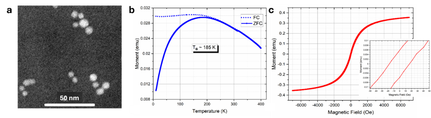
Figure 2. Structural and VSM magnetic characterization of SPIONs. (a) TEM image of the fabricated SPIONs sparsely dispersed on a TEM grid. (b) FC-ZFC curves collected under a magnetic field of 100 Oe. (c) Magnetization loop collected at 300 K over ± 7000 Oe range, with a zoomed-in view around the origin in the inset.
Lastly, the magnetization loop collected at 300 K in Figure 2c shows no visible hysteresis when plotted over a range of ± 7000 Oe. When zoomed-in over a ± 50 Oe range, a very small hysteresis, smaller than 20 Oe appears, but given the uncertainty of measurement, this hysteresis is considered negligible, and confirms superparamagnetism at 300 K.
Figure 3 shows magnetometry data for the SPIONs after being embedded in the microspheres. The FC-ZFC curves in Figure 3a suggests a much lower blocking temperature (TB ~ 75 K), which typically corresponds to what has been observed for isolated non-interacting monodisperse nanoparticles with average size around 8 nm [68, 69]. In addition, the value of the measured magnetic moment (on the y-axis) is approximately 100-fold smaller than the naked SPIONs in Figure 2b. These observations suggest that the SPIONs are well dispersed within the PLA microspheres, thus practically eliminating magnetic interparticle interactions. The magnetization loop collected at 300 K in Figure 3b shows no visible hysteresis when plotted over a field range of ± 7000 Oe. The zoomed in view over a ± 50 Oe in Figure 3c shows a very small hysteresis (less than 30 Oe), which given experimental uncertainty is also considered negligible, and thus the material is considered superparamagnetic at 300 K. In summary, the present SPIONs show no evidence of aggregation inside the PLA microspheres and exhibit superparamagnetic behavior at 300 K both in their naked form and when embedded in the microspheres.
Figure 3. VSM magnetic characterization of SPIONs embedded in microspheres. (a) FC-ZFC curves collected under a magnetic field of 100 Oe. (b) Magnetization loop collected at 300 K over ± 7000 Oe range (c) Zoomed-in view around the origin, over ± 50 Oe range.
3.2 Characterization of Particles Made by Double Emulsion
3.2.1 Visualization of Emulsions
Various experiments were performed to determine the mass ratios for the single and double emulsions which would minimize the size of the resulting microspheres. For the w/o emulsion, the ratios of 10:90, 5:95, and 2.5:97.5 w:o were examined. Light microscopy showed that the droplets decreased in diameter as the fraction of the first (inner) aqueous phase decreased in the w/o emulsion. Of the ratios tested, the w/o emulsion ratio which best minimized the diameter of the droplets was 2.5:97.5 w:o.
This primary, or inner emulsion, was then further emulsified with an outer aqueous phase containing surfactant. The light microscopy images of Figure 4 show that the "oil-phase" droplet size decreased as the fraction of w/o decreased in the w/o/w double emulsion, as expected.
The double emulsion ratio that minimized the diameter of the droplets in this study was found to be 20:80 w/o:w. Further decreasing the fraction of w/o in the double emulsion would likely further decrease the size of the final product particles; however, the 20:80 ratio was satisfactory to reach the goal of an average diameter below 2 µm in the product.
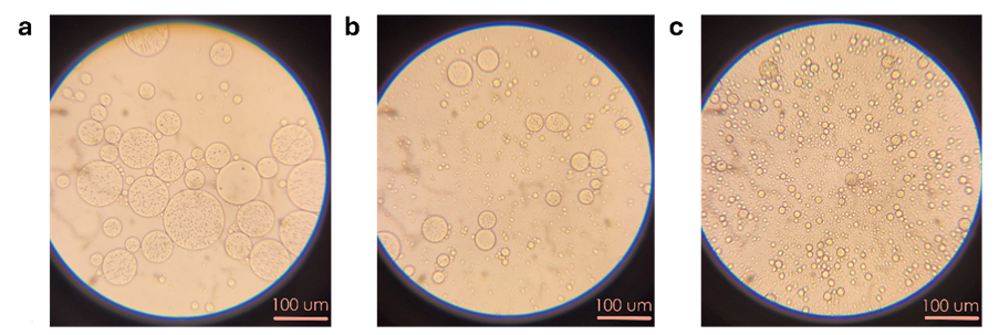
Figure 4. Visible light microscopy images of w/o/w emulsions with 1 wt% PVA in the outer water phase depicting the relative size of droplets in an emulsion ratio of a) 50:50 w/o:w, b) 40:60 w/o:w, c) 20:80 w/o:w. Scale bars are 100 µm.
3.2.2 Particle Quality
Emulsification methods were compared to determine the method which best minimized the final particle size. Experiments using a stir bar to emulsify the phases proved inadequate, producing w/o and w/o/w droplets observably macroscopic in size and too large for the purpose of this project. Experiments conducted using the Ultra Turrax emulsifier produced macroscopic w/o/w droplets much too large for this project. Experiments which utilized the sonicating probe at an amplitude of 15% instrument power (~1.5 W/cm2) to emulsify the phases produced microscopic w/o and w/o/w droplets. Because the target product size was obtained with the sonicator, all subsequent experiments in this study were conducted using the sonicating probe to emulsify the phases.
Surfactants PVA, SDS, and Brij O20 were used to determine which best minimized the average diameter and maximized the morphological uniformity of the product microspheres. Figure 5 provides a comparison of the final (dried) double emulsion product using each of these surfactants. Freeze drying was employed to capture the structure in the double emulsion without the evaporation process itself producing any possible agglomeration or deformation of particle shape.
The particles of Figure 5 show that double emulsions prepared with PVA as the surfactant produced spherical particles with an average diameter smaller than that of the other surfactants with some nonspherical, rigid product present. This rigid product was present only when the double emulsion with PVA surfactant was freeze dried, as will be discussed later in this study. Double emulsions prepared with Brij O20 as the surfactant produced mostly nonspherical, agglomerated product unsatisfactory for application by vascular delivery. Emulsions prepared with SDS as the surfactant produced large spherical particles entrapped by honeycomb-like matrices of solid. Furthermore, SDS produced the largest product particles with the larger particles nearing 100 µm in diameter. These and other SEM images showed that PVA surfactant produced the smallest and most uniformly spherical product up to 30 µm in diameter, while SDS produced microspheres up to 200 µm in diameter surrounded by a porous solid, and the use of Brij O20 produced nonspherical products. Therefore, PVA was selected for further studies.
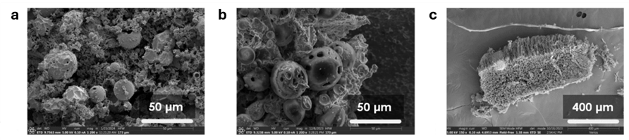
Figure 5. SEM images of dried particles with one candidate surfactant in both water phases (9 wt% surfactant in inner water phase and 1 wt% surfactant in outer water phase). These images are of freeze-dried product and prepared with various surfactants a) PVA, b) Brij O20, c) SDS. Scale bars for a, b, and c are 50 µm, 50 µm, and 400 µm respectively.
3.2.3 Particle Size
Macroscopic observation indicated that PVA concentration in the inner water phase was inversely related to the droplet diameter size. Thus, the saturation concentration of the PVA in water, 9 wt%, was used for the inner water phase for all experiments in this study. Trials were performed at different concentrations of PVA in the outer water phase to determine the effect of the surfactant on the product particle size, as shown in Figure 6. Associated SEM images of the product double emulsion microspheres are also included in Figure 6.
The SEM images in Figure 6 confirmed that the microspheres were distinct and suggested that the polydispersity index of the product was also inversely related to the concentration of PVA in the second water phase. The particles prepared with 2 wt% PVA in the outer water phase had the smallest and most uniform size distribution.
Several parameters relating to drying the double emulsions were examined, including the solvent evaporation method, vessel geometry, and drying time. The evaporation techniques include convection drying in a fume hood, convection drying in a fume hood with a stir bar, and freeze drying. The presence of a magnetic stir bar during solvent evaporation caused the droplets to collect on the stir bar due to their superparamagnetic properties, disrupting the necessary double emulsion mechanism for spherical product particles. Thus, magnetic stirring during evaporation could not be used.
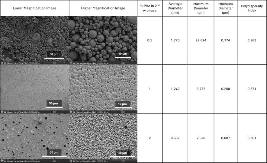
Figure 6. SEM images and data of dried double emulsion microspheres. SEM images have two scale bars of 50 and 10 µm. The concentration (wt%) of PVA in the second water phase is varied and the corresponding particle size information of each resulting formulation is given.
Figure 7 shows representative images that compare those products dried by convective evaporation and those prepared by freeze drying. Freeze drying resulted in a significant presence of solid debris in the final product, which was not present when dried by evaporation. With freeze drying, we hypothesize that during rapid cooling toward the freezing point, there was a sol-gel microphase separation in the liquid; and then during drying the surfactant and the small amount of polymer in the sol phase agglomerated and deposited on the particles. On the other hand, during evaporative drying, after the solvent evaporation and wash and water replacement, there was no free surfactant remaining to deposit on the surface of the particles.
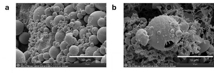
Figure 7. SEM images of double emulsion product particles with 1 wt% PVA in the outer water phase prepared with different drying methods a) convection drying, b) freeze drying. Scale bars are 10 µm.
The candidate vessels used for drying the product by convection in a fume hood were a petri dish and a watch glass. During the convective solvent removal process, there was sometimes a solid film (assumed to be polymer) observed to nucleate over the exposed surface of the double emulsion. However, diluting the sample with water before convective drying was observed to reduce the thickness of the film. This film was also observed to form in response to disturbance of the vessel in which the sample was drying. The film was observed to form when the sample was spread thinly across the vessel’s surface, suggesting that some nucleation process may have initiated film formation. If the film was physically pulled off from the sample’s surface, a new film was observed to form for as long as there was liquid remaining in the suspension. Additionally, this film sometimes formed over the entire course of the drying phase if the vessel was left undisturbed and unwashed. This film could be physically peeled back to reveal the final, dried product underneath. After many experiments and observations, we determined that for this drying process, a watch glass is preferable to a vessel of more uniform depth, such as a petri dish. The film formed by samples dried in a watch glass were observed to be significantly thinner than samples dried in a petri dish. Figure 8 compares the product particles when dried in a watch glass and in a petri dish.
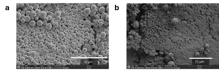
Figure 8. SEM images of double emulsion product particles with 1 wt% PVA in the outer water phase dried in a) a watch glass, b) a petri dish. Scale bars are 10 µm.
Microspheres produced by drying in a petri dish were also frequently observed to be less distinctly spherical, as shown in Figure 8b. Our experience suggests that drying the particles in a watch glass results in product particles significantly more spherical and individual in physical shape.
The most crucial parameter tested was the long drying time and replacing lost volume. It was found that redispersion of the double emulsion droplets in the water medium after completion of solvent evaporation phase was paramount to minimizing the size (diameter) of the product particles and to preserving spherical uniformity. Lost solvent mass was replaced with distilled water before redispersion and subsequent drying. For a sample 3 mL in volume, the presence of MeCl2 was no longer observable (by olfaction) after 48 hours, at which time the replacement water was added. The particles produced by similar processes with and without this additional dispersion step are compared in Figure 9. While the particle size distribution is not significantly different, the particles made by washing and replacement by water have a smoother surface and do not appear to reside on a solid matrix.
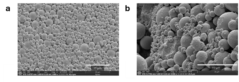
Figure 9. SEM images of double emulsion product particles with 1 wt% PVA in the outer water phase dried by convection: a) rinsing the double emulsion after solvent evaporation, replacing the mass of solvent lost by evaporation with distilled water, and redispersing the emulsion before drying; b) same process but without the additional wash, replacement, and redispersion step. Scale bars are 10 µm.
The results depicted by Figure 9 indicate that redispersion in water after solvent evaporation, followed by slow evaporation of water produced clean, smooth, spherical particles within the desired size range.
3.2.4 Optimal Formulation Procedure
To summarize the observations of the many parameters studied in particle formulation, the following method produces the smallest microspheres containing 5FU and SPIONs. The first water phase was prepared by dissolving 60 mg of 5FU and 500 mg of PVA in 5 mL of distilled water. The organic phase was prepared by dissolving 3.0 g of PLA in 17.0 mL (22.5 g) of MeCl2. The SPION dispersion was created by dispersing 10 mg of SPIONs in 20 mL (26.5 g) of MeCl2 by sonication in a sonicating bath for 1 hour. Then 1 g of the SPION dispersion was added to 4 g of the PLA/MeCl2, resulting in 0.075 µg of SPIONs per mg of organic phase and 0.094 mg of PLA per mg of organic phase. The resulting organic phase mixture was then vortexed for one minute.
The second (outer) water phase consisted of water and 2 wt% dissolved PVA.
The first emulsion (w/o) was formed by adding 0.125 g of the first water phase to 4.875 g of the organic phase in a 20 mL scintillation vial. The resulting emulsion was then sonicated using a sonicating probe at an amplitude of 15% power (1.5 W/cm2) for one minute in an ice bath, and then vortexed for one minute.
The second emulsion (w/o/w) was formed by adding 1 g of the first emulsion (w/o) to 4 g of the second water phase. The resulting emulsion was sonicated using the sonicating probe at the same amplitude of 15% for 3 minutes in an ice bath. The typical volume of double emulsion prepared was 5 mL.
This double emulsion in a scintillation vial was placed in a fume hood for 48 hours to evaporate the MeCl2. The remaining content was washed by magnetic collection and decanting three times using distilled water. Lost mass was replaced with distilled water. Then, the emulsion was redispersed using the sonicating probe at the same amplitude of 15% for one minute in an ice bath. After redispersion, the w/o/w emulsion was dried in a watch glass at room temperature in a fume hood for 48 hours until completely dry.
3.2.5 Drug Loading
Very low amounts of 5FU were observed in the outer water phase of the double emulsion, indicating that nearly all 5FU remained trapped inside the internal phase of the double emulsion. Calculations revealed a 5FU loading efficiency of 94% in all measurements made (n = 3). The concentration of 5FU in the dried product was 2.86 mg/g dried microspheres.
3.2.6 Drug Release
Release data was gathered in parallel trials (n = 3). As mentioned, these plots have error bars that represent the range of data collected while markers represent the mean of data in that range. Fraction release was calculated by dividing the cumulative release by the amount of 5FU loaded.
This part of the study examined how variations in formulation and release environment impacted the change in 5FU release from the base particle formation (PLA@2.0) using the standard procedure (PBS at pH of 7.4 at 37°C). This standard release profile is presented in Figure 10 with an insert showing data in the first 7 days. The first data point is at 1 hour.
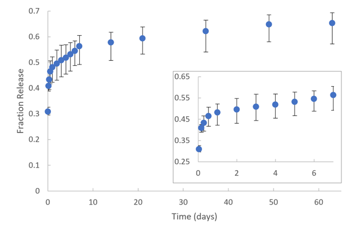
Figure 10. Fraction of 5FU released from standard microspheres with standard (unmodified) parameters: 2 wt% PVA in the outer water phase, PLA as the organic phase polymer, PBS with pH 7.4 as the release medium, and the release environment temperature of 37°C. The first datum point is at 1 hr. The bars represent the range of data, and the data points represent the mean of that range (n=3). The inset shows the release during the first 7 days.
The kinetics of drug release from PLA structures is highly dependent on the characteristics of the particles and is often described in prior literature as a sigmoidal shape due to polymer degradation [70, 71, 72]. This shape can be observed in Figure 10, with the caveat that our data showed a burst release in the first day (which is often observed in spherical PLA structures) replacing the normally observed increasing rate of release [73, 74]. The release kinetics can be described as follows: a burst release at 1 hr (the first point on Figure 10), a quick release through 1 day, a near linear release until 7 days, and a slower and constant rate of release through at least 9 weeks.
Comparison of drug release of 5FU from microspheres with varying concentrations of PVA in the outer emulsion during synthesis is presented in Figure 11. This plot includes the standard case of 2% PVA for comparison. The average sizes of these microspheres were 1.77 µm, 1.24 µm and 0.59 µm (standard) respectively (see Table 1).
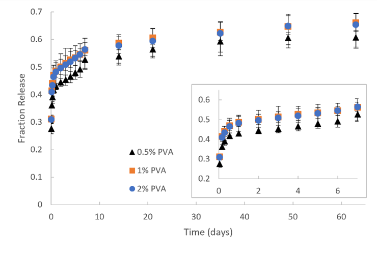
Figure 11. Fraction of 5FU released from microspheres with formulated 0.5, 1, and 2 wt% PVA in the outer water phase of the double emulsion. The organic phase polymer used during synthesis was PLA. The release medium was PBS with a pH of 7.4. The temperature of the release environment was 37°C. The first datum point is at 1 hr. The inset shows the release during the first 7 days.
In general, the pattern of release at other PVA concentrations (during the synthesis) was similar (see Figure 11): initially fast but slowing release rate in the first day, followed by one week of slower steady release, and finally weeks of constant release at an even slower rate. The PVA concentration of 0.5% was statistically different (p < 0.05) from the standard experiment only at time point 1 hr, while that of 1% was not statistically different (p > 0.05) from the standard experiment at all time points. Our particle size measurements show that the concentration of PVA in the outer water phase of the double emulsion microspheres was inversely related to the diameter of the resulting product microspheres. For example, Figure 6 shows that the samples prepared in this study with 0.5%, 1%, and 2% PVA had average diameters of 1.77, 1.24, and 0.70 µm, respectively. A decrease in diameter size suggests an increase in total surface area per mass of the spherical product. Furthermore, comparable literature shows that the particle size of PLA and PLGA structures is inversely related to the rate of drug release [70, 71, 75]. Therefore, we expected to see faster drug release in the samples prepared with a higher concentration of PVA. However, the release from these microspheres confirmed this hypothesis only for those formulated with 0.5% PVA and only at the first time point (1 hour).
Figure 12 compares the 5FU release at 37°C from microspheres prepared using either PLA or PLGA as the polymer. The largest difference occurs in the first day. The PLGA particles released about 60% of the 5FU in the first 24 hours, while the PLA particles released about 50% of its loading during the same time.
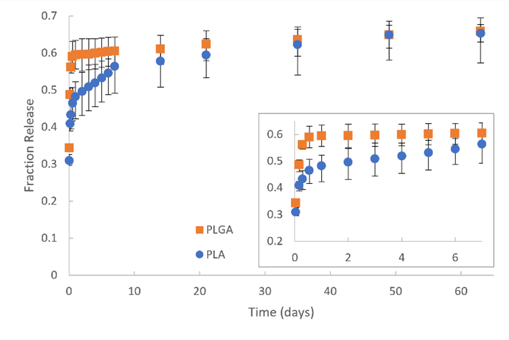
Figure 12. Fraction of 5FU released from microspheres with the organic phase polymer of PLA (blue dot) and PLGA (orange square). PVA concentration in the outer water phase of the double emulsion was 2%. The release environment was PBS at pH of 7.4 and 37°C. The first datum point is at 1 hr. The inset shows the release during the first 7 days.
Microspheres prepared using PLGA as the polymer released 5FU statistically faster (p < 0.05) than the standard experiment only up to 7 days (see Figure 12). This faster release is followed by a dramatically slower, sustained release as it converges with the release data from microspheres composed of PLA. The copolymer PLGA is usually less crystalline than PLA, purportedly leading to faster hydrolysis and subsequent more rapid polymer chain cleavage [72, 76]. Therefore, release behavior observed in Figure 12 is consistent with our expectation that PLA would degrade more slowly because it may be more crystalline than PLGA. Additionally, PLGA is less hydrophobic than PLA, so there may be more water diffusing into PLGA, which should produce faster degradation, as was observed.
Figure 13 compares the effect of the release medium on 5FU release, those being water at pH 7.0, and PBS at pH values of 5.4 and 7.4.
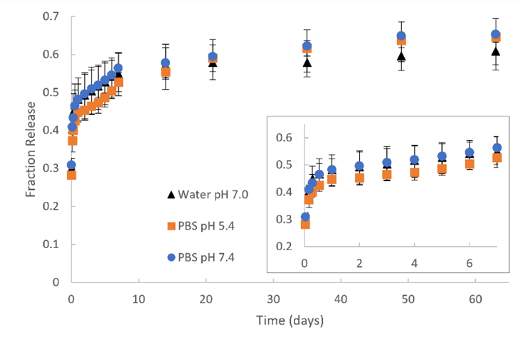
Figure 13. Fraction of 5FU released from microspheres into a release medium of distilled water at a pH of 7.0, PBS at a pH of 7.4, and PBS at a pH of 5.4. PVA concentration in the outer water phase of the double emulsion was 2%. The organic phase polymer used during synthesis was PLA. The release medium was PBS with a pH of 7.4. The temperature of the release environment was 37°C. The first datum point is at 1 hr. The inset shows the release during the first 7 days.
The 5FU release from these microspheres into various media (see Figure 13) is not statistically different at any time point. After two weeks, 5FU release into PBS regardless of the pH appears to be faster than 5FU release into water, but again the difference is not statistically significant in these experiments. We chose pH levels of 7.4 and 5.4 to simulate the typical pH level in the human body and tumor environments respectively. Additionally, we chose water at pH 7.0 as a comparison to PBS at pH 7.4 to determine if there is any significant impact of the high concentration of salts found in PBS and found in the body. Literature reports that a strongly acidic environment accelerates polymer degradation and that a slightly acidic pH environment (4-6 pH) decelerates polymer degradation compared to a neutral pH environment [31, 76, 77, 78]. Therefore, we expected to observe a slightly slower 5FU release in the pH 5.4 medium. Yet, the microspheres produced for this study did not release 5FU at a statistically different rate in the media evaluated. The slower release observed beyond 15 days into water than into PBS at pH values higher and lower than water pH (7.0) suggests that perhaps the ionic strength or ionic content may also contribute to the release of drug at long times.
The effect of the release environment temperature (21°C or 37°C) on 5FU release is compared in Figure 14.
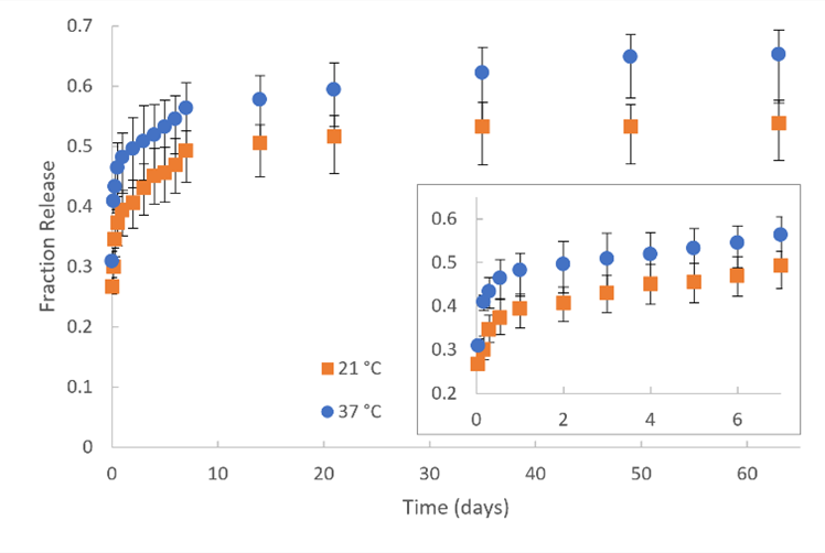
Figure 14. Fraction of 5FU released from microspheres with a release environment at the temperatures of 21°C and 37°C. PVA concentration in the outer water phase of the double emulsion was 2%. The organic phase polymer used during synthesis was PLA. The release medium was PBS with a pH of 7.4. The first datum point is at 1 hr. The inset shows the release during the first 7 days.
Drug release data regarding temperature (see Figure 14) indicate that 5FU release at 21 °C is statistically slower from the standard experiment at all time points (p < 0.05). 5FU release in an environment of 37 °C is significantly and consistently higher than that of 21 °C for the entire duration of the release. We chose the temperature of 37°C to simulate the typical temperature of the human body. The temperature of 21°C was selected because the reduction in hydrolysis of about 10%, calculated from data reported by Iñiguez-Franco et al. [79], corresponded to a temperature difference of 16°C and was expected to slow the release by a similar amount (10%), which we postulated would produce a measurable difference in our experiments. We found this to be the case. Additionally, literature reports of the effect of temperature on PLA degradation [78, 80, 81] is consistent with our observation of increased release.
3.2.7 Time-Concentration Comparison
The half-life of 5FU in the human body is 8-20 minutes in plasma circulation [82], which suggests that 5FU released into the blood would not remain in the body for the time scales of these release experiments. However, any 5FU taken up by cells in contact with or in proximity of the microspheres might be retained for hours. Whether the difference in release from PLA versus PLGA has a meaningful difference in a clinical situation is usually answered by AUC (area under the curve) analysis. In this research, the conventional AUC for drug in plasma was not measured in animal models. However, an analog to AUC can be defined for our experiments as the area under the fractional cumulative release curve for the first 24 hours. We call this the “relative AUC” and posit that this value would be proportional to the time-concentration exposure of nearby cells. In comparing the standard release from PLA with release from PLGA, the relative AUCs values were 10.2 and 12.8 hours, respectively, and were found to be statistically different (p = 0.003). This suggests that the drug exposure would be much higher with PLGA particles. We did similar comparisons for the other conditions of our study. The only other statistically different comparison is with the release at 21°C, at which the relative AUC was 11.1 hours (p = 0.002). All other comparisons of the relative AUC were found not to be statistically different.
3.2.8 Observation of Particle Degradation
Degradation of the microspheres was not yet completed in 63 days, likely precluding total release of the last fraction of sequestered 5FU. Figure 15 presents SEM images of PLA microspheres after 63 days of release into PBS pH 7.4 at 37°C, revealing a textured surface (no longer a polished, smooth surface), but without any evidence of cracks, flaking or crumbling. Supplementary materials (Figure S1) shows a micrograph of PLGA microspheres after 63 days of release which also have a rough surface compared to non-degraded particles.
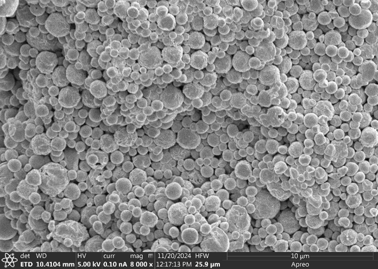
Figure 15. SEM image of PLA microspheres after 63 days of degradation in PBS pH 7.4 at 37°C (standard release case).
3.2.9 Consistent Absorbance Spectrum
Comparison of the absorbance spectra of 5FU before and after release showed no significant differences or new peaks, suggesting that 5FU does not undergo any discernable chemical change during the encapsulation and release process. See Figure S2 in supplementary materials.
3.2.10 Potency of 5FU
The MIC of 5FU toward S. aureus before and after release was found to be 16 µg/mL, suggesting that the formulation and release process does not change the drug potency. As mentioned, 5FU is cytotoxic to bacteria. For example, wild-type S. aureus is sensitive and susceptible to 5FU [83]. Our observations that neither the MIC nor the UV absorption spectrum of 5FU was altered by the encapsulation and release process supports our hypothesis that the release drug remains unchanged upon release.
4. DISCUSSION
4.1 SPIONs in Polymer Microspheres
Successful synthesis of these microspheres carries the potential to be spatially manipulated via the magnetic interaction between the entrapped SPIONs and a therapeutic magnetic field, such as a Halbach array [47]. These SPIONs are hydrophobic and thus are easily distributed throughout the polymer matrix of the microspheres. This non-aggregated distribution of SPIONs confers superparamagnetic properties to the microspheres, confirmed by FC/ZFC behavior and near-zero coercivity, therefore eliminating magnetic agglomeration between the microspheres when they are not in the presence of the magnetic array. The application of a Halbach array in tandem with these particles therefore presents the potential to precisely accumulate the microspheres in the capillaries of a tumor to release 5FU directly to the tumor microenvironment by preferential accumulation in or near the tumor tissue.
The use of double emulsion technology to produce microspheres containing drug is not a novel concept. However, the formulation presented in this study represents an optimized procedure to produce unique particles not heretofore reported. To our knowledge, this is the first report of spherical, non-aggregated solid PLA particles with a homogeneous distribution of hydrophobic SPIONs and hydrophilic anticancer drug with an average particle diameter less than 1 micron. Many studies have produced nonmagnetic w/o/w double emulsion PLA or PLGA microspheres loaded with drug with an average diameter above 10 µm [52, 53, 54, 55, 56, 57, 58]. State-of-the-art microspheres of a similar polyester composition have been reported with average diameters between 10 µm and 0.8 µm, but these were not magnetic [59, 60]. Delie et al. reports double emulsion formulations using PLA as the polymer and 25-mer-FITC-labelled oligonucleotide as the payload that have yielded particles as small as 0.45 µm in average diameter, but these particles were reportedly aspherical, aggregated, invaginated, and nonmagnetic [22]. Some of the smallest polyester particles loaded with a payload for biomedical application were reported by a classic study which produced porous PLGA particles loaded with proteins at an average diameter below 0.1 µm. However, these particles are not solid spheres, are not magnetic, and were prepared by phase inversion rather than double emulsion synthesis.
Many studies have produced magnetic microspheres with a microscopic Fe3O4 core and a PLA shell rather than a PLA microsphere with dispersed Fe3O4 nanoparticles and drug distributed throughout the structure. One such study produced microspheres over 50 µm in diameter with an Fe3O4 core and a PLA shell [62]. Experiments have produced microspheres with an Fe3O4 core and PLA shell under 2 µm in diameter using the complex method of electrohydrodynamic atomization [61]. Two studies produce nanospheres under 10 nm in diameter encapsulating hydrophobic anticancer drugs (curcumin and doxorubicin) within the PLA-PEG shell which coats the Fe3O4 core for controlled release and hyperthermic cancer therapy [64, 84].
A proof-of-concept study from which this work builds produced iron oxide by co-precipitation for dispersion in similar microspheres, but without determining its phase composition or magnetic purity [17]. This prior work produced monodisperse ~9 nm SPIONs synthesized via thermal decomposition and characterized by TEM and magnetometry. The reduction in blocking temperature upon encapsulation confirms that SPIONs remain dispersed within the polymer matrix rather than forming magnetic aggregates. Yet despite these favorable properties, the SEM images of these particles did not show distinct spherical particles. Individual non-aggregated particles are essential for capillary delivery so the capillary is not plugged or blocked.
Achieving the reported morphological and functional characteristics required extensive optimization of double-emulsion parameters, including polymer carrier composition, surfactant concentration, phase ratios, emulsification method, and drying conditions. The sensitivity of particle morphology to these variables underscores the nontrivial nature of the optimized process presented in this work. The resulting reproducible synthesis procedure provides a reliable foundation for future translational work, enabling the production of microspheres with consistent size, shape, and magnetic performance.
4.2 Efficient Drug Loading
Comparable literature reports drug loading efficiencies around 95% for double emulsion PLA microspheres containing bacterial antigens or proteins and without SPIONs [59, 60]. Our reported particles are comparable to this loading efficiency. A challenge in reporting drug loading efficiency with microspheres loaded with hydrophilic drug and composed of PLA is that polymer degradation begins as soon as the solid microspheres encounter the water or PBS medium and continues for months or more. Thus, measuring the loading by cumulative release at very long times can be problematic. Therefore, in our study encapsulation efficiency was determined by measuring the mass of 5FU in the aqueous phase decanted from the partially dried emulsion, rather than from the suspended solid product at long times. The remarkably high drug loading efficiency calculated herein could be attributed to the insolubility of 5FU in MeCl2, which apparently remained inside the aqueous phase when the water-in-organic emulsion was subsequently emulsified in the outer aqueous phase.
4.3 Drug Release
The long-term release behavior observed in this work provides a more extensive view of 5FU diffusion from PLA and PLGA microspheres than has been previously reported in the literature, revealing polymer dependent differences not apparent in short-term analyses. Because the microspheres produced in this work are engineered for higher uniform spherical morphology and narrow size distribution, variability arising from differences in particle geometry is minimized, allowing for the underlying effects of polymer chemistry on release rate to be more clearly observed. Particularly, PLA and PLGA exhibit distinct release rates and cumulative release profiles under identical conditions, demonstrating the potential for controlled modulation of release behavior based on copolymer composition.
Interestingly, about 40% of the 5FU was released in the first day and yet spherical PLA particle remained at 63 days and beyond. Thus, the rate of release greatly exceeded and preceded the polymer degradation. The drug release profiles from the particles presented in this study are similar to the release of hydrophobic drug from hydrophobic polymer systems over short times in that there is an initial burst release; however, these traditional release profiles are typically followed by another burst release likely caused by autoacceleration of polymer degradation [52, 57, 58, 85]. Yet the release in this study is more comparable to that of hydrophilic drug released from hydrophobic polymer systems in which there is an initial burst release followed by a slower, sustained release [34, 53]. Furthermore, the particles produced in this study are unique in that they have an average diameter under 2 µm, contain SPIONs distributed throughout the polymer structure, and are distinctly spherical in shape; however, there are no other reports that such changes in morphology affect the release behavior in any significant way when compared to hydrophilic drug released from hydrophobic polymer systems.
In clinical applications, the microspheres presented in this study are small enough to flow through the smallest capillaries of the human body. Additionally, small spherical particles have advantages over angular particles inside the circulatory system. Similar studies which synthesized distinctly spherical microparticles or nanoparticles containing a solid magnetic core likely produce particles with more capacity for magnetic interaction at the expense of higher drug loading. The microspheres produced in this study were observed to exhibit superparamagnetic properties. For the application of a Halbach array, the data presented by this study suggests the potential for a patient to rest in a Halbach array for less than 24 hours to achieve satisfactory particle collection and drug delivery at the site of a tumor.
The diffusion of water into the PLA apparently expanded its network and allowed sufficient solubilization of the 5FU that more than half was released within a day, and yet a given particle showed only a roughened surface after 63 days and not a fractured, crumbling, nor porous skeleton of the particle. We assume that the initial chain cleavage does not release large amounts of lactic acid since hydrolysis occurs anywhere on the polymer backbone, and not solely at the chain ends. Eventually PLA (and PLGA) oligomers will be freed from the particle surface and interior, and these oligomers will continue to circulate as they degrade into lactic (and glycolic) acid, are filtered through the kidneys into urine, or are captured by cell endocytosis or phagocytosis. Individual oligomers are not expected to clog capillaries in the organs of the body. PLA oligomers that are released from capillary walls in magnetic collection sites or were never trapped will continue to circulate as they degrade into products naturally eliminated by the body.
5. CONCLUSION
This work demonstrates the successful synthesis of sub-2-µm PLA and PLGA microspheres loaded with 5FU and 9 nm SPIONs, formulated using a double emulsion preparation method followed by slow solvent evaporation. From 5 mL of double emulsion, approximately 12 mg of dried product was recovered after drying for 96 total hours. These product particles had an average diameter as low as 0.697 µm with a polydispersity index of 0.501, were distinctly smooth and spherical in shape, and exhibited stable superparamagnetic properties, supporting their use in magnetic drug targeting applications. Because the microspheres were morphologically uniform and non-aggregated, variability in diffusion path lengths was minimized, elucidating polymer-dependent differences in release rate. The chemical integrity and biological activity of the released 5FU further validate the suitability of this platform for potential therapeutic use.
This study also evaluated controlled release of 5FU from PLA and PLGA microspheres of 0.7 to 2 µm in diameter loaded with SPIONs and 5FU at 93 µg and 22.48 mg respectively per gram of dried microspheres. Under standard conditions, the base-formulation microspheres (PVA@2.0) released 5FU with the following general characteristics: 1) a burst release during the first hr, 2) a fast release which continually slows through the first day, 3) a near constant release rate until day 7, 4) a slower but still fairly constant rate of release through the 63 days of this experiment. Cumulative release exceeded 65% in 63 days. Although PVA concentration strongly influenced particle size, its impact on 5FU fractional release was comparatively modest. Microspheres composed of PLGA released 5FU significantly faster than those composed of PLA. The release of 5FU was not statistically different in a slightly acidic environment of pH 5.4 or in water of pH 7.0. 5FU release at 37°C was statistically greater than release at 21°C for the entire duration of the study period. 5FU undergoes no apparent changes in chemical structure or potency throughout the formulation and release process.
The sub-2-µm microsphere size achieved in this work is ideal for application in intravascular magnetic cancer drug delivery to tumors because they are sufficiently small to be pulled onto the capillary walls within a tumor by an external magnetic field and release approximately 40% of their anticancer payload in the first 24 hours. Magnetic systems using Halbach arrays can further enhance targeting by generating high-gradient fields capable of pushing the microspheres to defined intravascular locations [47]. In this context, the demonstrated combination of uniform morphology, superparamagnetism, and long-term controlled release represents a meaningful advancement over prior studies of magnetic PLGA or PLA carriers. The release data reported in this study will support future predictive modeling, optimization, and translational development of magnetically guided chemotherapy systems.
ACKNOWLEDGEMENTS
We thank the Brigham Young University Harvey Fetcher Professorship (WGP and TPG), the Simmons Center for Cancer Research (TPG) and an Interdisciplinary Research grant from Brigham Young University (KC, RGH and RG) for funding this research; we also acknowledge the BYU electron microscopy facility for providing access to the equipment and expertise that allowed this project to be performed.
FUNDING:
This work was supported by the Simmons Center for Cancer Research at Brigham Young University.
AUTHOR CONTRIBUTIONS:
Tyler P. Green: Conceptualization, Investigation, Methodology, Writing – original draft, review, and editing. Ashley J. Spencer: Investigation, Writing – review and editing. Roger G. Harrison: Conceptualization, Writing – review and editing. Rajendra Gautam: Investigation, Writing – review and editing. Karine Chesnel: Conceptualization, Writing – review and editing. William G. Pitt: Conceptualization, Methodology, Funding, Project Administration, Writing – review and editing.
CONFLICT OF INTEREST:
The authors declare no conflict of interest.
Data availability statement:
Data supporting these findings are available at https://scholarsarchive.byu.edu/data/95/.
Supplementary materials:
The supplementary materials are available at https://scholarsarchive.byu.edu/data/95/.
REFERENCES


Tyler P. Green, Ashley J. Spencer, Roger G. Harrison, Rajendra P. Gautam, Karine Chesnel, William G. Pitt, Biodegradable PLA Double Emulsion Microspheres for Magnetically Guided Drug Delivery of 5-Fluorouracil, Int. J. of Pharm. Sci., 2026, Vol 4, Issue 1, 3191-3220. https://doi.org/10.5281/zenodo.18397126
 10.5281/zenodo.18397126