

We use cookies to ensure our website works properly and to personalise your experience. Cookies policy
1PG Student Department of Pharmaceutics, Sarojini Naidu Vanita Pharmacy Maha Vidyalaya.
2Assistant Professor Department of Pharmaceutics, Sarojini Naidu Vanita Pharmacy Maha Vidyalaya.
The present study focused on the formulation and evaluation of ciprofloxacin hydrochloride-loaded niosomal gel for ocular drug delivery. Niosomes were prepared by the thin-film hydration method using different non-ionic surfactants, including Tween 20, Tween 60, Tween 80, and Span 60, along with cholesterol. Nine formulations (F1–F9) were developed and evaluated to identify the formulation with better drug content, entrapment efficiency, and drug-release characteristics. Based on the evaluation results, F6 was selected as the optimized formulation. It showed a drug content of 73.36 ± 0.05%, an entrapment efficiency of 62.57 ± 0.02%, and cumulative drug release of 96.55 ± 0.03% at 24 hours. Further characterization of F6 showed a particle size of 159.4 nm, a polydispersity index (PDI) of 0.293, and a zeta potential of ?24.3 mV. FTIR studies were also carried out on the pure drug, physical mixture, and optimized formulation to examine possible interactions between the drug and the formulation ingredients. The optimized niosomal suspension was then incorporated into a Carbopol-based gel. The prepared gel was examined for its appearance, homogeneity, pH, viscosity, spreadability, and in-vitro drug release. The gel had a smooth and homogeneous appearance, a pH of 7.3 ± 0.5, a viscosity of 155 ± 10 cP, and a spreadability of 6.67 g·cm/s. During comparative in-vitro release testing, the niosomal gel released 80.65% of the drug at 24 hours, whereas the optimized niosomal suspension released 96.55%. Overall, the results suggest that incorporating the optimized niosomes into a gel can modify the drug-release pattern. Further studies are needed to establish its suitability for ocular use.
Anatomy And Physiology Of The Eye
The eye is a highly specialized sensory organ consisting of several anatomical structures that work together to provide vision and protect the internal ocular environment. Major structures include the cornea, conjunctiva, sclera, iris, anterior chamber, lens, vitreous body, retina, choroid, and optic nerve. The cornea forms the transparent anterior portion of the eye and represents an important pathway and barrier for topically administered drugs [1].
The cornea consists of five principal layers: epithelium, Bowman's membrane, stroma, Descemet's membrane, and endothelium. The corneal epithelium contains tight junctions and provides substantial resistance to hydrophilic substances, whereas the stromal layer is relatively hydrophilic and can restrict highly lipophilic compounds. These differences in physicochemical characteristics influence the penetration and distribution of drugs administered to the ocular surface [2].
The tear film, conjunctiva, aqueous humor, and ocular blood circulation also influence drug absorption and elimination. Continuous tear secretion, blinking, and drainage through the nasolacrimal duct contribute to rapid removal of topical formulations from the ocular surface [3].
NIOSOMES
Niosomes are closed bilayer vesicles composed mainly of non-ionic surfactants, cholesterol, and an aqueous phase, formed by the self-assembly of amphiphilic molecules [4].
The unique structure of niosomes consists of a hydrophilic aqueous core surrounded by a hydrophobic bilayer, allowing incorporation of both water-soluble and lipid-soluble drugs. This property makes niosomes suitable carriers for antibiotics, anticancer drugs, proteins, peptides, and other therapeutic molecules [5].
Niosomes are classified based on vesicle structure, composition, and preparation approaches, including conventional niosomes, proniosomes, and advanced surface-modified niosomal systems. Recent advances have expanded their role in controlled and targeted drug delivery applications[11].
Compared with liposomes, niosomes offer advantages such as better chemical stability, reduced susceptibility to oxidation, lower production cost, and greater flexibility in formulation design due to the use of non-ionic surfactants [6,7].
The performance of niosomes depends on various formulation factors, including surfactant type, hydrophilic–lipophilic balance (HLB), phase transition temperature, cholesterol concentration, and preparation method. These variables influence vesicle size, membrane rigidity, entrapment efficiency, and drug release behaviour [4].
Niosomes have shown significant potential in ocular drug delivery by improving drug retention, enhancing corneal penetration, and providing prolonged release of ophthalmic drugs [10].
They have also been investigated for transdermal delivery, where their flexible vesicular structure improves penetration through the skin barrier and enhances drug deposition [8].
In cancer therapy, niosomes provide an effective platform for improving the delivery of anticancer agents by enhancing drug accumulation at target sites, reducing systemic toxicity, and providing controlled release [9].
Although niosomes demonstrate remarkable potential, challenges such as physical instability, drug leakage, scale-up difficulties, batch-to-batch variation, and regulatory issues remain major barriers for clinical translation [12].
Therefore, continuous research is required to optimize formulation strategies, improve stability, and establish standardized manufacturing approaches for successful clinical application of niosomal systems.
Molecular Design Of Niosomes
The molecular design of niosomes plays a crucial role in determining their physicochemical characteristics, stability, drug encapsulation efficiency, and biological performance. The formation and properties of niosomes are mainly influenced by the type of non-ionic surfactant, cholesterol content, surfactant-to-cholesterol ratio, surface charge, and preparation conditions [4,6].
Niosomes are formed through the self-assembly of amphiphilic non-ionic surfactant molecules in an aqueous environment. The hydrophilic head groups orient toward the aqueous phase, while the hydrophobic tails associate inward to form a bilayer structure [4].
The incorporation of cholesterol between surfactant molecules improves membrane rigidity, reduces permeability, and enhances vesicle stability [5].
The selection of surfactant is one of the most important molecular design parameters. Non-ionic surfactants such as Span and Tween series are commonly used because they provide good biocompatibility and flexibility in controlling vesicle characteristics [6].
Surfactants with suitable hydrophilic–lipophilic balance (HLB) values promote stable bilayer formation and influence drug entrapment and release behaviour [13].
The molecular packing of surfactants, critical packing parameter, and interaction between surfactant and cholesterol determine vesicle morphology, membrane fluidity, and drug retention properties. Optimization of these parameters is essential for developing stable niosomal formulations [19].
The phase transition temperature (Tc) of the surfactant affects membrane fluidity and vesicle stability. Surfactants with higher phase transition temperatures generally produce more rigid vesicles with slower drug release, whereas lower transition temperature surfactants form more flexible membranes [4,6].
Cholesterol acts as a membrane stabilizer by filling gaps between surfactant molecules. It improves mechanical strength, prevents leakage of encapsulated drug, and regulates membrane permeability. However, excessive cholesterol concentration may reduce drug entrapment by decreasing available space within the bilayer [5,7].
Surface charge modification is another important strategy in niosomal design. Charge-inducing agents such as dicetyl phosphate (negative charge) or stearylamine (positive charge) can be incorporated to increase electrostatic repulsion between vesicles and reduce aggregation [7].
Advanced molecular designs include surface-modified and targeted niosomes. Attachment of ligands, polymers, antibodies, or other targeting molecules can enhance tissue-specific delivery and improve therapeutic outcomes [16,17]
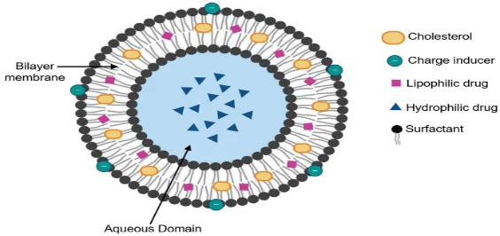
Figure 1: Structure of Noisome
Components Of Niosomes
Niosomes are primarily composed of non-ionic surfactants, cholesterol, charge-inducing agents, and an aqueous hydration medium. Each component contributes to vesicle formation, stability, drug loading, and release characteristics.
Table 1: Components of Niosomes
| S.no | Component | Examples | Function |
|---|---|---|---|
| 1 | Non-ionic surfactants | Span 20, Span 60, Span 80, Tween 20, Tween 60, Tween 80 | Formation of bilayer structure and vesicle formation |
| 2 | Cholesterol | Cholesterol | Improves membrane rigidity and stability |
| 3 | Charge-inducing agents | Diacetyl phosphate, Stearylamine |
Enhances zeta potential & prevents aggregation |
| 4 | Hydration medium | Water, PBS | Provides aqueous environment for vesicle formation |
| 5 | Targeting molecules | Ligands, antibodies, polymers | Improves site-specific drug delivery |
Preparation Methods Of Niosomes
The preparation method strongly influences the physicochemical characteristics of niosomes, including vesicle size, morphology, drug entrapment efficiency, lamellarity, and stability. Selection of an appropriate preparation technique depends on the nature of the drug, surfactant properties, desired vesicle size, and intended route of administration [4,5].
In the preparation of niosomes, formulation optimization using statistical experimental designs such as Box–Behnken design and response surface methodology helps evaluate the influence of formulation variables, including surfactant concentration, cholesterol ratio, and processing conditions, on critical quality attributes [15].
Several conventional and advanced techniques have been developed for niosome preparation, including thin-film hydration, reverse-phase evaporation, ether injection, sonication, microfluidization, and proniosome-based methods [4].
Characterization Of Niosomes
Characterization of niosomes is essential to evaluate their physicochemical properties, stability, drug-loading capacity, and suitability as vesicular drug delivery systems. The major characterization parameters include particle size, polydispersity index (PDI), zeta potential, morphology, entrapment efficiency, vesicle structure, drug–excipient compatibility, in vitro drug release, and stability studies. These parameters influence drug delivery performance, bioavailability, and therapeutic efficacy of niosomal formulations [23].
Particle size and polydispersity index are critical quality attributes because they influence stability, cellular uptake, tissue penetration, and drug release behaviour [20].
Optimization of niosomal formulations requires systematic evaluation of particle size, polydispersity index, entrapment efficiency, and drug release behaviour to establish the relationship between formulation variables and performance characteristics [4].
Table 2: Characterization of Niosomes
| S.no | Parameter | Instrument | Significance |
|---|---|---|---|
| 1 | Vesicle size | Dynamic light scattering | Determines particle size and predicts drug delivery behaviour |
| 2 | Size distribution | PDI measurement by DLS | Evaluates uniformity of vesicle population |
| 3 | Zeta potential | Zeta potential analyser | Determines surface charge and physical stability |
| 4 | Morphology | TEM, SEM | Confirms vesicle shape, structure, and integrity |
| 5 | Entrapment efficiency | Centrifugation, dialysis, ultrafiltration | Determines amount of drug incorporated inside vesicles |
| 6 | Drug loading | UV spectroscopy/HPLC | Measures drug content in formulation |
| 7 | Drug release | Dialysis membrane/Franz diffusion cell | Evaluates sustained and controlled release |
| 8 | Release kinetics | Mathematical modelling | Determines mechanism of drug release |
| 9 | FTIR analysis | Fourier transform infrared Spectroscopy | Evaluates drug-excipient compatibility |
| 10 | Stability study | Storage under different conditions | Determines shelf life and formulation stability |
Therapeutic Applications Of Niosomes
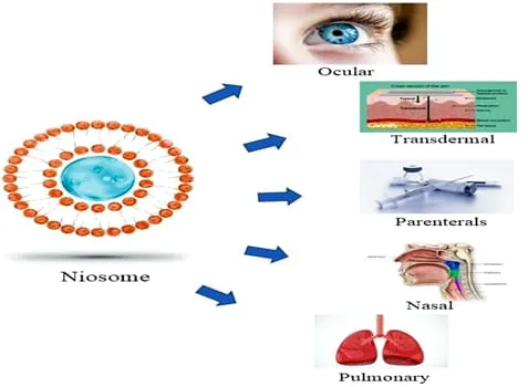
Figure 2: Applications of Niosomes
a. Ocular Drug Delivery
Ocular drug delivery is challenging because of physiological barriers such as tear dilution, blinking, nasolacrimal drainage, and limited corneal permeability, which result in poor drug bioavailability from conventional eye drops [10].
Niosomes provide an effective approach for ocular delivery by increasing precorneal residence time, improving drug penetration, and enabling sustained release of ophthalmic drugs [10,14].
b. Transdermal Drug Delivery
The stratum corneum acts as a major barrier limiting penetration of many therapeutic agents through the skin. Niosomes improve transdermal delivery by enhancing drug partitioning into skin layers and acting as penetration-enhancing vesicular carriers [8,21].
c. Topical Drug Delivery
Topical delivery aims to achieve localized drug action while minimizing systemic exposure. Niosomes incorporated into gels and creams improve drug deposition within skin layers and provide prolonged drug release [5,8].
d. Oral Drug Delivery
Oral administration remains the preferred route due to convenience and patient compliance; however, poor solubility, enzymatic degradation, and gastrointestinal barriers limit the bioavailability of many drugs [5].
Niosomes can improve oral drug delivery by protecting drugs from degradation, enhancing intestinal absorption, and providing sustained release [4,5].
e. Cancer Drug Delivery
Cancer therapy is one of the most explored applications of niosomes because conventional chemotherapy often suffers from poor selectivity and systemic toxicity [9,18].
f. Vaccine Delivery
Niosomes have gained interest as vaccine delivery carriers because they can protect antigens from degradation and enhance immune responses [5].
g. Gene and Biological Molecule Delivery
Recent research has expanded niosomal applications toward delivery of biological molecules such as DNA, RNA, peptides, and proteins [5,17].
Niosomes protect genetic materials from enzymatic degradation and improve cellular uptake through vesicular transport mechanisms [17,22].
Niosomal Gel
Niosomal gel is a semisolid formulation in which drug-loaded niosomes are incorporated into a suitable gel matrix. This approach combines the vesicular characteristics of niosomes with the increased viscosity and residence characteristics of a gel. The niosomal vesicles can act as drug carriers, while the gel matrix can influence residence time, drug diffusion, and release characteristics [24].
Incorporation of niosomes into a gel base may provide a more structured formulation and can modify the rate at which the drug diffuses from the vesicles and subsequently through the gel matrix. The combined system may therefore provide prolonged drug release compared with a conventional drug-loaded gel, depending on the properties of both the vesicles and the gel base[25].
For ocular application, a niosomal gel should possess suitable pH, viscosity, homogeneity, spreadability, drug content, physical stability, and drug-release characteristics. The formulation components should also be selected with consideration of ocular tolerability and compatibility with the intended site of administration [26].
MATERIALS AND METHODS
List Of Materials And Equipments Used In Preparation Of Ciprofloxacin HCl Niosomal Gel:
Table 3: List of Materials
| S.No | Materials | Purpose | Company Name |
|---|---|---|---|
| 1 | Ciprofloxacin HCl | Antibacterial drug | DCL (Drug Control Laboratory) Hyderabad, Govt Of Telangana. |
| 2 | Tween 20 | Non-ionic surfactant | SDFCL (s d fine-chem limited), Mumbai, India. |
| 3 | Tween 60 | Non-ionic surfactant | SDFCL (s d fine-chem limited), Mumbai, India. |
| 4 | Tween 80 | Non-ionic surfactant | Thomas Baker, pvt Limited, Mumbai |
| 5 | Span 60 | Non-ionic surfactant | SDFCL (s d fine-chem limited), Mumbai, India. |
| 6 | Cholesterol | Membrane stabilizer | Molychem, Mumbai, India |
| 7 | Carbopol 934 | Gelling agent | SDFCL (s d fine-chem limited), Mumbai, India. |
| 8 | Triethanolamine | Neutralizing and PH-adjusting agent | SDFCL (s d fine-chem limited), Mumbai, India. |
| 9 | Propylene Glycol | Co-solvent | Molychem, Mumbai, India. |
| 10 | Chloroform | Organic solvent | Sisco Research Laboratories Pvt. Ltd, Mumbai, India. |
| 11 | Methanol | Organic solvent | Rankem, Avantor Performance Materials India Pvt. Ltd. Maharashtra, India. |
| 12 | Saline Water | Hydration medium | Aculife Manufactured in India by: Aculife Healthcare Pvt. Ltd. Gujarat, India. |
METHODOLOGY
Pre-Formulation Studies:
These investigations are carried out prior to formulation in order to confirm that the drug material's physical and chemical characteristics match those of the excipients.
i. Organoleptic properties: A variety of organoleptic characteristics, such as the drug substance's appearance, color, and odor, are noted.
ii. Solubility studies: Drug dissolution in various solvents, such as water, saline water, phosphate buffer saline pH-7.4, methanol, ethanol, and chloroform, is known as a solubility study. Additionally, we are aware that ciprofloxacin HCl is a BCS III medication (high solubility and limited permeability).
Preparation Of Standard Curve Of Ciprofloxacin HCl In Phosphate Buffer Saline 7.4
Buffer Preparation:
8.0 g of sodium chloride (NaCl), 0.2 g of potassium chloride (KCl), 1.44 g of disodium hydrogen phosphate (Na₂HPO₄), and 0.24 g of potassium dihydrogen phosphate (KH₂PO₄) were dissolved in around 800 mL of distilled water to create PBS with pH 7.4. And stirred continuously until it dissolves completely. The solution’s pH was tuned to 7.4 ± 0.05 by using dilute Hydrochloric acid and it was assessed calibrated digital pH meter. The total volume was adjusted to 1000 mL using distilled water and the solution was mixed thoroughly. The finalized PBS pH 7.4 was utilized as the dissolution medium for creating standard solutions and for later drug release investigations.
Table 4: Composition of PBS 7.4
| S.no | Component | Quantity for 1L |
|---|---|---|
| 1 | NaCl | 8.0g |
| 2 | KCl | 0.2g |
| 3 | Na2HPO4 | 1.44g |
| 4 | KH2PO4 | 0.24g |
| 5 | Distilled water | q.s to 1000ml |
Determination of λmax of Ciprofloxacin HCl:
The wavelength of maximum absorption (λmax) of Ciprofloxacin Hydrochloride was estimated by UV–Visible spectrophotometric analysis. 10 µg/mL standard solution of Ciprofloxacin HCl was prepared in phosphate buffered saline (PBS)7.4. UV-Visible spectrophotometer is employed to analyze the prepared solution using PBS 7.4 as blank within the wavelength range of 200–400 nm. Ciprofloxacin.HCl exhibited its maximum absorbance at 272 nm.
Standard graph of Ciprofloxacin HCl:
Using PBS 7.4 solvent a standard stock solution of Ciprofloxacin HCl was prepared. Prepare a set of Std solutions with concentraions of 2,4,6,8,10,12 and 14 µg/ml. The UV–Visible spectrophotometric analysis was conducted on the prepared standard solutions at a wavelength of 272 nm (λmax) against PBS 7.4 as blank, and absorbance values of each std solutions were noted. Construction of standard calibration curve by taking the concentrations of drug against absorbance. For the evaluation of the prepared niosomal formulations this developed calibration curve is used.
FORMULATION DEVELOPMENT OF CIPROFLOXACIN HCl NIOSOMAL GEL:
Preparation Of Ciprofloxacin HCl Niosomes:
Thin film hydration technique is used for developing the Ciprofloxacin HCl niosomal suspension loaded with non-ionic surfactants and cholesterol.
Step-1 Weighing of ingredients: The required quantities of Ciprofloxacin Hydrochloride, Non-ionic surfactant and cholesterol were accurately weighed according to the composition of each formulation.
Step-2 Preparation of organic phase: The weighed drug, surfactants, and cholesterol were transferred into a suitable round-bottom flask. A mixture of chloroform and methanol in a 2:1 ratio was added as the organic solvent system. The contents were mixed adequately to obtain a homogeneous solution.
Step-4 Formation of thin film: Fix the flask to the rotary evaporator, set the water bath temperature at 45°C and 100 rpm, evaporate the solvent under reduced pressure. Rotate for 30-40 mints until the thin dry lipid film is produced on the inner surface of the flask.
Step-5 Removal of residual solvent: Removal of residual organic solvent present in the flask to get completely dry film by keeping it under vacuum for 30 mints (stop heating & rotation, only keep the flask connected to the vacuum pump)
Step-6 Preparation of drug solution: Specified quantity of pure Ciprofloxacin HCl was dissolved in 20ml of saline water and warm the solution at 60°C.
Step-7 Hydration: The dried lipid film was hydrated using warmed drug solution prepared by normal saline water. The temperature of water bath was set to 45°C and 100 rpm for 30 to 45 minutes with continuous rotation to facilitate detachment of lipid film from the walls of the flask and form milky niosomal suspension containing probably large vesicles.
Step-8 Probe sonication: The developed niosomal suspension was subjected to probe sonication in pulse mode for 5 minutes to reduce the vesicle size and improve the uniformity. The heat generated was reduced by using cooling bath.
Step-9 Maturation: For complete vesicle formation store the suspension at 4°C overnight and observe under digital microscope.
Step-11 Storage: The prepared niosomal formulations were stored in a clean labelled container in refrigerator until further characterization and evaluation.
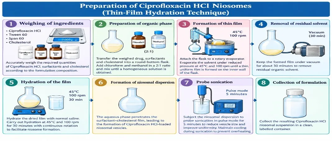
Figure 3: Pictorial Representation of Preparation of Ciprofloxacin HCl Niosomes
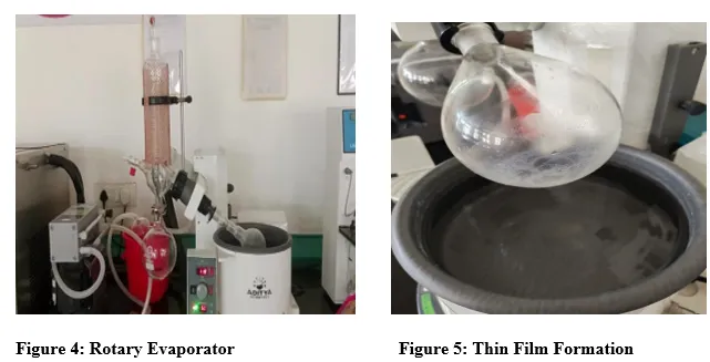
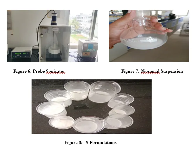
Table 5: Formulation of 20ml Ciprofloxacin HCl Niosomal Suspension
| S.no | Ingredients | F1 | F2 | F3 | F4 | F5 | F6 | F7 | F8 | F9 |
|---|---|---|---|---|---|---|---|---|---|---|
| 1 | Drug | 40mg | 40mg | 40mg | 40mg | 40mg | 40mg | 40mg | 40mg | 40mg |
| 2 | Cholesterol | 40mg | 40mg | 40mg | 40mg | 40mg | 40mg | 40mg | 40mg | 40mg |
| 3 | Tween 20 | 40mg | - | - | - | - | - | - | - | - |
| 4 | Tween 60 | - | 40mg | - | - | 30mg | 20mg | 10mg | 24mg | 16mg |
| 5 | Tween 80 | - | - | 40mg | - | - | - | - | - | - |
| 6 | Span 60 | - | - | - | 40mg | 10mg | 20mg | 30mg | 16mg | 24mg |
Evaluation Of Ciprofloxacin HCl Niosomal Suspension
i. Visual Appearance and Clarity:
The color, clarity and homogeneity of developed niosomal formulations are tested. Turbidity or non-uniformity of the suspensions was tested against white and black backgrounds under light[28].
ii. Drug Content:
Take 1 mL of the niosomal suspension in a 100mL volumetric flask, and add 20ml of methanol, sonicate the suspension to disrupt the vesicles, the volume was adjusted to 100ml with PBS 7.4. 1 mL of this solution was diluted to 10 mL with PBS pH 7.4. The absorbance was measured at 272nm[29].

iii. Entrapment Efficiency (EE%):
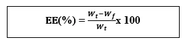
The entrapment efficiency of Ciprofloxacin HCl niosomes was determined using the centrifugation method. The concentration of free Ciprofloxacin HCl was determined using UV-visible spectrophotometry and the established calibration curve. The niosomal suspension was taken in centrifuge tube, set the speed and start the centrifugation, Separate the supernatant and observe in uv calculate the amount of free drug , Subtract the amount of free drug from the total amount of drug to get amount entrapped drug [30].
Where:
Wt = total amount of drug initially added.
Wf= amount of unentrapped drug present in the supernatant.
iv. The in-vitro drug-release study of the Ciprofloxacin HCl niosomal suspension was carried out using a dialysis pouch method:
Clean and boil the gelatin sheet in water at 45℃ for half and hour. Niosomal suspension, equivalent to a known amount of drug, was taken into the pouch and sealed.
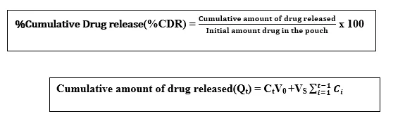
Now immerse the pouch the beaker containing magnetic bead & 100ml of PBS 7.4 maintained at 37±0.5℃, place the beaker on the magnetic stirrer at 100 rpm. At specific time intervals Samples were taken and same volume of fresh, pre-warmed PBS 7.4 was added after each withdrawal. Samples were analyzed by UV-visible spectrophotometer at 272nm. The %CDR was calculated and plotted against time [31].
Where,
Qt= Cumulative amount of drug released upto time point t
Ct= Current sample concentration
V0= Total volume release medium
VS= Volume of sample withdrawn at each interval
Ci= released Concentration of previous sample
v. Determination of Particle Size, Polydispersity Index (PDI), Zeta Potential Of Optimized niosomal suspension
DLS is used to determine the particle size and PDI and the results were expressed as the Z-average particle size and the PDI was obtained from the particle-size distribution analysis. PDI tells about the uniformity. Lower PDI values generally indicate a narrower particle-size distribution. Zeta potential of optimized formulation was measured by Zeta potential analyzer. A suitably diluted sample was transferred into the appropriate measurement cell and analyzed according to the instrument procedure. The surface charge and stability are know by zeta potential[30].
FTIR Spectroscopic Characterization Of The Optimized Niosomal Suspension
FTIR analysis was done to identify the characteristic functional groups of drug (ciprofloxacin HCl) and to understand about drug–excipient compatibility. The range of the spectra observed was 400-4000 cm-1. FTIR spectrophotometer is used to analyze the optimized formulation. The FTIR of pure drug displays absorption peaks corresponding to functional groups this spectrum was used to be compared with the optimized formulation spectrum [32].
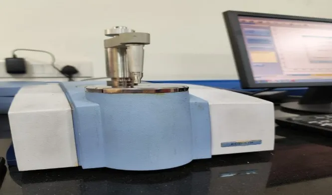
Figure 10: FTIR Spectrophotometry
Preparation Ciprofloxacin HCl Niosomal Gel:
Step 1: In 4.0 g of purified water while slowly sprinkle 0.10 g of Carbopol 934. Allow it to hydrate completely.
Step 2: To the dispersion now add 0.5g of PEG and mix.
Step 3: Now slowly add 5.0g of optimized Ciprofloxacin HCl niosomal suspension to the dispersion and Continue the mixing till the niosomal suspension is completely incorporated into gel base.
Step 4: Add triethanolamine q.s. dropwise with gentle stirring to obtain the desired gel consistency and adjust the pH to the target value established for the formulation.
Step 5: Add sufficient purified water to make the final batch weight 10 g.
Step 6: Mix gently until a smooth and homogeneous Ciprofloxacin HCl niosomal gel is obtained. Don’t disturb the formulation until the air bubbles are removed. Now store the 10g niosomal gel for further evaluation.
Table 6: Formulation of 10G Ciprofloxacin HCl Niosomal Gel
| S.no | Ingredients | Quantity For 10g |
|---|---|---|
| 1 | Optimized Ciprofloxacin HCl Niosomal Suspension (F6) | 5ml |
| 2 | Carbopol 934 | 0.1g |
| 3 | Propylene glycol | 0.5g |
| 4 | Triethanolamine | q.s. |
| 5 | Purified water | q.s. to 10g |
Evaluation Of Optimized Ciprofloxacin HCl Niosomal Gel
i. Visual Appearance and Homogeneity
The prepared niosomal gel was visually examined for colour, appearance, consistency and uniformity. The formulation was inspected for visible lumps, aggregates, air bubbles and phase separation to assess its physical homogeneity [27].
ii. Determination of pH
The pH meter was 1st calibrated using standard buffer solutions. Now take 1g of gel and mix in distilled water. The readings of the pH meter were noted after the reading was stabilized by immersing the electrode in the sample [33].
iii. Viscosity
Brookfield viscometer to determine the viscosity of developed formulation to know its resistance to flow. Immerse the suitable spindle in a beaker containg 5g of gel. Units are centipoise (cP) or mPa·s.
Viscosity is an important parameter for ocular gel formulations because it influences consistency, spreadability, drug release and potential residence time on the ocular surface [27].
iv. Spreadability
gel was placed between two glass slides. A 100 g weight was applied for 60 seconds, and measure the distance and also Measure the widest distance from one edge of the gel to the opposite edge. Calculate the spreadability [33].
v. In-vitro Diffusion Studies Using a Franz Diffusion Cell
Franz diffusion cell is used to determine the invitro drug release of developed formulation. PBS7.4 is taken in receptor compartment and membrane was placed between two compartments.
In donor compartment known amount of gel was taken. To maintain the sink conditions the same volume of fresh buffer is to be added after withdrawal of samples at predetermined time intervals.
Dilute the withdrawn samples with the PBS 7.4 and analyze using UV-visible spectrophotometer at 272nm. %CDR was calculated [27].
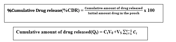
%Cumulative Drug release(%CDR) = x 100 Cumulative amount of drug released(Qt) = CtV0 +VS
Where,
Qt= Cumulative amount of drug released upto time point t
Ct= Current sample concentration
V0= Total volume release medium
VS= Volume of sample withdrawn at each interval
Ci= released Concentration of previous sample
RESULTS AND DISCUSSION
Pre-Formulation Studies Of Ciprofloxacin HCl:
Drug characters are recorded and results compiles with pharmacopoeia values.
Table 7: Pre-Formulation Studies
| S.no | Parameter | Report |
|---|---|---|
| 1 | Organoleptic properties | Pale yellow to light yellow crystalline odourless powder |
| 2 | Solubility | Highly soluble in water, PBS 7.4, Saline water, Slightly soluble in ethanol, insoluble in chloroform |
Standard Crve Of Ciprofloxacin HCl In Phosphate Buffer Saline 7.4
Stock solution 1 was prepared by adding 100mg of Ciprofloxacin HCl in small amount of PBS 7.4 and volume was adjusted to 100ml in volumetric flask to develop 1000 µg/ml from this 10ml was pipetted out and taken 100ml volumetric flask and make the volume with PBS-7.4, now 2-14 µg/ml series of standard solutions are prepared. Absorbance was measured for each standard solutions at 272nm using PBS 7.4 as blank. To determine R2 value, plot a graph by taking concentration (µg/mL) on X-axis and absorbance on Y-axis.
Table 8: Calibration Curve of Ciprofloxacin HCl
| Concentration(µg/ml) | Absorbance |
|---|---|
| 2 | 0.118 |
| 4 | 0.227 |
| 6 | 0.345 |
| 8 | 0.442 |
| 10 | 0.553 |
| 12 | 0.643 |
| 14 | 0.788 |
[Figure - see the formatted PDF]
Figure 11: Standard Calibration Curve of Ciprofloxacin HCl
Evaluation Of Ciprofloxacin HCl Niosomal Suspension
Visual appearance and Clarity
Table 9: Physical Appearance of Ciprofloxacin HCl Niosomal Suspension (N=3, Mean ±SD)
| Formulation code | Visual appearance | Clarity |
|---|---|---|
| F1 | Milky white | Clear |
| F2 | Milky white | Clear |
| F3 | Milky white | Clear |
| F4 | Milky white | Clear |
| F5 | Milky white | Clear |
| F6 | Milky white | Clear |
| F7 | Milky white | Clear |
| F8 | Milky white | Clear |
| F9 | Milky white | Clear |
All the values represent mean± standard deviation (SD) n=3
Drug Content
Tells about how much amount of drug available in the Formulation. Among 9 formulations F6 shows highest percentage of drug content value that is 73.36±0.05 and lowest percentage of drug content value is shown by F1 that is 48.93±0.05.
Table 10: Drug Content Data (N=3, Mean ±SD)
| Formulation code | Absorbance | %Drug Content |
|---|---|---|
| F1 | 0.060 | 48.93±0.02 |
| F2 | 0.068 | 56.34±0.06 |
| F3 | 0.064 | 52.61±0.03 |
| F4 | 0.070 | 58.34±0.12 |
| F5 | 0.073 | 61.63±0.01 |
| F6 | 0.086 | 73.36±0.05 |
| F7 | 0.076 | 64.25±0.33 |
| F8 | 0.080 | 67.83±0.04 |
| F9 | 0.083 | 70.59±0.09 |
All the values represent mean± standard deviation (SD) n=3
Entrapment Efficiency (EE):
Tells about how much amount of drug was entrapped by the niosomal vesicles. Among 9 formulations F6 shows highest %EE that is 62.57±0.02 and lowest %EE is shown by F1 that is 36.2±0.05.
Table 11: Entrapment Efficiency Data (N=3, Mean ±SD)
| Formulation code | %EE |
|---|---|
| F1 | 36.20±0.05 |
| F2 | 41.14±0.03 |
| F3 | 38.30±0.01 |
| F4 | 45.60±0.06 |
| F5 | 51.30±0.02 |
| F6 | 62.57±0.02 |
| F7 | 55.36±0.05 |
| F8 | 57.48±0.03 |
| F9 | 58.32±0.01 |
All the values represent mean± standard deviation (SD) n=3
[Figure - see the formatted PDF]
Figure 12: %Entrapment Efficiency
The in-vitro drug-release study of the Ciprofloxacin HCl niosomal suspension was carried out using a dialysis pouch method:
F1 and F3 niosomal formulation showed lesser release of Ciprofloxacin HCl among 9 formulations. F6 niosomal formulation shows sustained & accelerated release approximately 96.55% ±0.03 within 24 hours.
Table 12: %Cumulative Drug Release Data (N=3, Mean ±SD)
| Time | F1 | F2 | F3 | F4 | F5 | F6 | F7 | F8 | F9 |
|---|---|---|---|---|---|---|---|---|---|
| 30 mins | 6.69± 0.01 |
7.33± 0.02 |
6.97± 0.05 |
7.88± 0.01 |
8.42± 0.02 |
9.52± 0.03 |
9.06± 0.04 |
9.34± 0.01 |
8.52± 0.02 |
| 1 hr | 16.50±0.03 | 17.63±0.01 | 16.79±0.08 | 17.93±0.03 | 18.41±0.01 | 19.65±0.02 | 19.35±0.03 | 19.46±0.02 | 18.87±0.01 |
| 2 hr | 20.41±0.02 | 20.77±0.03 | 20.52±0.03 | 21.08±0.02 | 21.58±0.03 | 23.52±0.01 | 22.85±0.02 | 23.05±0.03 | 22.07±0.03 |
| 3 hr | 26.01±0.03 | 27.02±0.01 | 26.41±0.01 | 27.44±0.04 | 28.24±0.05 | 31.00±0.04 | 30.010.66± | 30.41±0.05 | 30.12±0.04 |
| 4 hr | 32.40±0.06 | 33.55±0.02 | 33.01±0.04 | 33.80±0.01 | 34.18±0.06 | 37.34±0.02 | 36.99±0.03 | 36.73±0.08 | 35.87±0.06 |
| 5 hr | 40.50±0.08 | 41.61±0.04 | 40.85±0.02 | 41.87±0.01 | 42.54±0.01 | 45.38±0.03 | 44.92±0.05 | 44.11±0.03 | 43.22±0.04 |
| 6 hr | 46.94±0.02 | 48.27±0.05 | 47.57±0.05 | 49.45±0.02 | 50.33±0.03 | 53.47±0.01 | 52.63±0.02 | 52.32±0.04 | 51.03±0.03 |
| 7 hr | 56.15±0.01 | 58.09±0.06 | 57.27±0.06 | 58.69±0.02 | 59.60±0.02 | 62.78±0.01 | 61.99±0.03 | 61.21±0.01 | 60.23±0.04 |
| 8 hr | 65.91±0.03 | 67.38±0.02 | 66.44±0.03 | 68.27±0.03 | 69.04±0.03 | 72.16±0.02 | 71.80±0.01 | 70.16±0.04 | 69.69±0.05 |
| 12 hr | 76.76±0.06 | 78.74±0.01 | 77.95±0.08 | 80.22±0.01 | 80.84±0.02 | 84.80±0.06 | 83.88±0.02 | 83.07±0.03 | 82.59±0.01 |
| 24 hr | 82.01±0.05 | 86.83±0.03 | 85.16±0.01 | 87.34±0.03 | 89.61±0.01 | 96.55±0.03 | 94.50±0.01 | 92.39±0.02 | 91.53±0.06 |
All the values represent mean± standard deviation (SD) n=3
[Figure - see the formatted PDF]
Figure 13: %CDR
Determination of Particle Size, Polydispersity Index (PDI), Zeta Potential Of Optimized niosomal suspension
For optimized niosomal suspension
Z-average =159.4nm
PDI value = 0.293
Zeta potential = -24.3mV
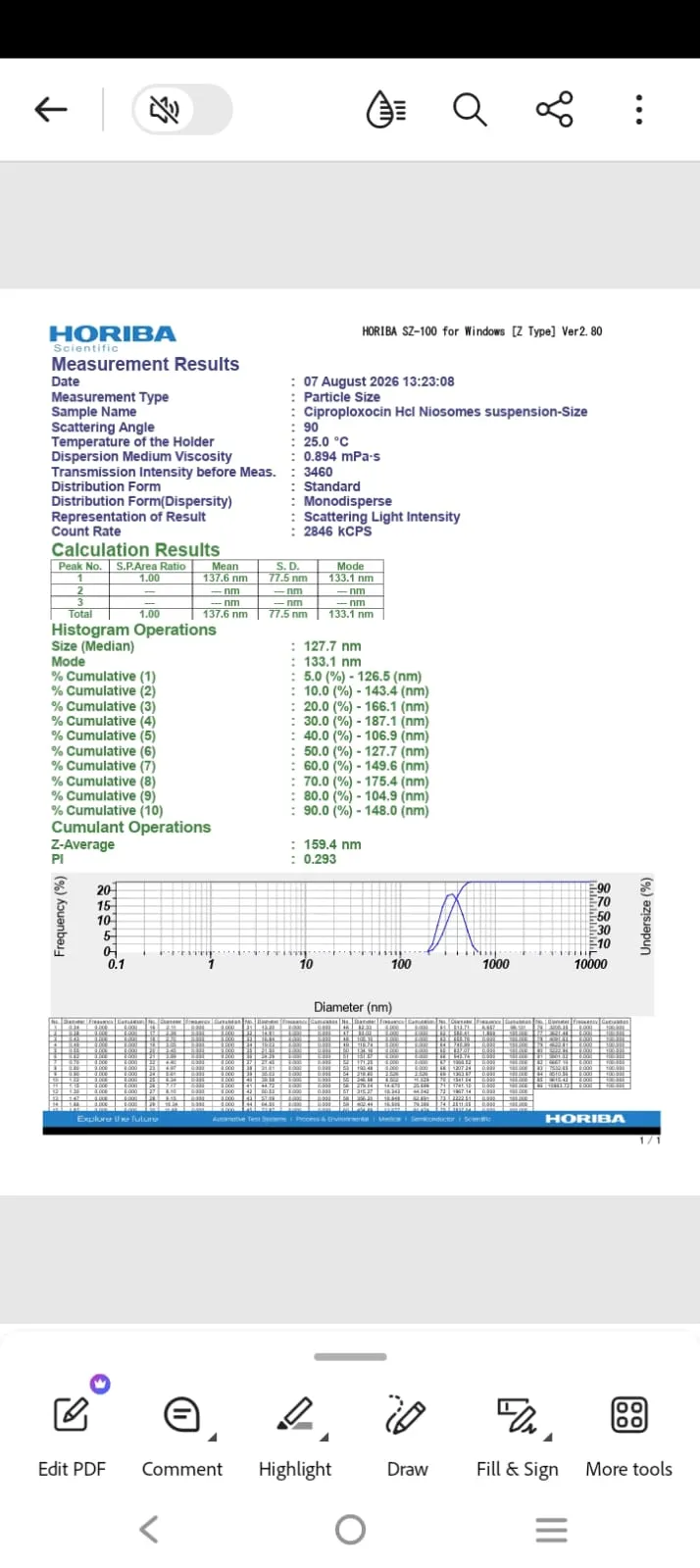
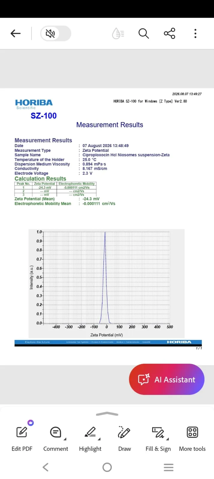
Figure 14: Particle Size & PDI Figure 15: Zeta Potential
FTIR Spectroscopic Characterization Of The Optimized Niosomal Suspension
The pure Ciprofloxacin HCl spectrum showed characteristic peaks corresponding to its major functional groups, such as O–H/N–H, C–H, C=O, C–N, and C–F . The spectra of the physical mixture and optimized formulation also showed several bands in similar regions, although some differences in peak positions and intensities were observed.
[Figure - see the formatted PDF]
Figure 16: FTIR of Pure Drug Ciprofloxacin HCl Figure
[Figure - see the formatted PDF]
17: FTIR Of Physical Mixtu
The presence of characteristic drug-related peaks in the optimized formulation indicates that the major functional groups of Ciprofloxacin HCl were retained during formulation.
[Figure - see the formatted PDF]
Figure 18: FTIR of Optimized Formulation (F6)
Table 13: FTIR Spectral Analysis of Ciprofloxacin HCl, Physical Mixture & Optimized Formulation
| S.no | Functional groups | Range of ṽ(cm-1) | ṽ of pure drug (cm-1) | ṽ of Physical mixture(cm-1) | ṽ of optimized formulation (cm-1) |
|---|---|---|---|---|---|
| 1 | C-N Stretching | 1000-1250 | 1024 | 1103 | 1054 |
| 2 | C=O Stretching | 1650-1750 | 1702 | 1735 | 1690 |
| 3 | C-H Stretching (aliphatic) | 2850-3000 | 2920 | 2916 | 2920 |
| 4 | C-F Stretching | 1000-1400 | 1308 | 1348 | 1378 |
| 5 | O-H Stretching | 3200-3600 | 3526 | 3525 | 3362 |
| 6 | N-H Stretching | 3100-3500 | 3376 | 3350 | 3362 |
| 7 | C=C Stretching | 1000-1400 | 1267 | 1250 | 1080 |
| 8 | Carboxylate vibration (COO-) | 1350-1650 | 1619 | 1622 | 1637 |
Evaluation Of Optimized Ciprofloxacin HCl Niosomal Gel
i. Visual appearance and homogeneity:
The gel was smooth with uniform appearance and it is homogenous because no lumps/ aggregates were observed.
ii. pH, Viscosity, Spreadability of the niosomal gel
pH =7.3±0.5
Viscosity =155±10 cp.
Spreadability = 6.67 g. cm/s.
iii. In-vitro diffusion studies for niosomal gel by Franz diffusion cell
The niosomal gel shows more sustained release of Ciprofloxacin HCl then that of the optimized F6 niosomal suspension that is only 80.65%±0.03 was released for 24 hours where as for F6 96.55%±0.03 was released for 24 hours.
Table 14: %Cumulative Drug Release Data (N=3, Mean ±SD)
| Time | %CDR of F6 | %CDR of Gel |
|---|---|---|
| 30 mins | 9.52±0.03 | 6.36 |
| 1 hr | 19.65±0.02 | 10.22 |
| 2 hr | 23.52±0.01 | 14.58 |
| 3 hr | 31.00±0.04 | 20.42 |
| 4 hr | 37.34±0.02 | 26.94 |
| 5 hr | 45.38±0.03 | 32.54 |
| 6 hr | 53.47±0.01 | 42.02 |
| 7 hr | 62.78±0.01 | 48.71 |
| 8 hr | 72.16±0.02 | 55.83 |
| 12 hr | 84.80±0.06 | 70.01 |
| 24 hr | 96.55±0.03 | 80.65 |
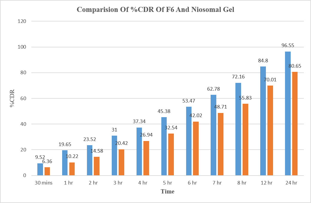
Figure 19: Comparision of %CDR of F6 and Niosomal Gel
CONCLUSION
The present investigation successfully formulated ciprofloxacin hydrochloride-loaded niosomes by thin-film hydration technique, to detect the optimized formulation, 9 formulations were evaluated. Out of them best overall results were observed in F6, that is drug content=73.36 ± 0.05%, %EE 62.57 ± 0.02%, and 96.55 ± 0.03% %CDR at 24 hours. The characteristics of the optimized formulation was obtained by the particle size, PDI, zeta potential and FTIR.
Gel was developed by incorporating the optimized F6 niosomal into a Carbopol-934 and evaluated for its physical properties and in-vitro drug release. The gel showed a smooth, homogeneous appearance and released 80.65% of the drug at 24 hours, compared with 96.55% from the niosomal suspension. These results suggest that incorporating the niosomes into a gel modified the drug-release profile.
However, other studies, like ocular tolerability, ex-vivo permeation, and stability testing, are required to perform before its suitability for ocular application can be established.
REFERENCES


Lattupally Prashanthi Reddy, P. M. Sameera, Comparative Formulation, Optimization & Evaluation of Non-Ionic Surfactants Based Ciprofloxacin HCl Niosomal Gel for Enhanced Ocular Drug Delivery, Int. J. of Pharm. Sci., 2026, Vol 4, Issue 11, 1850-1868. https://doi.org/10.5281/zenodo.24153215
 10.5281/zenodo.24153215